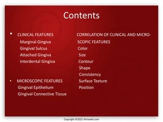
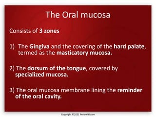
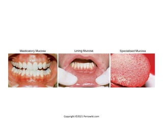
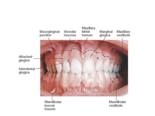
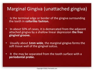
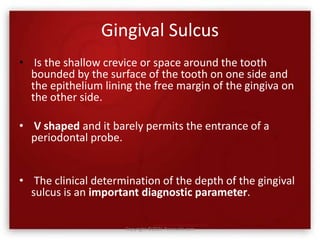
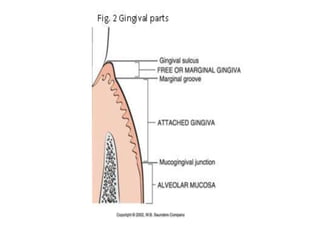
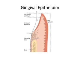
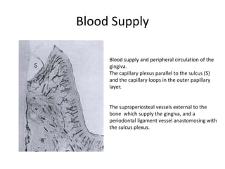
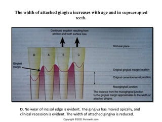
More Related Content Similar to The gingiva (20) More from Periowiki.com (19) The gingiva2. Contents
• CLINICAL FEATURES CORRELATION OF CLINICAL AND MICRO-
Marginal Gingiva SCOPIC FEATURES
Gingival Sulcus Color
Attached Gingiva Size
Interdental Gingiva Contour
Shape
Consistency
• MICROSCOPIC FEATURES Surface Texture
Gingival Epithelium Position
Gingival Connective Tissue
Copyright ©2021 Periowiki.com
3. The Oral mucosa
Consists of 3 zones
1) The Gingiva and the covering of the hard palate,
termed as the masticatory mucosa.
2) The dorsum of the tongue, covered by
specialized mucosa.
3) The oral mucosa membrane lining the reminder
of the oral cavity.
Copyright ©2021 Periowiki.com
5. Gingiva
Is the part of the oral mucosa that covers the
alveolar processes of the jaws and surrounds
the necks of the teeth.
In an adult, normal gingiva covers the alveolar
bone and the tooth root to a level just coronal
to the cementoenamel junction.
Copyright ©2021 Periowiki.com
6. Gingiva
• The gingiva is divided anatomically into:-
marginal, attached and interdental areas.
• Although each type gingiva exhibits considerable
variation in differentiation, histology and
thickness according to its functional demands,
all types are specially structured to
function appropriately against mechanical and
microbial damage.
Copyright ©2021 Periowiki.com
7. Gingiva
• That is, the specific structure of different
gingiva reflects its effectiveness as a barrier to
the penetration by microbes and noxious
agents into the deeper tissue.
Copyright ©2021 Periowiki.com
9. Marginal Gingiva (unattached gingiva)
• Is the terminal edge or border of the gingiva surrounding
the teeth in collarlike fashion.
• In about 50% of cases, it is demarcated from the adjacent
attached gingiva by a shallow linear depression the free
gingival groove.
• Usually about 1mm wide, the marginal gingiva forms the
soft tissue wall of the gingival sulcus.
• It the may be separated from the tooth surface with a
periodontal probe.
Copyright ©2021 Periowiki.com
10. Gingival Sulcus
• Is the shallow crevice or space around the tooth
bounded by the surface of the tooth on one side and
the epithelium lining the free margin of the gingiva on
the other side.
• V shaped and it barely permits the entrance of a
periodontal probe.
• The clinical determination of the depth of the gingival
sulcus is an important diagnostic parameter.
Copyright ©2021 Periowiki.com
12. Gingival Sulcus
• Under absolutely normal or ideal conditions,
the depth of the gingival sulcus is 0mm or
close to 0mm.
• These strict conditions of normalcy can be
produced experimentally only in germ-free
animals or after intense, prolonged plaque
control.
Copyright ©2021 Periowiki.com
13. Gingival Sulcus
• The depth of this sulcus in histologic sections
has been reported as 1.8mm,1.5mm and
0.69mm (0 to 6mm).
• The clinical evaluation used to determine the
depth of the sulcus involves the introduction
of a metallic instrument, periodontal probe
and the estimation of the distance it
penetrates.
Copyright ©2021 Periowiki.com
14. Gingival Sulcus
• The histologic depth of a sulcus does not
need to be exactly equal to the depth of
penetration of the probe.
• The so-called probing depth of a clinically
normal gingival sulcus in humans is 2 to 3mm.
Copyright ©2021 Periowiki.com
15. Attached Gingiva
• Is continuous with the marginal gingiva.
• Is firm, resilient and tightly bound to the
underlying periosteum of alveolar bone.
• The facial aspect of the attached gingiva
extends to the relatively loose and movable
alveolar mucosa and is demarcated by the
mucogingival junction.
Copyright ©2021 Periowiki.com
16. Attached Gingiva
• The width of the attached gingiva is the
distance between the mucogingival junction
and projection on the external surface of the
bottom of the gingival sulcus or the
periodontal pocket.
• It should not be confused with the width of
the keratinized gingiva because the latter
includes the marginal gingiva.
Copyright ©2021 Periowiki.com
17. Attached Gingiva
• The width of the attached gingiva on the facial
aspect differs in different areas of the mouth.
• Maxilla:-
incisor region – 3.5 to 4.5 mm
1st premolars – 1.9mm
• Mandible:-
incisor region – 3.3 to 3.9mm
1st premolars – 1.8mm
Copyright ©2021 Periowiki.com
18. Attached Gingiva
• Because the MGJ remains stationary
throughout the life, the changes in the width
of the AG are caused by the modifications in
the position of its coronal portion.
• The width of the AG increases with age and in
supraerupted teeth.
Copyright ©2021 Periowiki.com
19. Attached Gingiva
• On the lingual aspect of the mandible, the AG
terminates at the junction of the lingual
alveolar mucosa, which is continuous with the
mucous membrane lining the floor of the
mouth.
• The palatal surface of the attached gingiva in
the maxilla blends imperceptibly with the
equally firm and resilient palatal mucosa.
Copyright ©2021 Periowiki.com
20. Interdental Gingiva
• Occupies the gingival embrasure, which is the
interproximal space beneath the area of tooth
contact.
• pyramidal shape - located immediately beneath
the contact point; or
• "col" shape - valleylike depression that connects a
facial and lingual papilla and conforms to the
shape of the interproximal contact
Copyright ©2021 Periowiki.com
21. Interdental Gingiva
• Shape of the gingiva in a given interdental
space depends on the contact point between
the two adjoining teeth and the presence or
absence of some degree of recession.
Copyright ©2021 Periowiki.com
22. Interdental Gingiva
• The facial and lingual surfaces are tapered
toward the interproximal contact area, and the
mesial and distal surfaces are slightly concave.
• The lateral borders and tips of the interdental
papillae are formed by a continuation of the
marginal gingiva from the adjacent teeth. The
intervening portion consists of attached gingiva.
Copyright ©2021 Periowiki.com
24. MICROSCOPIC FEATURES
• The gingiva consists of :-
- a central core of connective tissue covered by
- stratified squamous epithelium.
Copyright ©2021 Periowiki.com
25. Gingival Epithelium
• Although it constitutes a continuous lining of
stratified squamous epithelium, three different
areas can be defined from the morphologic and
functional points of view:
the oral or outer epithelium,
sulcular epithelium,
and junctional epithelium.
Copyright ©2021 Periowiki.com
27. Gingival Epithelium
• Principal cell type of the gingival epithelium,
as well as of other stratified squamous
epithelia, is the keratinocyte.
• Other cells found in the epithelium are the
clear cells or nonkeratinocytes, which include
the Langerhans cells, Merkel cells, and
melanocytes.
Copyright ©2021 Periowiki.com
28. Gingival Epithelium
• Main function of the gingival epithelium is to
protect the deep structures while allowing a
selective interchange with the oral
environment.
• This is achieved by proliferation and
differentiation of the keratinocyte.
Copyright ©2021 Periowiki.com
29. Gingival Epithelium
• Proliferation of keratinocytes –
• takes place by mitosis in the basal layer and
less frequently in the suprabasal layers,
• where a small proportion of cells remain as a
proliferative compartment while a larger
number begins to migrate to the surface.
Copyright ©2021 Periowiki.com
30. Gingival Epithelium
• Differentiation involves:-
the process of keratinization, which consists of
a sequence of biochemical and morphologic
events that occur in the cell as it migrates
from the basal layer.
Copyright ©2021 Periowiki.com
31. Gingival Epithelium
• The main morphologic change is a progressive
flattening of the cell with an increasing
prevalence of tonofilaments and intercellular
junctions coupled to the production of
keratohyaline granules and the
disappearance of the nucleus.
Copyright ©2021 Periowiki.com
32. Gingival Epithelium
Orthokeratinized epithelium :-
Complete keratinization process.
no nuclei in the stratum corneum
and a well-defined stratum granulosum.
• Only some areas of the outer gingival epithelium are
orthokeratinized;
• the other gingival areas are covered by parakeratinized
• or nonkeratinized epithelium considered to beat intermediate
stages of keratinization. These areas can progress to maturity or
dedifferentiate under different physiologic or pathologic conditions.
Copyright ©2021 Periowiki.com
33. Gingival Epithelium
Parakeratinized epithelia,
• the stratum corneum retains pyknotic nuclei and
• the keratohyalin granules are dispersed, not giving rise to a
stratum granulosum.
Nonkeratinized epithelium (although cytokeratins are the major
component, as in all epithelia)
• has neither granulosum
• nor corneum strata, and
• superficial cells have viable nuclei.
Copyright ©2021 Periowiki.com
34. Gingival Epithelium
• Keratin proteins:-
- Immunohistochemistry, gel electrophoresis, and
immunoblot techniques have made identification of the
characteristic pattern of cytokeratins possible in each
epithelial type.
- The keratin proteins are composed of different polypeptide
subunits characterized by their isoelectric points and
molecular weights.
Copyright ©2021 Periowiki.com
35. Gingival Epithelium
• Keratin proteins:-
- They are numbered in a sequence contrary to their
molecular weight.
- Generally, basal cells begin synthesizing lower molecular
weight keratins (such as K19, 40 kd) and express other,
higher molecular weight keratins as they migrate to the
surface.
- K1 keratin polypeptide of 68 kd is the main component of
the stratum corneum.
Copyright ©2021 Periowiki.com
36. Gingival Epithelium
• keratolinin and involucrin:-
- Unrelated to keratins and are synthesized during the
maturation process.
- They are precursors of a chemically resistant structure (the
envelope) located below the cell membrane.
Copyright ©2021 Periowiki.com
37. Gingival Epithelium
• Filaggrin:-
- Precursors of which are packed into the keratohyalin
granules.
- In the sudden transition to the horny layer, the
keratohyalin granules disappear and give rise to
filaggrin, which forms the matrix of the most
differentiated epithelial cell, the corneocyte.
Copyright ©2021 Periowiki.com
38. Gingival Epithelium
• Thus in the fully differentiated state, the
Corneocytes are mainly formed by bundles of
keratin tonofilaments embedded in an
amorphous matrix of filaggrin and surrounded
by a resistant envelope under the cell
membrane.
Copyright ©2021 Periowiki.com
39. Gingival Epithelium
The immunohistochemical patterns of the
different keratin types, envelope proteins, and
filaggrin, change under normal or pathologic
stimuli, modifying the keratinization process
Copyright ©2021 Periowiki.com
40. Gingival Epithelium
Desmosomes:-
Electron microscopy reveals that keratinocytes are
interconnected by structures on the cell periphery called
desmosomes.
Structure consists of two dense attachment plaques into
which tonofibrils insert and an intermediate, electron-
dense line in the extracellular compartment.
Tonofilaments, which are the morphologic expression of the
cytoskeleton of keratin proteins, radiate in brushlike fashion
from the attachment plaques into the cytoplasm of the
cells. Copyright ©2021 Periowiki.com
41. Desmosome Structure
- Intermediate filaments:-
Example:- Keratin.
-Extracellular portions of
Desmocollins and
Desmogleins from opposing
cells interact with each other
In the localized area of
Desmosome, forming the
“Cadherin Zipper”
42. Gingival Epithelium
The space between the cells shows cytoplasmic projections
resembling microvilli that extend into the intercellular
space and often interdigitate.
Less frequently observed forms of epithelial cell connections
are tight junctions (zonae occludens), where the
membranes of the adjoining cells are believed to be fused.
Evidence suggests that these structures allow ions and
small molecules to pass from one cell to another.
Copyright ©2021 Periowiki.com
43. Gingival Epithelium
Cytoplasmic organelle:-
• Concentration varies among different epithelial strata.
• Mitochondria are more numerous in deeper strata and decrease toward
the surface of the cell.
• Accordingly, histochemical demonstration of succinic dehydrogenase,
nicotinamide-adenine dinucleotide, cytochrome oxidase, and other
mitochondrial enzymes revealed a more active tricarboxylic cycle in basal
and parabasal cells, where the proximity of the blood supply facilitates
energy production through aerobic glycolysis.
Copyright ©2021 Periowiki.com
44. Gingival Epithelium
• Conversely, enzymes of the pentose shunt (an alternative
pathway of glycolysis), such as glucose-6-phosphatase,
increase their activity towards the surface.
• This pathway produces a larger amount of intermediate
products for the production of ribonucleic acid (RNA),
which in turn can be used for the synthesis of keratinization
proteins.
• This histochemical pattern is in accordance with the
increased volume and amount of tonofilaments observed in
cells reaching the surface, and the intensity of activity is
proportional to the degree of differentiation.
Copyright ©2021 Periowiki.com
45. Gingival Epithelium
• The uppermost cells of the stratum spinosum contain
numerous dense granules, keratinosomes or Odland
bodies, which are modified lysosomes.
• They contain a large amount of acid phosphatase, an
enzyme involved in the destruction of organelle
membranes, which occurs suddenly between the
granulosum and corneum strata and during the
intercellular cementation of cornified cells.
• Thus acid phosphatase is another enzyme closely
related to the degree of keratinization.
Copyright ©2021 Periowiki.com
46. Gingival Epithelium
Nonkeratinocyte cells are present in gingival epithelium as in other
malpighian epithelia.
Melanocytes are dendritic cells located in the basal and spinous layers
of the gingival epithelium. They synthesize melanin in organelles
called premelanosomes or melanosomes.
These contain tyrosinase, which hydroxylates tyrosine to dihydroxy-
phenylalanine (dopa), which in turn is progressively converted to
melanin.
Melanin granules are phagocytosed and contained within other cells of
the epithelium and connective tissue, called melanophages or
melanophores.
Copyright ©2021 Periowiki.com
47. Gingival Epithelium
• Langerhans cells:-
- Are dendritic cells located among keratinocytes at all
suprabasal levels.
• They belong to the mononuclear phagocyte system
(reticuloendothelial system) as modified monocytes
derived from the bone marrow.
• They contain elongated granules and are considered
macrophages with possible antigenic properties.
Copyright ©2021 Periowiki.com
48. Gingival Epithelium
• Langerhans cells:-
- Have an important role in the immune reaction as antigen-
presenting cells for lymphocytes.
- They contain g-specific granules (Birbeck's granules) and have
marked adenosine triphosphatase activity.
- They are found in oral epithelium of normal gingiva and in
smaller amounts in the sulcular epithelium; they are probably
absent from the junctional epithelium of normal gingiva.
Copyright ©2021 Periowiki.com
49. Gingival Epithelium
• Merkel cells are located in the deeper layers of the
epithelium, harbor nerve endings, and are connected to
adjacent cells by desmosomes. They have been identified
as tactile perceptors.
Copyright ©2021 Periowiki.com
50. Gingival Epithelium
• Basal Lamina:-
- The epithelium is joined to the underlying connective tissue
by a basal lamina 300 to 400 A thick, lying approximately
400 A beneath the epithelial basal layer.
- The basal lamina consists of lamina lucida and lamina
densa.
- Hemidesmosomes of the basal epithelial cells abut the
lamina lucida, which is mainly composed of the
glycoprotein laminin.
Copyright ©2021 Periowiki.com
52. Gingival Epithelium
• The lamina densa is composed of type IV collagen. The
basal lamina, clearly distinguishable at the ultrastructural
level, is connected to a reticular condensation of the under-
lying connective tissue fibrils (mainly collagen type IV) by
the anchoring fibrils.
• The complex of basal lamina and fibrils is the periodic acid-
Schiff (paS) positive and argyrophilic line observed at the
optical level. The basal lamina is permeable to fluids but
acts as a barrier to particulate matter.
Copyright ©2021 Periowiki.com
53. Oral or Outer epithelium
• The oral or outer epithelium covers the crest and outer surface of
the marginal gingiva and the surface of the attached gingiva. It is
keratinized or parakeratinized or presents various combinations of
these conditions.
• The prevalent surface, however, is parakeratinized.
• The degree of gingival keratinization diminishes with age and the
onset of menopause but is not necessarily related to the different
phases of the menstrual cycle.
• Keratinization of the oral mucosa varies in different areas in the
following order: palate (most keratinized), gingiva, ventral aspect of
the tongue, and cheek (least keratinized).
Copyright ©2021 Periowiki.com
54. Oral or Outer epithelium
• Keratins K1, K2, and K10 to K12, which are specific of
epidermal-type differentiation, are immunohistochemically
expressed with high intensity in orthokeratinized areas and
less intensity in parakeratinized areas.
• K6 and K16, characteristic of highly proliferative epithelia,
and K5 and K14, stratification-specific cytokeratins, also are
present.
• Parakeratinized areas express K19, which is usually absent
from orthokeratinized normal epithelia.
Copyright ©2021 Periowiki.com
55. Oral or Outer epithelium
• Glycogen can accumulate intracellularly when it is not
completely degraded by any of the glycolytic pathways.
• Thus its concentration in normal gingiva is inversely related
to the degree of keratinization and inflammation.
Copyright ©2021 Periowiki.com
56. Sulcular Epithelium
• Lines the gingival sulcus.
• It is a thin, nonkeratinized stratified squamous epithelium
without rete pegs.
• Extends from the coronal limit of the junctional epithelium
to the crest of the gingival margin.
• It usually shows many cells with hydropic degeneration.
• Contains K4 and K13, the so-called esophageal type
cytokeratins. It also expresses K19.
• Normally does not contain Merkel cells.
Copyright ©2021 Periowiki.com
57. Sulcular Epithelium
• Lacks granulosum and corneum strata and K1, K2,
and K10-K12 cytokeratins.
• Histochemical studies of enzymes:-
- revealed a lower degree of activity than in the
outer epithelium, particularly in the case of
enzymes related to keratinization.
- Glucose-6-phosphate dehydrogenase expressed a
faint and homogeneous reaction in all strata,
unlike the increasing gradient toward the surface
observed in cornified epithelia.
Copyright ©2021 Periowiki.com
58. Sulcular Epithelium
• Histochemical studies of enzymes:-
- Acid phosphatase staining is negative, although lysosomes
have been described in exfoliated cells .
• Despite these morphologic and chemical characteristics,
the sulcular epithelium has the potential to keratinize if:-
(1) it is reflected and exposed to the oral cavity or
(2) the bacterial flora of the sulcus is totally eliminated.
Copyright ©2021 Periowiki.com
59. Sulcular Epithelium
• Conversely, the outer epithelium loses its keratinization
when it is placed in contact with the tooth .
• These findings suggest that the local irritation of the sulcus
prevents sulcular keratinization.
• The sulcular epithelium is extremely important because it
may act as a semipermeable membrane through which
injurious bacterial products pass into the gingiva and tissue
fluid from the gingiva seeps into the sulcus.
Copyright ©2021 Periowiki.com
60. Junctional Epithelium
• Consists of a collarlike band of stratified squamous
nonkeratinizing epithelium.
• It is three to four layers thick in early life, but the number
of layers increases with age to 10 or even 20 layers.
• These cells can be grouped in two strata: basal and
suprabasal.
• The length of the junctional epithelium ranges from 0.25
to 1.35 mm.
Copyright ©2021 Periowiki.com
61. Junctional Epithelium
• Formed by the confluence of the oral epithelium and the
reduced enamel epithelium during tooth eruption.
• However, the reduced enamel epithelium is not essential
for its formation.
• In fact, junctional epithelium is completely restored after
pocket instrumentation or surgery and forms around an
implant.
Copyright ©2021 Periowiki.com
62. Junctional Epithelium
• Keratin polypeptides of junctional epithelium:-
- Expresses :- K5, 13, 14, 19.
- Another particular behavior is the lack of expression of K6
and K16, which is usually linked to highly proliferative
epithelia, although the turnover of the cells is very high.
• Similar to sulcular epithelium, junctional epithelium
exhibits lower glycolytic enzyme activity than outer
epithelium and lacks acid phosphatase activity.
Copyright ©2021 Periowiki.com
63. Junctional Epithelium
• Is attached to the tooth surface (epithelial attachment) by
means of an internal basal lamina and to the gingival
connective tissue by an external basal lamina that has the
same structure as other epithelial-connective tissue
attachments elsewhere in the body.
• The internal basal lamina consists of a lamina densa
(adjacent to the enamel) and a lamina lucida to which
hemidesmosomes are attached. Organic strands from the
enamel appear to extend into the lamina densa.
Copyright ©2021 Periowiki.com
64. Junctional Epithelium
• Attaches to afibrillar cementum present on the crown (usually
restricted to an area within 1 mm of the cementoenamel
junction) and root cementum in a similar manner.
• Data also have shown that the basal lamina of the junctional
epithelium resembles that of endothelial and epithelial cells
in its laminin content but differs in its internal basal lamina,
which has no type IV collagen.
• These findings indicate that the cells of the junctional
epithelium are involved in the production of laminin and play
a key role in the adhesion mechanism.
Copyright ©2021 Periowiki.com
65. Junctional Epithelium
• The attachment of the junctional epithelium to the tooth is
reinforced by the gingival fibers, which brace the marginal
gingiva against the tooth surface.
• For this reason, the junctional epithelium and the gingival
fibers are considered a functional unit, referred to as the
dentogingival unit.
Copyright ©2021 Periowiki.com
66. Junctional Epithelium
• Development of the Gingival Sulcus:-
- When the tooth penetrates the oral mucosa, the
reduced enamel epithelium unites with the oral
epithelium and transforms into the junctional
epithelium.
- As the tooth erupts, this united epithelium
condenses along the crown, and the ameloblasts,
which form the inner layer of the reduced enamel
epithelium gradually become squamous epithelial
cells.
Copyright ©2021 Periowiki.com
67. Junctional Epithelium
- The transformation of reduced enamel
epithelium into a junctional epithelium
proceeds in an apical direction without
interrupting the attachment to the tooth.
- According to Schroeder and Listgarten, this
process takes between 1 and 2 years.
Copyright ©2021 Periowiki.com
68. Junctional Epithelium
• Is a continually self renewing structure with mitotic activity
occurring in all cell layers.
• The regenerating epithelial cells move toward the tooth
surface and along it in a coronal direction to the gingival
sulcus, where they are shed".
• The migrating daughter cells provide a continuous
attachment to the tooth surface.
Copyright ©2021 Periowiki.com
70. Groeger S, Meyle J. Oral Mucosal Epithelial Cells. Frontiers of Immunology 2019;10:1-22.
71. Renewal of Gingival Epithelium
• The oral epithelium undergoes continuous renewal. Its
thickness is maintained by a balance between new cell
formation in the basal and spinous layers and the shedding
of old cells at the surface.
• The mitotic activity exhibits a 24-hour periodicity, with
highest and lowest rates occurring in the morning and
evening, respectively.
• The mitotic rate is higher in nonkeratinized areas and is
increased in gingivitis, without significant gender
differences.
Copyright ©2021 Periowiki.com
72. Renewal of Gingival Epithelium
• Opinions differ as to whether the mitotic rate is increased
or decreased with age.
• The following have been reported as turnover times for
different areas of the oral epithelium in experimental
animals:
- palate, tongue, and cheek, 5 to 6 days;
- gingiva, 10 to 12 days, with the same or more time
required with age;
- junctional epithelium, 1 to 6 days.
Copyright ©2021 Periowiki.com
73. Cuticular Structures on the Tooth
• The term cuticle describes a thin, acellular structure with a
homogeneous matrix, sometimes enclosed within clearly
demarcated, linear borders.
• Listgarten has classified cuticular structures into coatings
of developmental origin and acquired coatings .
• Acquired coatings include those of exogenous origin, such
as saliva, bacteria, calculus, and surface stains.
Copyright ©2021 Periowiki.com
74. Cuticular Structures on the Tooth
• Coatings of developmental origin are those normally
formed as part of tooth development. They include the
reduced enamel epithelium, coronal cementum and dental
cuticle.
• After enamel formation is completed, the ameloblastic
epithelium is reduced to one or two layers of cells that
remain attached to the enamel surface by hemi-
desmosomes and a basal lamina.
Copyright ©2021 Periowiki.com
75. Cuticular Structures on the Tooth
• This reduced enamel epithelium consists of postsecretory
ameloblasts and cells from the stratum intermedium of the
enamel organ.
• Electron microscopy has shown a dental cuticle consisting
of a layer of homogeneous organic material of variable
thickness (approximately 0.25 micron) overlying the
enamel surface.
Copyright ©2021 Periowiki.com
76. Cuticular Structures on the Tooth
• It is nonmineralized and not always present.
• In some instances near the cementoenamel junction, it is
deposited over a layer of afibrillar cementum, which in turn
overlies enamel.
• The cuticle may be present between the junctional
epithelium and the tooth.
Copyright ©2021 Periowiki.com
77. Cuticular Structures on the Tooth
• Ultrastructural histochemical studies have shown the
dental cuticle to be of proteinaceous nature and it may be
an accumulation of tissue fluid components.
Copyright ©2021 Periowiki.com
78. Gingival Fluid (Sulcular Fluid)
• Contains a fluid that seeps into it from the gingival
connective tissue through the thin sulcular epithelium.
• The gingival fluid is believed to:-
1) Cleanse material from the sulcus,
2) Contain plasma proteins that may improve adhesion of the
epithelium to the tooth,
3) Possess antimicrobial properties, and
4) Exert antibody activity to defend the gingiva.
Copyright ©2021 Periowiki.com
79. Gingival Connective Tissue
• The connective tissue of the gingiva is known as the lamina
propria and consists of two layers:
1) a papillary layer subjacent to the epithelium, which
consists of papillary projections between the epithelial
rete pegs,
2) a reticular layer contiguous with the periosteum of the
alveolar bone. Connective tissue has a cellular and an
extracellular compartment composed of fibers and
ground substance.
Copyright ©2021 Periowiki.com
80. Gingival Connective Tissue
• The ground substance fills the space between fibers and
cells, is amorphous, and has a high content of water.
• It is composed of proteoglycans, mainly hyaluronic acid and
chondroitin sulfate, and glycoproteins, mainly fibronectin.
• Glycoproteins account for the faint pas positive reaction of
the ground substance.
Copyright ©2021 Periowiki.com
81. Gingival Connective Tissue
• Fibronectin binds fibroblasts to the fibers and many other
components of the intercellular matrix, helping mediate
cell adhesion and migration.
• Laminin is another glycoprotein found in the basal laminae,
which serves to attach it to epithelial cells.
• The three types of connective tissue fibers are collagen,
reticular, and elastic. Collagen type I forms the bulk of the
lamina propria and provides the tensile strength to the
gingival tissue.
Copyright ©2021 Periowiki.com
82. Gingival Connective Tissue
• Type IV collagen (argyrophilic reticulum fiber) branches
between the collagen type I bundles and is continuous with
fibers of the basement membrane and blood vessel walls.
• The elastic fiber system is composed of oxytalan, elaunin,
and elastin fibers distributed among collagen fibers .
Copyright ©2021 Periowiki.com
83. Gingival Fibers
• The connective tissue of the marginal gingiva is densely
collagenous, containing a prominent system of collagen
fiber bundles called the gingival fibers.
• They consist of type I collagen.
• The gingival fibers have the following functions:
1. To brace the marginal gingiva firmly against the tooth.
2. To provide the rigidity necessary to withstand the forces of
mastication without being deflected away from the tooth
surface.
Copyright ©2021 Periowiki.com
84. Gingival Fibers
3. To unite the free marginal gingiva with the cementum of
the root and the adjacent attached gingiva.
• The gingival fibers are arranged in three groups:
gingivodental
circular
transseptal.
Copyright ©2021 Periowiki.com
85. Gingival Fibers
• GINGIVODENTAL GROUP –
- The gingivodental fibers are those on the facial, lingual and interproximal
surfaces.
- They are embedded in the cementum just beneath the epithelium at the
base of the gingival sulcus.
- On the facial and lingual surfaces, they project from the cementum in
fanlike conformation toward the crest and outer surface of the marginal
gingiva, terminating short of the epithelium.
- They also extend externally to the periosteum of the facial and lingual
alveolar bones and terminate in the attached gingiva or blend with the
periosteum of the bone. Interproximally, the gingivodental fibers extend
toward the crest of the interdental gingiva.
Copyright ©2021 Periowiki.com
86. Gingival Fibers
• CIRCULAR GROUP –
- The circular fibers course through the connective tissue of
the marginal and interdental gingivae and encircle the
tooth in ringlike fashion.
Copyright ©2021 Periowiki.com
87. Gingival Fibers
• TRANSSEPTAL GROUP –
- Located interproximally, the transseptal fibers form
horizontal bundles that extend between the cementum of
approximating teeth into which they are embedded.
- They lie in the area between the epithelium at the base of
the gingival sulcus and the crest of the interdental bone
and are sometimes classified with the principal fibers of the
periodontal ligament.
Copyright ©2021 Periowiki.com
88. Gingival Fibers
• Page and co-workers also have described :-
(1) a group of semicircular fibers, which attach at the
proximal surface of a tooth, immediately below the
cementoenamel junction, go around the facial or lingual
marginal gingiva of the tooth, and attach on the other
proximal surface of the same tooth.
Copyright ©2021 Periowiki.com
89. Gingival Fibers
(2) A group of transgingival fibers, that attach in the proximal
surface of one tooth, traverse the interdental space
diagonally, go around the facial or lingual surface of the
adjacent tooth,
again traverse diagonally the interdental
space, and attach in the proximal surface of the next tooth.
Copyright ©2021 Periowiki.com
90. Cellular Elements
• The preponderant cellular element in the gingival
connective tissue is the fibroblast.
• Numerous fibroblasts are found between the fiber bundles.
• As in connective tissue elsewhere in the body, fibroblasts
synthesize collagen and elastic fibers as well as the
glycoproteins and glycosaminoglycans of the amorphous
intercellular substance.
• Fibroblasts also regulate collagen degradation.
Copyright ©2021 Periowiki.com
91. Cellular Elements
• Mast cells, which are distributed throughout the body, are
numerous in the connective tissue of the oral mucosa and
the gingiva.
• Fixed macrophages and histiocytes are present in the
gingival connective tissue as components of the
mononuclear phagocyte system (reticuloendothelial
system) and are derived from blood monocytes.
• Adipose cells and eosinophils, although scarce, also are
present in the lamina propria.
Copyright ©2021 Periowiki.com
92. Cellular Elements
• In clinically normal gingiva, small foci of plasma cells and
lymphocytes are found in the connective tissue near the
base of the sulcus.
• Neutrophils can be seen in relatively high numbers in both
the gingival connective tissue and the sulcus.
• These inflammatory cells usually are present in small
amounts in clinically normal gingiva.
Copyright ©2021 Periowiki.com
93. Cellular Elements
• Speculations about whether small amounts of leukocytes
should be considered a normal component of the gingiva or
an incipient inflammatory infiltrate without clinical
expression are of theoretic rather than practical
importance.
• Lymphocytes are absent when gingival normalcy is judged
by very strict clinical criteria or under very special
experimental conditions but are practically constant in
healthy normal gingiva, even before complete tooth
eruption.
Copyright ©2021 Periowiki.com
94. Cellular Elements
• Immunohistochemical studies using monoclonal antibodies
have identified the different lymphocyte subpopulations.
• The infiltrate in the area below the junctional epithelium of
healthy gingiva in recently erupted teeth in children is
mainly composed of T-lymphocytes (helper, cytotoxic,
suppressor and natural killer) and thus could be interpreted
as a normal lymphoid tissue involved in the early defense
recognition system.
Copyright ©2021 Periowiki.com
95. Cellular Elements
• As time elapses, B-lymphocytes and plasma cells appear in
greater proportions to elaborate specific antibodies against
already recognized antigens that are always present in the
sulcus of clinically normal gingiva.
Copyright ©2021 Periowiki.com
96. Blood Supply, Lymphatics, and Nerves
• Blood vessels are easily evidenced in tissue sections by
means of immunohistochemical reactions against proteins
of endothelial cells (factor VIII and adhesion molecules).
• Before these techniques were developed, vascularization
patterns of periodontal tissues had been described using
histoenzymatic reactions for alkaline phosphatase and
adenosine triphosphatase owing to the great activity of
these enzymes in endothelial cells.
Copyright ©2021 Periowiki.com
97. Blood Supply, Lymphatics, and Nerves
• In experimental animals, the perfusion with India ink also
was used to study vascular distribution. The injection and
subsequent demonstration of peroxidase allows blood
vessel identification and permeability studies.
• The paS reaction also outlines vascular walls by a positive
line in their basal membrane. Endothelial cells also express
5-nucleotidase activity. Scanning electron microscopy can
be used after injection of plastic into the vessels through
the carotid artery, followed by corrosion of the soft tissues.
Copyright ©2021 Periowiki.com
98. Blood Supply
1) Supraperiosteal arterioles along the facial and lingual
surfaces of the alveolar bone, from which capillaries
extend along the sulcular epithelium and between the
rete pegs of the external gingival surface.
Occasional branches of the arterioles pass
through the alveolar bone to the periodontal ligament or
run over the crest of the alveolar bone.
Copyright ©2021 Periowiki.com
99. Blood Supply
2) Vessels of the periodontal ligament, which extend into the
gingiva and anastomose with capillaries in the sulcus area.
3) Arterioles, which emerge from the crest of the interdental
septa and extend parallel to the crest of the bone to
anastomose with vessels of the periodontal ligament, with
capillaries in the gingival crevicular areas and vessels that
run over the alveolar crest.
Copyright ©2021 Periowiki.com
100. Blood Supply
Blood supply and peripheral circulation of the
gingiva.
The capillary plexus parallel to the sulcus (S)
and the capillary loops in the outer papillary
layer.
The supraperiosteal vessels external to the
bone which supply the gingiva, and a
periodontal ligament vessel anastomosing with
the sulcus plexus.
101. Blood Supply
• The loops are sometimes linked by cross- communications
and flattened capillaries serve as reserve vessels when the
circulation is increased in response to irritation .
• Along the sulcular epithelium, capillaries are arranged in a
flat anastomosing plexus that extends parallel to the
enamel from the base of the sulcus to the gingival margin.
• In the col area, a mixed pattern of anastomosing capillaries
and loops occurs.
Copyright ©2021 Periowiki.com
102. The lymphatic drainage o f the gingiva
• Brings in the lymphatics of the connective tissue papillae.
• It progresses into the collecting network external to the
periosteum of the alveolar process and then to the regional
lymph nodes (particularly the submaxillary group).
• In addition, lymphatics just beneath the junctional
epithelium extend into the periodontal ligament and
accompany the blood vessels.
Copyright ©2021 Periowiki.com
103. Gingival innervation
• Is derived from fibers arising from nerves in the periodontal
ligament and from the labial, buccal, and palatal nerves.
• The following nerve structures are present in the connective
tissue:
- a meshwork of terminal argyrophilic fibers, some of which
extend into the epithelium;
- Meissner-type tactile corpuscles; Krausetype end bulbs, which
are temperature receptors; and encapsulated spindles.
Copyright ©2021 Periowiki.com
104. III) CORRELATION OF CLINICAL AND
MICROSCOPIC FEATURES
1. Color :-
Attached gingiva and marginal gingiva generally
described as – “coral pink”.
* Coral is reddish or pinkish shade of orange.
Copyright ©2021 Periowiki.com
105. III) CORRELATION OF CLINICAL AND
MICROSCOPIC FEATURES
1. Color :-
Is produced by :-
. Vascular supply . degree of epithelium
keratinization
. presence of pigmenting cells
Copyright ©2021 Periowiki.com
106. III) CORRELATION OF CLINICAL AND
MICROSCOPIC FEATURES
1. Color :-
It varies among different persons and appears to
be correlated with the cutaneous
pigmentation.
Copyright ©2021 Periowiki.com
107. III) CORRELATION OF CLINICAL AND
MICROSCOPIC FEATURES
1. Color :-
It is lighter in blond individuals with fair
complexion then in swarthy (dark
complexioned), dark haired individuals.
Copyright ©2021 Periowiki.com
108. III) CORRELATION OF CLINICAL AND
MICROSCOPIC FEATURES
1. Color :-
The attached gingiva is demarcated from the
adjacent alveolar mucosa on the buccal
aspect by a clearly defined mucogingival line.
Copyright ©2021 Periowiki.com
109. III) CORRELATION OF CLINICAL AND
MICROSCOPIC FEATURES
1. Color :-
The alveolar mucosa is red, smooth and shiny
rather than pink and stippled.
Copyright ©2021 Periowiki.com
110. III) CORRELATION OF CLINICAL AND
MICROSCOPIC FEATURES
1. Color :-
Comparison of the microscopic structure of the
attached gingiva with that of the alveolar
mucosa provides an explanation for the
difference in appearance.
Copyright ©2021 Periowiki.com
111. III) CORRELATION OF CLINICAL AND
MICROSCOPIC FEATURES
1. Color :-
Alveolar mucosa –
Epithelium is thinner, nonkeratinized with no
rete pegs.
Connective tissue is loosely arranged and blood
vessels are more numerous.
Copyright ©2021 Periowiki.com
112. III) CORRELATION OF CLINICAL AND
MICROSCOPIC FEATURES
1. Color :-
Physiologic pigmentation (Melanin)-
• Melanin, a non–hemoglobin-derived brown
pigment, is responsible for the normal
pigmentation of the skin, gingiva, and remainder
of the oral mucous membrane.
• It is present in all normal individuals, often not in
sufficient quantities to be detected clinically, but
is absent or severely diminished in albinos.
Copyright ©2021 Periowiki.com
113. III) CORRELATION OF CLINICAL AND
MICROSCOPIC FEATURES
1. Color :-
Physiologic pigmentation (Melanin)-
• Melanin pigmentation in the oral cavity is
prominent in black individuals.
• Ascorbic acid directly downregulates melanin
pigmentation in gingival tissues.
Copyright ©2021 Periowiki.com
114. III) CORRELATION OF CLINICAL AND
MICROSCOPIC FEATURES
1. Color :-
• According to Dummett (1959) –
The distribution of oral pigmentation
in black individuals is as follows:
gingiva, 60%;
hard palate, 61%;
mucous membrane, 22%; and
tongue, 15%.
Copyright ©2021 Periowiki.com
115. III) CORRELATION OF CLINICAL AND
MICROSCOPIC FEATURES
1. Color :-
• According to Dummett (1959) –
Gingival pigmentation occurs as a
diffuse, deep-purplish discoloration or as
irregularly shaped, brown and light-brown
patches.
It may appear in the gingiva as
early as 3 hours after birth and often is the
only evidence of pigmentation.
Copyright ©2021 Periowiki.com
116. III) CORRELATION OF CLINICAL AND
MICROSCOPIC FEATURES
1. Color :-
• Oral repigmentation refers to :-
the clinical reappearance of melanin
pigment after a period of clinical depigmentation
of the oral mucosa resulting from chemical,
thermal, surgical, pharmacologic, or idiopathic
factors.
Information on the repigmentation of
oral tissues after surgical procedures is extremely
limited, and no definitive treatment is offered at
this time.
Copyright ©2021 Periowiki.com
117. III) CORRELATION OF CLINICAL AND
MICROSCOPIC FEATURES
2. Size :-
The size of the gingiva corresponds with the sum
total of the bulk of cellular and intercellular
elements and their vascular supply.
Alteration in size is a common feature of gingival
disease.
Copyright ©2021 Periowiki.com
118. III) CORRELATION OF CLINICAL AND
MICROSCOPIC FEATURES
3. Contour (or shape of the gingiva):-
Varies considerably and
depends on the:-
.shape of the teeth and their alignment in the arch,
.the location and size of the area of proximal contact,
and
.the dimensions of the facial and lingual gingival
embrasures.
Copyright ©2021 Periowiki.com
119. III) CORRELATION OF CLINICAL AND
MICROSCOPIC FEATURES
4. Shape :-
The shape of the interdental gingiva is governed by
the contour of the proximal tooth surfaces and
the location and shape of gingival embrasures.
*Gingival embrasure is the interproximal space beneath the area of
tooth contact.
Copyright ©2021 Periowiki.com
120. III) CORRELATION OF CLINICAL AND
MICROSCOPIC FEATURES
4. Shape :-
When the proximal surfaces of the crowns are
relatively flat faciolingually, the roots are close
together, the interdental bone is thin
mesiodistally, and the gingival embrasures and
interdental gingiva are narrow mesiodistally.
Copyright ©2021 Periowiki.com
121. III) CORRELATION OF CLINICAL AND
MICROSCOPIC FEATURES
4. Shape :-
Conversely, with proximal surfaces that flare
away from the area of contact, the mesiodistal
diameter of the interdental gingiva is broad.
Copyright ©2021 Periowiki.com
122. III) CORRELATION OF CLINICAL AND
MICROSCOPIC FEATURES
4. Shape :-
• The height of the interdental gingiva varies
with the location of the proximal contact.
• Thus, in the anterior region of the dentition,
the interdental papilla is pyramidal in form,
whereas
• the papilla is more flattened in a buccolingual
direction in the molar region.
Copyright ©2021 Periowiki.com
123. III) CORRELATION OF CLINICAL AND
MICROSCOPIC FEATURES
5. Consistency:-
• The gingiva is firm and resilient and with the
exception of the movable free margin, tightly
bound to the underlying bone.
Copyright ©2021 Periowiki.com
124. III) CORRELATION OF CLINICAL AND
MICROSCOPIC FEATURES
5. Consistency:-
• The collagenous nature of the lamina propria
and its contiguity with the mucoperiosteum of
the alveolar bone determine the firmness of
the attached gingiva.
Copyright ©2021 Periowiki.com
125. III) CORRELATION OF CLINICAL AND
MICROSCOPIC FEATURES
5. Consistency:-
• The gingival fibers contribute to the firmness
of the gingival margin.
Copyright ©2021 Periowiki.com
126. III) CORRELATION OF CLINICAL AND
MICROSCOPIC FEATURES
6. Surface texture:-
• The gingiva presents a textured surface similar
to an orange peel and is referred to as being
stippled.
• Stippling is best viewed by drying the gingiva.
Copyright ©2021 Periowiki.com
127. III) CORRELATION OF CLINICAL AND
MICROSCOPIC FEATURES
6. Surface texture:-
• The attached gingiva is stippled; the marginal
gingiva is not.
• The central portion of the interdental papillae
is usually stippled, but the marginal borders
are smooth.
Copyright ©2021 Periowiki.com
128. III) CORRELATION OF CLINICAL AND
MICROSCOPIC FEATURES
6. Surface texture:-
• The pattern and extent of stippling vary
among individuals and different areas of the
same mouth.
• Stippling is less prominent on lingual than
facial surfaces and may be absent in some
persons.
Copyright ©2021 Periowiki.com
129. III) CORRELATION OF CLINICAL AND
MICROSCOPIC FEATURES
6. Surface texture:-
• Microscopically, stippling is produced by- alternate
rounded protuberances and depressions in the
gingival surface.
• The papillary layer of the connective tissue projects
into the elevations, and the elevated and depressed
areas are covered by stratified squamous epithelium.
• The degree of keratinization and the prominence of
stippling appear to be related.
Copyright ©2021 Periowiki.com
130. III) CORRELATION OF CLINICAL AND
MICROSCOPIC FEATURES
6. Surface texture:-
• Scanning electron microscopy has shown
considerable variation in shape, but a relatively
constant depth of stippling.
• At low magnification, a rippled surface is seen,
interrupted by irregular depressions 50 μm in
diameter.
• At higher magnification, cell micropits are seen.
Copyright ©2021 Periowiki.com
131. III) CORRELATION OF CLINICAL AND
MICROSCOPIC FEATURES
6. Surface texture:-
• Stippling is a form of adaptive specialization or
reinforcement for function.
• It is a feature of healthy gingiva, and reduction or
loss of stippling is a common sign of gingival disease.
• When the gingiva is restored to health after
treatment, the stippled appearance returns.
Copyright ©2021 Periowiki.com
132. III) CORRELATION OF CLINICAL AND
MICROSCOPIC FEATURES
6. Surface texture:-
• The surface texture of the gingiva is also related to
the presence and degree of epithelial keratinization.
• Keratinization is considered a protective adaptation
to function.
• It increases when the gingiva is stimulated by
toothbrushing.
Copyright ©2021 Periowiki.com
133. III) CORRELATION OF CLINICAL AND
MICROSCOPIC FEATURES
6. Surface texture:-
• However, research on free gingival grafts has shown
that when connective tissue is transplanted from a
keratinized area to a nonkeratinized area, it becomes
covered by a keratinized epithelium.
• This finding suggests a connective tissue–based
genetic determination of the type of epithelial
surface.
Copyright ©2021 Periowiki.com
134. III) CORRELATION OF CLINICAL AND
MICROSCOPIC FEATURES
7. Position:-
• The position of the gingiva refers to the level at
which the gingival margin is attached to the tooth.
• When the tooth erupts into the oral cavity, the
margin and sulcus are at the tip of the crown;
• as eruption progresses, they are seen closer to the
root.
Copyright ©2021 Periowiki.com
135. III) CORRELATION OF CLINICAL AND
MICROSCOPIC FEATURES
7. Position:-
• During this eruption process, as described earlier, the
junctional epithelium, oral epithelium, and reduced
enamel epithelium undergo extensive alterations and
remodeling while maintaining the shallow
physiologic depth of the sulcus.
Copyright ©2021 Periowiki.com
136. III) CORRELATION OF CLINICAL AND
MICROSCOPIC FEATURES
7. Position:-
• Without this remodeling of the epithelia, an
abnormal anatomic relationship between the
gingiva and the tooth would result.
Copyright ©2021 Periowiki.com
137. III) CORRELATION OF CLINICAL AND
MICROSCOPIC FEATURES
7. Position:-
Continuous tooth eruption-
• According to the concept of continuous
eruption,
eruption does not cease when teeth
meet their functional antagonists but
continues throughout life.
Copyright ©2021 Periowiki.com
138. III) CORRELATION OF CLINICAL AND
MICROSCOPIC FEATURES
7. Position:-
Continuous tooth eruption-
• Eruption consists of an active and a passive
phase. Active eruption is the movement of the
teeth in the direction of the occlusal plane,
whereas passive eruption is the
exposure of the teeth by apical migration of
the gingiva.
Copyright ©2021 Periowiki.com
139. III) CORRELATION OF CLINICAL AND
MICROSCOPIC FEATURES
7. Position:-
Continuous tooth eruption-
• This concept distinguishes between the anatomic
crown (portion of the tooth covered by enamel)
and the anatomic root (portion of the tooth
covered by cementum) and between
the clinical crown (part of the tooth
that has been denuded of its gingiva and projects
into the oral cavity) and clinical root (portion of
the tooth covered by periodontal tissues).
Copyright ©2021 Periowiki.com
140. III) CORRELATION OF CLINICAL AND
MICROSCOPIC FEATURES
7. Position:-
Continuous tooth eruption-
• When the teeth reach their functional
antagonists, the gingival sulcus and junctional
epithelium are still on the enamel, and the
clinical crown is approximately two-thirds of
the anatomic crown.
Copyright ©2021 Periowiki.com
141. III) CORRELATION OF CLINICAL AND
MICROSCOPIC FEATURES
7. Position:-
Continuous tooth eruption-
• Gottlieb and Orban(1933) believed that :-
active and passive eruption proceed together.
Active eruption is coordinated
with attrition; the teeth erupt to compensate
for tooth substance worn away by attrition.
Copyright ©2021 Periowiki.com
142. III) CORRELATION OF CLINICAL AND
MICROSCOPIC FEATURES
7. Position:-
Continuous tooth eruption-
• Attrition reduces the clinical crown and
prevents it from becoming disproportionately
long in relation to the clinical root, thus
avoiding excessive leverage on the periodontal
tissues.
Copyright ©2021 Periowiki.com
143. III) CORRELATION OF CLINICAL AND
MICROSCOPIC FEATURES
7. Position:-
Continuous tooth eruption-
• the rate of active eruption keeps pace with
tooth wear, preserving the vertical dimension
of the dentition.
Copyright ©2021 Periowiki.com
144. III) CORRELATION OF CLINICAL AND
MICROSCOPIC FEATURES
7. Position:-
Continuous tooth eruption-
• As teeth erupt, cementum is deposited at the apices and
furcations of the roots and bone is formed along the
fundus of the alveolus and at the crest of the alveolar
bone.
In this way, part of the tooth substance lost
by attrition is replaced by lengthening of the root, and
socket depth is maintained to support the root.
Copyright ©2021 Periowiki.com
145. III) CORRELATION OF CLINICAL AND
MICROSCOPIC FEATURES
7. Position:-
Continuous tooth eruption-
Although originally thought to be a normal
physiologic process, passive eruption is now
considered a pathologic process.
Copyright ©2021 Periowiki.com
146. III) CORRELATION OF CLINICAL AND
MICROSCOPIC FEATURES
7. Position:-
Continuous tooth eruption-
• Passive eruption is divided into the following
four stages :-
Stage 1: The teeth reach the line of occlusion.
The junctional epithelium and base of the
gingival sulcus are on the enamel.
Copyright ©2021 Periowiki.com
147. III) CORRELATION OF CLINICAL AND
MICROSCOPIC FEATURES
7. Position:-
Continuous tooth eruption-
• Passive eruption is divided into the following
four stages :-
• Stage 2: The junctional epithelium proliferates
so that part is on the cementum and part is on
the enamel. The base of the sulcus is still on
the enamel.
Copyright ©2021 Periowiki.com
148. III) CORRELATION OF CLINICAL AND
MICROSCOPIC FEATURES
7. Position:-
Continuous tooth eruption-
• Passive eruption is divided into the following four
stages :-
• Stage 3: The entire junctional epithelium is on the
cementum, and the base of the sulcus is at the
cementoenamel junction.
As the junctional epithelium
proliferates from the crown onto the root, it does not
remain at the cementoenamel junction any longer than
at any other area of the tooth.
Copyright ©2021 Periowiki.com
149. III) CORRELATION OF CLINICAL AND
MICROSCOPIC FEATURES
7. Position:-
Continuous tooth eruption-
• Passive eruption is divided into the following four stages :-
• Stage 4: The junctional epithelium has proliferated farther on
the cementum. The base of the sulcus is on the cementum, a
portion of which is exposed.
Proliferation of the junctional epithelium onto the
root is accompanied by degeneration of gingival and
periodontal ligament fibers and their detachment from the
tooth.
The cause of this degeneration is not understood.
At present, it is believed to be the result of chronic
inflammation and therefore a pathologic process.
Copyright ©2021 Periowiki.com
150. III) CORRELATION OF CLINICAL AND
MICROSCOPIC FEATURES
7. Position:-
Continuous tooth eruption-
• As noted, apposition of bone accompanies
active eruption.
• The distance between the apical end of the
junctional epithelium and the crest of the
alveolus remains constant throughout
continuous tooth eruption (1.07 mm).
Copyright ©2021 Periowiki.com
151. III) CORRELATION OF CLINICAL AND
MICROSCOPIC FEATURES
7. Position:-
Continuous tooth eruption-
• Exposure of the tooth by the apical migration of
the gingiva is called gingival recession, or atrophy.
• According to the concept of continuous eruption,
the gingival sulcus may be located on the crown,
cementoenamel junction, or root, depending on
the age of the patient and stage of eruption.
Copyright ©2021 Periowiki.com
152. III) CORRELATION OF CLINICAL AND
MICROSCOPIC FEATURES
7. Position:-
Continuous tooth eruption-
• Therefore some root exposure with age would be
considered normal and referred to as physiologic
recession.
Again, this concept is not accepted at
present.
Excessive exposure is termed pathologic
recession.
Copyright ©2021 Periowiki.com
153. Ainamo & Talari et al 1976, 1978 :
The width of attached gingiva increases with age and in supraerupted
teeth.
A, Normal relationship with the gingival
margin 1 to 2 mm above the cementoenamel
junction.
Copyright ©2021 Periowiki.com
154. The width of attached gingiva increases with age and in supraerupted
teeth.
B, Wear of the incisal edge and continued tooth eruption. The gingival
margin remains in the same position as shown in A. Therefore, the
root surface is exposed, and clinical recession is evident. The width of
the attached gingiva has not changed.
Copyright ©2021 Periowiki.com
155. The width of attached gingiva increases with age and in supraerupted
teeth.
C, Wear of the incisal edge and continued tooth eruption. The gingival margin
has moved with the tooth; therefore, the entire dentogingival complex has
moved coronally, with a resulting increase in the width of the attached gingiva.
Copyright ©2021 Periowiki.com
156. The width of attached gingiva increases with age and in supraerupted
teeth.
D, No wear of incisal edge is evident. The gingiva has moved apically, and
clinical recession is evident. The width of attached gingiva is reduced.
Copyright ©2021 Periowiki.com
157. • The reasons for increased AGW with age are a
concomitant reduction in sulcus depth and
there is a constant eruption of teeth
throughout one's lifespan to compensate the
occlusal surface attrition leading to the
coronal migration of cementoenamel junction,
while the mucogingival junction remains
unchanged.
Ainamo J, Talari A. The increase with age of the width of attached
gingiva. J Periodontal Res 1976;11:182-8.
Copyright ©2021 Periowiki.com
158. References
. Joseph P. Fiorellini, David M. Kim and Satoshi O. Ishikawa , Ainamo J, Talari
A. THE GINGIVA. In: Carranza’s Clinical Periodontology 9th edition.
. Groeger S, Meyle J. Oral Mucosal Epithelial Cells. Frontiers of Immunology
2019;10:1-22.
. The increase with age of the width of attached gingiva. J Periodontal Res
1976;11:182-8.
Periowiki.com holds copyright of this power point presentation only.
Photographs, flowcharts credit – google, textbooks and journal articles
(details mentioned in references section).
Copyright ©2021 Periowiki.com