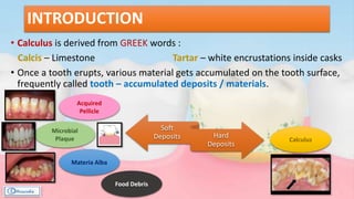
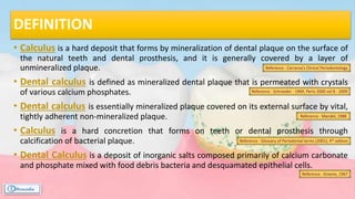
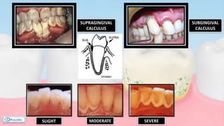
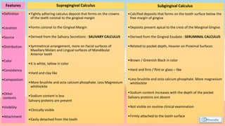
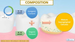
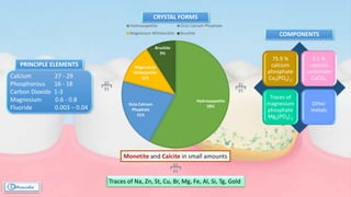
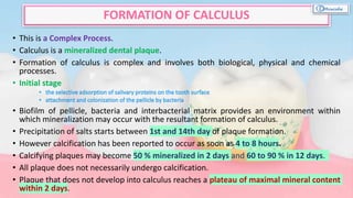
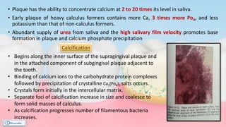
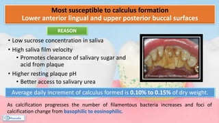
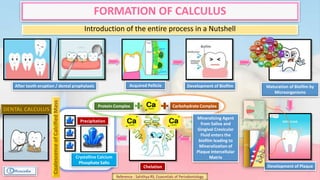
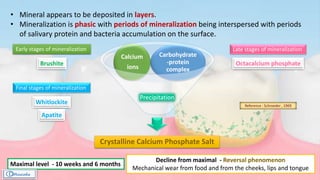
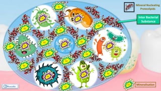
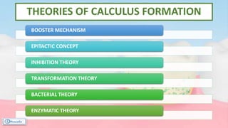
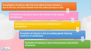
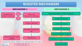
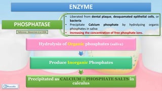
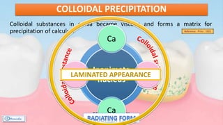
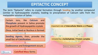
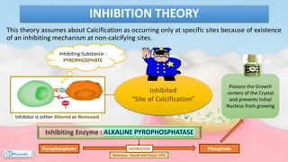
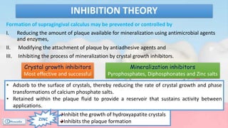
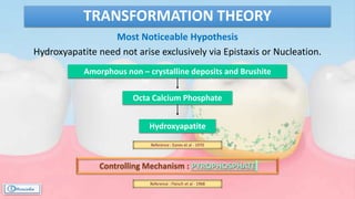
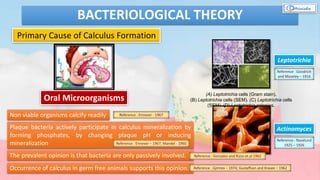
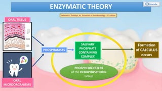
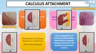
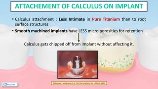
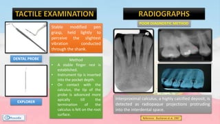
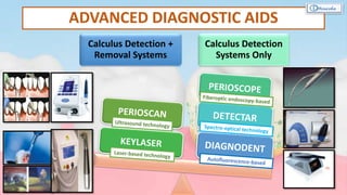
DR. RINISHASINHA
MDS PARTI
DEPARTMENT OF PERIODONTOLOGY
DENTAL
CALCULUS
CONTENTS
• Introduction
• Definition
• Synonyms
• History
• Classification
• Composition
• Structure of Calculus
• Formation of Calculus
• Theories of Calculus
Formation
• Calculus Attachment
• Attachment of Calculus on Implant
• Microbiology of Dental Calculus
• Prevalence
• Assessment and Evaluation of
Calculus
• Advanced Diagnostic Aids
• Etiological Significance of Calculus in
Periodontal Disease
• Conclusion
• Reference
INTRODUCTION
• Calculus is derived from GREEK words :
Calcis – Limestone Tartar – white encrustations inside casks
• Once a tooth erupts, various material gets accumulated on the tooth surface,
frequently called tooth – accumulated deposits / materials.
Soft
Deposits Hard
Deposits
Acquired
Pellicle
Microbial
Plaque
Materia Alba
Food Debris
Calculus
DEFINITION
• Calculus is a hard deposit that forms by mineralization of dental plaque on the surface of
the natural teeth and dental prosthesis, and it is generally covered by a layer of
unmineralized plaque.
• Dental calculus is defined as mineralized dental plaque that is permeated with crystals
of various calcium phosphates.
• Dental calculus is essentially mineralized plaque covered on its external surface by vital,
tightly adherent non-mineralized plaque.
• Calculus is a hard concretion that forms on teeth or dental prosthesis through
calcification of bacterial plaque.
• Dental Calculus is a deposit of inorganic salts composed primarily of calcium carbonate
and phosphate mixed with food debris bacteria and desquamated epithelial cells.
Reference : Carranza’s Clinical Periodontology
Reference : Schroeder - 1969; Perio 2000 vol 8 - 2009
Reference : Glossary of Periodontal terms (2001); 4th edition
Reference : Mandel, 1988
Reference : Greene, 1967
SYNONYMS
• Tartar
• Disambiguation
• Calcis
• Odontolithiasis
• Fossilized plaque
Reference : Mandel et. Al; Calculus revisited : A Review - 2013
• Recognized as far as back in the 10th Century.
• Hippocrates (460 – 337 BC)
Found the association of dental deposits and oral disease.
The deleterious effects of the teeth and gums of pituita (calculus).
• Albucasis (936 – 1013 AD)
Arabian physician and surgeon
Explained relationship between calculus and disease.
First to design a set of 14 scalers for the thorough cleaning of
teeth.
• Paracelsus (1535)
Swiss German physician and alchemist
Introduced term “tartar” as a designation for a variety of stony
concretions that form in humans.
Tartaric disease
• For nearly 5000 years, Calculus was considered to be the Prime Etiologic Agent in
the Periodontal disease.
• Microorganisms in Tartar were initially referred to as Animalcules.
• There was a change in perception, that became most apparent in 1960’s, mainly as
a response to two lines of investigation –
Reference : Van Leeuwenhoek, 1967
Experimental and electron microscopic
studies of developing plaque and
calculus demonstrated that supra and
subgingival calculus was mineralized plaque
covered by an unmineralized bacterial layer.
The experimental demonstration in humans
that plaque, allowed to develop in the
absence of oral hygiene, results in a
gingivitis which is reversible on the
resumption of tooth cleaning.
Reference : Mandei et al – 1957; Muhleman and Schroeder –
1964; Oshrain et al - 1968
Reference : Loe et al – 1965
CLASSIFICATION
According to Source of Mineralization
Salivary calculus
Serumal calculus
Reference : Jenkins, Stewart – 1966Reference : Carranza’s Clinical Periodontology
According to Location on the tooth surface as related
to the adjacent Free Gingival Margin :
Supragingival calculus
Subgingival Calculus
According to initiation and rate of accumulation,
calculus formers are classified as :
Non calculus formers
Slight calculus formers
Moderate calculus formers
Heavy calculus formers
Reference : Mandel JD et al - 1976
According to Surface
Exogenous
Endogenous
Reference : Melz – 1950
SUPRAGINGIVAL
CALCULUS
SLIGHT MODERATE SEVERE
SUBGINGIVAL
CALCULUS
Features
•Definition
•Location
•Source
•Distribution
•Color
•Consistency
•Composition
•Other
contents
•Visibility
•Attachment
Supragingival Calculus Subgingival Calculus
•Tightly adhering calculus deposit that forms on the crowns
of the teeth coronal to the gingival margin
•Forms coronal to the Gingival Margin
•Derived from the Salivary Secretions : SALIVARY CALCULUS
•Symmetrical arrangement, more on Facial surfaces of
Maxillary Molars and Lingual surfaces of Mandibular
Anterior teeth
•It is white, tallow in color
•Hard and clay like
•More brushite and octa calcium phosphate. Less Magnesium
whitlockite
•Sodium content is less
Salivary proteins are present
•Clinically visible
•Easily detached from the tooth
•Calcified deposits that forms on the tooth surface below the
free margin of gingiva
•Deposits present apical to the crest of the Marginal Gingiva
•Derived from the Gingival Exudate : SERUMINAL CALCULUS
•Related to pocket depth, Heavier on Proximal Surfaces
•Brown / Greenish Black in color
•Hard and firm / flint or glass – like
•Less brushite and octa calcium phosphate. More magnesium
whitlockite
•Sodium content increases with the depth of the pocket
Salivary proteins are absent
•Not visible on routine clinical examination
•Firmly attached to the tooth surface
COMPOSITION
Inorganic
Content
Organic
Content
Mature
Supragingival
Calculus
70 – 90
percent
20 - 30
percent
Reference : Little et al – 1963; Lundberg et al – 1966; Schroeder – 1969
2/3
CRYSTALLINE
CRYSTAL FORMS
Hydroxyapetite
58%Octa Calcium
Phophate
21%
Magnesium
Whitelockite
12%
Brushite
9%
Hydroxyapetite Octa Calcium Phophate
Magnesium Whitelockite Brushite
Calcium 27 - 29
Phosphorous 16 - 18
Carbon Dioxide 1-3
Magnesium 0.6 - 0.8
Fluoride 0.003 – 0.04
PRINCIPLE ELEMENTS
75.9 %
calcium
phosphate
Ca3(PO4) 2
3.1 %
calcium
carbonate
CaCO3
Traces of
magnesium
phosphate
Mg3(PO4) 2
Other
metals
Traces of Na, Zn, St, Cu, Br, Mg, Fe, Al, Si, Tg, Gold
COMPONENTS
Monetite and Calcite in small amounts
CO-RELATIVE ANALYSIS
Supragingival Calculus :
Hydroxyapatite + Octa Calcium Phosphate
Subgingival Calculus :
Large amount of Magnesium whitlockite
Same Hydroxyapetite
Presence of alkaline and anaerobic conditions
and concomitant presence of magnesia (or Zn
and CO3)
Mandibular Anterior Region :
Brushite (more common)
Posterior Region :
Magnesium whitlockite (more common)
Reference : Sundberg & Friskopp 1985
ORGANIC CONTENT
CALCULU
S
Desquamated
epithelial cells
Leukocytes
Various
Microorganisms
Protein – polysaccharide
complexes
STRUCTURE OF CALCULUS
MINERAL CRYSTALS are deposited in an organic matrix
composed of
• degraded microorganisms, which are often
filamentous,
• embedded in a finely granular, fibrillar or amorphous
ground substance and derived from organisms .
The CALCULI are divided by incremental lines which
tend to follow the tooth surface ,
• usually horizontal in supragingival calculus and
• vertically in subgingival calculus.
This stratification is considered to be indicative of
calculus deposits growing by apposition of new layers
of calcifying plaque.
• A number of different crystals types can be seen between and within the bacterial
remnants, varying as identified by electron diffraction, as:
• The matrix of subgingival calculus appears to be homogenous than that of
supragingival calculus which frequently exhibit areas of non-decalcified material
appearing at all levels.
• Both types of deposits are covered by a layer of unmineralized plaque.
• Recently Tan et al (2004), concluded that viable aerobic and anaerobic bacteria
may be present in supragingival calculus, specifically within internal channels and
lacunae.
• Clinically this is important , since incomplete removal of calculus may expose these
reservoirs of possible pathogenic bacteria and be a factor in recurrence of
periodontal disease after treatment.
STRUCTURE OF CALCULUS
 Small needle shaped crystals : HYDROXYAPATITE
 Long ribbon like crystals : OCTA CALCIUM PHOSPHATES (bundles and rosettes)
 Large crystalline aggregates: BRUSHITE
• This is a Complex Process.
• Calculus is a mineralized dental plaque.
• Formation of calculus is complex and involves both biological, physical and chemical
processes.
• Initial stage
• Biofilm of pellicle, bacteria and interbacterial matrix provides an environment within
which mineralization may occur with the resultant formation of calculus.
• Precipitation of salts starts between 1st and 14th day of plaque formation.
• However calcification has been reported to occur as soon as 4 to 8 hours.
• Calcifying plaques may become 50 % mineralized in 2 days and 60 to 90 % in 12 days.
• All plaque does not necessarily undergo calcification.
• Plaque that does not develop into calculus reaches a plateau of maximal mineral content
within 2 days.
FORMATION OF CALCULUS
• Plaque has the ability to concentrate calcium at 2 to 20 times its level in saliva.
• Early plaque of heavy calculus formers contains more Ca, 3 times more Po4, and less
potassium than that of non-calculus formers.
• Abundant supply of urea from saliva and the high salivary film velocity promotes base
formation in plaque and calcium phosphate precipitation
• Begins along the inner surface of the supragingival plaque and
in the attached component of subgingival plaque adjacent to
the tooth.
• Binding of calcium ions to the carbohydrate protein complexes
followed by precipitation of crystalline Ca3(Po4)2 salts occurs.
• Crystals form initially in the intercellular matrix.
• Separate foci of calcification increase in size and coalesce to
form solid masses of calculus.
• As calcification progresses number of filamentous bacteria
increases.
Calcification
Most susceptible to calculus formation
Lower anterior lingual and upper posterior buccal surfaces
• Low sucrose concentration in saliva
• High saliva film velocity
• Promotes clearance of salivary sugar and
acid from plaque
• Higher resting plaque pH
• Better access to salivary urea
REASON
Average daily increment of calculus formed is 0.10% to 0.15% of dry weight.
As calcification progresses the number of filamentous bacteria increases and foci of
calcification change from
FORMATION OF CALCULUS
Introduction of the entire process in a Nutshell
After tooth eruption / dental prophylaxis Acquired Pellicle Development of Biofilm Maturation of Biofilm by
Microorganisms
Development of Plaque
Mineralizing Agent
from Saliva and
Gingival Crevicular
Fluid enters the
biofilm leading to
Mineralization of
Plaque Intercellular
Matrix
Chelation
Carbohydrate ComplexProtein Complex
Crystalline Calcium
Phosphate Salts
Precipitation
CoalescenceofCalcifiedMass
Reference : Sahithya RS; Essesntials of Periodontology
• Mineral appears to be deposited in layers.
• Mineralization is phasic with periods of mineralization being interspersed with periods
of salivary protein and bacteria accumulation on the surface.
Crystalline Calcium Phosphate Salt
Carbohydrate
-protein
complex
Calcium
ions
Precipitation
Early stages of mineralization
Brushite
Late stages of mineralization
Octacalcium phosphate
Final stages of mineralization
Whitlockite
Apatite
Reference : Schroeder , 1969
Decline from maximal - Reversal phenomenon
Mechanical wear from food and from the cheeks, lips and tongue
Maximal level - 10 weeks and 6 months
Mineral Nucleating
Proteolipids
Inter Bacterial
Substance
M
M
M
M
M
M
M
M
M
M
M
M
M Mineralisation
M
M
M
M
M M
M
THEORIES OF CALCULUS FORMATION
BOOSTER MECHANISM
EPITACTIC CONCEPT
INHIBITION THEORY
TRANSFORMATION THEORY
BACTERIAL THEORY
ENZYMATIC THEORY
Precipitation of calcium salts from the saliva to form calculus is
due to the loss of carbon dioxide from the saliva (increase in pH)
Formation of calculus is due to the activity of the enzyme
phosphatase
Formation of calculus is due to a colloidal type of
precipitation
Formation of calculus is due to seeding agents inducing
small foci of calcification
Formation of calculus is due to the presence and activity
of bacteria
BOOSTER MECHANISM
MECHANISM 1 MECHANISM 2
Saliva – CO2
Tension = 54 – 65 mm Hg
CO2
CO2Atmosphere CO2
Tension = 0.3 mm Hg
Increase in Salivary pH
Dissociation of Phosphoric
Acid
Phosphate ions
Calcium Phosphate Crystal
formation
Dental Plaque Bacteria / Protein Precipitation
Formation of Ammonia
Loss of Carbon Dioxide
Increased pH of the Saliva
Lowered precipitation constant
Precipitation of Calcium Phosphate salts
Calculus Formation
Presence of carbonic anhydrase was demonstrated in the saliva
Reference : Rapp - 1946
• Liberated from dental plaque, desquamated epithelial cells, or
bacteria
• Precipitate Calcium phosphate by hydrolyzing organic
phosphates in saliva
• Increasing the concentration of free phosphate ions.
ENZYME
Hydrolysis of Organic phosphates (saliva)
Produce Inorganic Phosphates
Precipitated as CALCIUM – PHOSPHATE SALTS in
calculus
PHOSPHATASE
Reference : Wasserman et al 1958
Colloidal substances in saliva became viscous and forms a matrix for
precipitation of calculus.
COLLOIDAL PRECIPITATION
Reference : Prinz - 1921
Inanimate
Nucleus
Inanimate
nucleus
Ca
Mg
Ca
MgLAMINATED APPEARANCE
The term “Epitactic” refers to crystal formation through Seeding by another compound
(similar to hydroxyapatite crystals), leading to precipitation of Calcium salts from the
metastable solution of Saliva.
EPITACTIC CONCEPT
Once, Initial Seed or Nucleus is formed
Certain ions, like Calcium and
Phosphate present in Saliva promote
the growth of Hydroxyapatite crystals
Seeding Agents, then, provoke the
small foci of Calcification
Coalescence and Enlargement occurs
Initiated by a Carbohydrate / Protein complex
Important role of Intercellular Matrix and Plaque
Calcified Mass forms
Reference : booskey et al - 1981
Poisons the Growth
centers of the Crystal
and prevents Initial
Nucleus from growing
This theory assumes about Calcification as occurring only at specific sites because of existence
of an inhibiting mechanism at non-calcifying sites.
INHIBITION THEORY
Inhibited
“Site of Calcification”
Inhibitor is either Altered or Removed
Inhibiting Enzyme : ALKALINE PYROPHOSPHATASE
Pyrophosphate Phosphate
Reference : Russell and Fleisch 1970
Inhibiting Substance :
PYROPHOSPHATE
HYDROLYSIS
Formation of supragingival calculus may be prevented or controlled by
I. Reducing the amount of plaque available for mineralization using antimicrobial agents
and enzymes,
II. Modifying the attachment of plaque by antiadhesive agents and
III. Inhibiting the process of mineralization by crystal growth inhibitors.
INHIBITION THEORY
Mineralization inhibitors
Pyrophosphates, Diphosphonates and Zinc salts
Crystal growth inhibitors
Most effective and successful
• Adsorb to the surface of crystals, thereby reducing the rate of crystal growth and phase
transformations of calcium phosphate salts.
• Retained within the plaque fluid to provide a reservoir that sustains activity between
applications.
Inhibit the growth of hydroxyapatite crystals
Inhibits the plaque formationZINC
Most Noticeable Hypothesis
Hydroxyapatite need not arise exclusively via Epistaxis or Nucleation.
TRANSFORMATION THEORY
Amorphous non – crystalline deposits and Brushite
Octa Calcium Phosphate
Hydroxyapatite
Reference : Eanes et al - 1970
Controlling Mechanism : PYROPHOSPHATE
Reference : Fleisch et al - 1968
BACTERIOLOGICAL THEORY
Primary Cause of Calculus Formation
Oral Microorganisms
(A) Leptotrichia cells (Gram stain).
(B) Leptotrichia cells (SEM). (C) Leptotrichia cells
(SEM). (D) Leptotrichia colonies.
Leptotrichia
Actinomyces
Non viable organisms calcify readily Reference : Ennever - 1967
Plaque bacteria actively participate in calculus mineralization by
forming phosphates, by changing plaque pH or inducing
mineralization Reference : Ennever – 1967; Mandel - 1960
The prevalent opinion is that bacteria are only passively involved. Reference : Gonzales and Rizzo et.al 1962
Occurrence of calculus in germ free animals supports this opinion. Reference : Gjmreo – 1974; Gustaffson and Krasee – 1962
Reference : Goodrich
and Moseley – 1916
Reference : Naselund
1925 – 1926
ENZYMATIC THEORY
PHOSPHATASES
ORAL TISSUE
ORAL
MICROORGANISMS
SALIVARY
PHOSPHATE
CONTAINING
COMPLEX
PHOSPHERIC ESTERS
of the HEXOPHOSPHORIC
Group
Formation
of CALCULUS
occurs
Reference : Sahithya, RS. Essentials of Periodontology – 1st Edition
CALCULUS ATTACHMENT
Attachment by means of
Organic Pellicle on
Enamel
Mechanical Interlocking
in Cemental Resorption
Lacunae
Penetration of Calculus
Bacteria in Cementum
(Not acknowledged)
Close adaptation of
Calculus undersurface
depressions to gently
sloping mounds on the
unaltered cementum
surface
Reference : Carranza’s Clinical Periodontology
Reference : Zander - 1953
Shroff (1955)
Type of calculus attachment depends on the length of time the calculus
has been on the tooth.
the organic matrix of the deposit adhered in some manner to the
tooth,
there may be changes in the cementum underlying the calculus that
might change the type of attachment perhaps by bacterial or
chemical means
• Bacteria were important in the attachment of calculus.
• Calculus attachment : Less Intimate in Pure Titanium than to root
surface structures
• Smooth machined implants have LESS micro porosities for retention
Calculus gets chipped off from implant without affecting it.
ATTACHEMENT OF CALCULUS ON IMPLANT
Reference : Matarasso et al; Clin Oral Implants Res – March 1996
MICROBIOLOGY OF DENTAL CALCULUS
• Average microscopic count of Bacteria in Unmineralized Plaque
= 2.1 × 10 mg wet weight.
• Supragingival Calculus  Viable Aerobic and Anerobic Bacteria
detected
• Subgingival Calculus  Excellent environment for Microbial
Adhesion and Growth
• Periopathogens :
 Aggregatibacter actinomycetemcomitans
 Porphyromonas gingivalis
 Treponema denticola
• Bacteria are not essential for Calculus formation, but they
enable its development.
• Hence, high amount of Calculus indicates Poor Oral Hygiene
for months or even years. Reference : Carranza’s Clinical Periodontology
Reference : Zander et al. 1960, Schroeder 1969
PREVALENCE
Calculus is prevalent in populations throughout the world.
 Two national surveys by have
provided data on the prevalence of calculus.
The prevalence of supragingival calculus only and the mean proportion of
teeth affected did not increase with age.
In contrast, the prevalence of subgingival calculus, with or without
supragingival calculus, showed a slight but consistent increase with age for
both sexes.
The prevalence of both types of calculus was approximately 3% higher in
boys than girls.
Approximately 8% of teeth had supragingival calculus only and 4% had
subgingival deposits.
They studied two populations comprising
A group of with no access to dental care and
practicing no oral hygiene.
A group of who performed twice-daily tooth brushing and
received regular dental care.
All Sri Lankans and most Norwegians (93%) had some calculus.
 However, only 6% of teeth were calculus free in Sri Lankans compared with 74% in
Norwegians.
Less than 1% of Sri Lankans had only supragingival calculus compared with
56% of Norwegians.
All Sri Lankans had subgingival calculus on almost all teeth, whereas only 36%
of Norwegians had subgingival deposits involving an average of 9% of teeth.
Anerud et al (1991)
National Health and Nutrition
Examination Survey
(NHANES Ι Ι Ι)
They evaluated 9689 adults in United States between 1988 and 1994 out of
which
91.8 % of subjects had detectable calculus and
55.1% had subgingival calculus.
ASSESSMENT AND EVALUATION OF CALCULUS
SUPRAGINGIVAL CALCULUS
• Usually by Direct Vision
• Unstained small amount of calculus is
invisible when they are wet with saliva.
SUBGINGIVAL CALCULUS
• Visually when the gingival margin is pushed
open by Blowing Air down the Gingival
Crevice or retracted by a dental instrument.
• A brownish to black calcified hard mass
with a rough surface may become visible at
or beneath the gingival margin that shine
from through the gingival margin ( due to
its dark color )
Gentle air blast
Transillumination
Gingival tissue
color change
DENTAL PROBE
EXPLORER
Stable modified pen
grasp, held lightly to
perceive the slightest
vibration conducted
through the shank.
Method
• A stable finger rest is
established.
• Instrument tip is inserted
into the pocket depth.
• On contact with the
calculus, the tip of the
probe is advanced more
apically till the
termination of the
calculus is felt on the root
surface.
Interproximal calculus, a highly calcified deposit, is
detected as radiopaque projections protruding
into the interdental space.
POOR DIAGNOSTIC METHOD
Reference : Buchanan et al, 1987
ADVANCED DIAGNOSTIC AIDS
Calculus Detection +
Removal Systems
Calculus Detection
Systems Only
• Strong associations between calculus deposits and periodontitis have been demonstrated in
experimental and epidemiologic studies.
• It has to be realized that calculus is always covered by an unmineralized layer of viable bacterial
plaque.
• Calculus always has a layer of plaque on its outer surface and is difficult to separate the effects of
these two on the periodontal disease.
• It has been debated whether or not calculus may exert a detrimental effect on the soft tissues owing
to its rough surface.
• It has clearly been established that surface roughness alone does not initiate gingivitis.
• It has been demonstrated that autoclaved calculus may be encapsulated in connective tissue
without inducing marked inflammation or abscess formation.
• These studies clearly exclude the possibility of dental calculus being a primary cause of periodontal
diseases.
• The effect of Calculus seems to be secondary by providing an ideal surface configuration conducive
to further plaque accumulation and subsequent mineralization.
• Calculus may amplify the effects of bacterial plaque by keeping the bacterial deposits in close
contact with the tissue surface, thereby influencing both bacterial ecology and tissue response.
ETIOLOGICAL SIGNIFICANCE OF CALCULUS IN PERIODONTAL DISEASE
Reference : Wrhaug - 1952, 1955; Lovdal et al. - 1958
Reference : Wrhaug 1956
Reference : Allen & Kerr 1965
Reference : Friskopp & Hammarstrom 1980
Reference : Mandel - 1990
• Damage to the gingival margin is due to immunologic and enzymatic effects of plaque
microorganisms. The process is however enhanced by supragingival calculus which
provides further retention and plaque accumulation.
• Calculus + plaque provokes a greater reaction than plaque alone.
• Microscopically mineralized parts of supra and subgingival calculus are not in contact with
the periodontal tissues and calculus is covered by plaque that is in contact with epithelial
cells of gingival sulcus.
• A study of daily use of chlorhexidine for 2 years found gingiva remained healthy even in
presence of calculus.
• Electronic microscopic study in monkeys treated with CHX revealed a normal junctional
epithelium attached to subgingival calculus.
• Autoclaved calculus may be encapsulated in connective tissue without causing marked
inflammation.
• These studies clearly exclude the possibility of dental calculus being a primary cause of
periodontal diseases.
by providing an ideal surface configuration
conducive to further plaque accumulation and subsequent mineralization
Reference : Schroeder - 1969
Reference : Mandel - 1986
Reference : Waerhaug – 1953
Reference : Loe et al – 1976
Reference : Listgarten and Ellegaro – 1973
Reference : Allen and Kerr - 1965
VARIOUS STUDIES
BUT THEN…
Calculus may amplify the effects of bacterial plaque by keeping the
bacterial deposits in close contact with the tissue surface, thereby
influencing both bacterial ecology and tissue response.
Reference : Friskopp and Hammarstrom – 1980
• Dental calculus represents mineralized bacterial plaque.
• It is always covered by unmineralized viable bacterial plaque, and
hence, does not directly come into contact with the gingival tissues.
• Calculus, therefore, is a secondary etiologic factor for periodontitis.
CONCLUDING
REFERENCES
 Carranza’s Clinical
Periodontology :
10th Edition
 Periobasics : A
textbook of
Periodontics and
Implantology
 Journal of
Pharmaceuticals
and Scientific
Innovation
• Calculus
revisited: A review
• Mandel,
Gaffar – 1986
 Journal of
Health Science
and Research
• A Comprehensive
Review on Dental
Calculus : Review
Article
• Aghanashini
S et al - 2016
 Periodontology
2000
• Supraginigival
dental calculus
• Stanley.P
Hazen. -
1995
• The natural history
and clinical course
of calculus
formation in man
Ånerud, A., Lo¨e,
H. & Boysen, H. -
1991
• Supragingival
calculus and
periodontal
disease
• M Robin et
al - 1997
"Your tartar is your calcified hate. Not only the microflora in your
oral cavity but also your muddled thoughts, your obstinate squinting
backward, the way you regress when you mean to progress, in other
words, the tendency of your diseased gums to form germ catching
pockets, all that, the sum of dental picture and psyche, betrays you,
it is stored up violence, full of murderous designs"
Gunter Grass.
THANK YOU

Dental calculus

  • 1.
    DR. RINISHASINHA MDS PARTI DEPARTMENTOF PERIODONTOLOGY DENTAL CALCULUS
  • 2.
    CONTENTS • Introduction • Definition •Synonyms • History • Classification • Composition • Structure of Calculus • Formation of Calculus • Theories of Calculus Formation • Calculus Attachment • Attachment of Calculus on Implant • Microbiology of Dental Calculus • Prevalence • Assessment and Evaluation of Calculus • Advanced Diagnostic Aids • Etiological Significance of Calculus in Periodontal Disease • Conclusion • Reference
  • 3.
    INTRODUCTION • Calculus isderived from GREEK words : Calcis – Limestone Tartar – white encrustations inside casks • Once a tooth erupts, various material gets accumulated on the tooth surface, frequently called tooth – accumulated deposits / materials. Soft Deposits Hard Deposits Acquired Pellicle Microbial Plaque Materia Alba Food Debris Calculus
  • 4.
    DEFINITION • Calculus isa hard deposit that forms by mineralization of dental plaque on the surface of the natural teeth and dental prosthesis, and it is generally covered by a layer of unmineralized plaque. • Dental calculus is defined as mineralized dental plaque that is permeated with crystals of various calcium phosphates. • Dental calculus is essentially mineralized plaque covered on its external surface by vital, tightly adherent non-mineralized plaque. • Calculus is a hard concretion that forms on teeth or dental prosthesis through calcification of bacterial plaque. • Dental Calculus is a deposit of inorganic salts composed primarily of calcium carbonate and phosphate mixed with food debris bacteria and desquamated epithelial cells. Reference : Carranza’s Clinical Periodontology Reference : Schroeder - 1969; Perio 2000 vol 8 - 2009 Reference : Glossary of Periodontal terms (2001); 4th edition Reference : Mandel, 1988 Reference : Greene, 1967
  • 5.
    SYNONYMS • Tartar • Disambiguation •Calcis • Odontolithiasis • Fossilized plaque Reference : Mandel et. Al; Calculus revisited : A Review - 2013
  • 6.
    • Recognized asfar as back in the 10th Century. • Hippocrates (460 – 337 BC) Found the association of dental deposits and oral disease. The deleterious effects of the teeth and gums of pituita (calculus). • Albucasis (936 – 1013 AD) Arabian physician and surgeon Explained relationship between calculus and disease. First to design a set of 14 scalers for the thorough cleaning of teeth. • Paracelsus (1535) Swiss German physician and alchemist Introduced term “tartar” as a designation for a variety of stony concretions that form in humans. Tartaric disease
  • 7.
    • For nearly5000 years, Calculus was considered to be the Prime Etiologic Agent in the Periodontal disease. • Microorganisms in Tartar were initially referred to as Animalcules. • There was a change in perception, that became most apparent in 1960’s, mainly as a response to two lines of investigation – Reference : Van Leeuwenhoek, 1967 Experimental and electron microscopic studies of developing plaque and calculus demonstrated that supra and subgingival calculus was mineralized plaque covered by an unmineralized bacterial layer. The experimental demonstration in humans that plaque, allowed to develop in the absence of oral hygiene, results in a gingivitis which is reversible on the resumption of tooth cleaning. Reference : Mandei et al – 1957; Muhleman and Schroeder – 1964; Oshrain et al - 1968 Reference : Loe et al – 1965
  • 8.
    CLASSIFICATION According to Sourceof Mineralization Salivary calculus Serumal calculus Reference : Jenkins, Stewart – 1966Reference : Carranza’s Clinical Periodontology According to Location on the tooth surface as related to the adjacent Free Gingival Margin : Supragingival calculus Subgingival Calculus According to initiation and rate of accumulation, calculus formers are classified as : Non calculus formers Slight calculus formers Moderate calculus formers Heavy calculus formers Reference : Mandel JD et al - 1976 According to Surface Exogenous Endogenous Reference : Melz – 1950
  • 9.
  • 10.
    Features •Definition •Location •Source •Distribution •Color •Consistency •Composition •Other contents •Visibility •Attachment Supragingival Calculus SubgingivalCalculus •Tightly adhering calculus deposit that forms on the crowns of the teeth coronal to the gingival margin •Forms coronal to the Gingival Margin •Derived from the Salivary Secretions : SALIVARY CALCULUS •Symmetrical arrangement, more on Facial surfaces of Maxillary Molars and Lingual surfaces of Mandibular Anterior teeth •It is white, tallow in color •Hard and clay like •More brushite and octa calcium phosphate. Less Magnesium whitlockite •Sodium content is less Salivary proteins are present •Clinically visible •Easily detached from the tooth •Calcified deposits that forms on the tooth surface below the free margin of gingiva •Deposits present apical to the crest of the Marginal Gingiva •Derived from the Gingival Exudate : SERUMINAL CALCULUS •Related to pocket depth, Heavier on Proximal Surfaces •Brown / Greenish Black in color •Hard and firm / flint or glass – like •Less brushite and octa calcium phosphate. More magnesium whitlockite •Sodium content increases with the depth of the pocket Salivary proteins are absent •Not visible on routine clinical examination •Firmly attached to the tooth surface
  • 11.
    COMPOSITION Inorganic Content Organic Content Mature Supragingival Calculus 70 – 90 percent 20- 30 percent Reference : Little et al – 1963; Lundberg et al – 1966; Schroeder – 1969 2/3 CRYSTALLINE
  • 12.
    CRYSTAL FORMS Hydroxyapetite 58%Octa Calcium Phophate 21% Magnesium Whitelockite 12% Brushite 9% HydroxyapetiteOcta Calcium Phophate Magnesium Whitelockite Brushite Calcium 27 - 29 Phosphorous 16 - 18 Carbon Dioxide 1-3 Magnesium 0.6 - 0.8 Fluoride 0.003 – 0.04 PRINCIPLE ELEMENTS 75.9 % calcium phosphate Ca3(PO4) 2 3.1 % calcium carbonate CaCO3 Traces of magnesium phosphate Mg3(PO4) 2 Other metals Traces of Na, Zn, St, Cu, Br, Mg, Fe, Al, Si, Tg, Gold COMPONENTS Monetite and Calcite in small amounts
  • 13.
    CO-RELATIVE ANALYSIS Supragingival Calculus: Hydroxyapatite + Octa Calcium Phosphate Subgingival Calculus : Large amount of Magnesium whitlockite Same Hydroxyapetite Presence of alkaline and anaerobic conditions and concomitant presence of magnesia (or Zn and CO3) Mandibular Anterior Region : Brushite (more common) Posterior Region : Magnesium whitlockite (more common) Reference : Sundberg & Friskopp 1985
  • 14.
  • 15.
    STRUCTURE OF CALCULUS MINERALCRYSTALS are deposited in an organic matrix composed of • degraded microorganisms, which are often filamentous, • embedded in a finely granular, fibrillar or amorphous ground substance and derived from organisms . The CALCULI are divided by incremental lines which tend to follow the tooth surface , • usually horizontal in supragingival calculus and • vertically in subgingival calculus. This stratification is considered to be indicative of calculus deposits growing by apposition of new layers of calcifying plaque.
  • 16.
    • A numberof different crystals types can be seen between and within the bacterial remnants, varying as identified by electron diffraction, as: • The matrix of subgingival calculus appears to be homogenous than that of supragingival calculus which frequently exhibit areas of non-decalcified material appearing at all levels. • Both types of deposits are covered by a layer of unmineralized plaque. • Recently Tan et al (2004), concluded that viable aerobic and anaerobic bacteria may be present in supragingival calculus, specifically within internal channels and lacunae. • Clinically this is important , since incomplete removal of calculus may expose these reservoirs of possible pathogenic bacteria and be a factor in recurrence of periodontal disease after treatment. STRUCTURE OF CALCULUS  Small needle shaped crystals : HYDROXYAPATITE  Long ribbon like crystals : OCTA CALCIUM PHOSPHATES (bundles and rosettes)  Large crystalline aggregates: BRUSHITE
  • 17.
    • This isa Complex Process. • Calculus is a mineralized dental plaque. • Formation of calculus is complex and involves both biological, physical and chemical processes. • Initial stage • Biofilm of pellicle, bacteria and interbacterial matrix provides an environment within which mineralization may occur with the resultant formation of calculus. • Precipitation of salts starts between 1st and 14th day of plaque formation. • However calcification has been reported to occur as soon as 4 to 8 hours. • Calcifying plaques may become 50 % mineralized in 2 days and 60 to 90 % in 12 days. • All plaque does not necessarily undergo calcification. • Plaque that does not develop into calculus reaches a plateau of maximal mineral content within 2 days. FORMATION OF CALCULUS
  • 18.
    • Plaque hasthe ability to concentrate calcium at 2 to 20 times its level in saliva. • Early plaque of heavy calculus formers contains more Ca, 3 times more Po4, and less potassium than that of non-calculus formers. • Abundant supply of urea from saliva and the high salivary film velocity promotes base formation in plaque and calcium phosphate precipitation • Begins along the inner surface of the supragingival plaque and in the attached component of subgingival plaque adjacent to the tooth. • Binding of calcium ions to the carbohydrate protein complexes followed by precipitation of crystalline Ca3(Po4)2 salts occurs. • Crystals form initially in the intercellular matrix. • Separate foci of calcification increase in size and coalesce to form solid masses of calculus. • As calcification progresses number of filamentous bacteria increases. Calcification
  • 19.
    Most susceptible tocalculus formation Lower anterior lingual and upper posterior buccal surfaces • Low sucrose concentration in saliva • High saliva film velocity • Promotes clearance of salivary sugar and acid from plaque • Higher resting plaque pH • Better access to salivary urea REASON Average daily increment of calculus formed is 0.10% to 0.15% of dry weight. As calcification progresses the number of filamentous bacteria increases and foci of calcification change from
  • 20.
    FORMATION OF CALCULUS Introductionof the entire process in a Nutshell After tooth eruption / dental prophylaxis Acquired Pellicle Development of Biofilm Maturation of Biofilm by Microorganisms Development of Plaque Mineralizing Agent from Saliva and Gingival Crevicular Fluid enters the biofilm leading to Mineralization of Plaque Intercellular Matrix Chelation Carbohydrate ComplexProtein Complex Crystalline Calcium Phosphate Salts Precipitation CoalescenceofCalcifiedMass Reference : Sahithya RS; Essesntials of Periodontology
  • 21.
    • Mineral appearsto be deposited in layers. • Mineralization is phasic with periods of mineralization being interspersed with periods of salivary protein and bacteria accumulation on the surface. Crystalline Calcium Phosphate Salt Carbohydrate -protein complex Calcium ions Precipitation Early stages of mineralization Brushite Late stages of mineralization Octacalcium phosphate Final stages of mineralization Whitlockite Apatite Reference : Schroeder , 1969 Decline from maximal - Reversal phenomenon Mechanical wear from food and from the cheeks, lips and tongue Maximal level - 10 weeks and 6 months
  • 22.
  • 23.
    THEORIES OF CALCULUSFORMATION BOOSTER MECHANISM EPITACTIC CONCEPT INHIBITION THEORY TRANSFORMATION THEORY BACTERIAL THEORY ENZYMATIC THEORY
  • 24.
    Precipitation of calciumsalts from the saliva to form calculus is due to the loss of carbon dioxide from the saliva (increase in pH) Formation of calculus is due to the activity of the enzyme phosphatase Formation of calculus is due to a colloidal type of precipitation Formation of calculus is due to seeding agents inducing small foci of calcification Formation of calculus is due to the presence and activity of bacteria
  • 25.
    BOOSTER MECHANISM MECHANISM 1MECHANISM 2 Saliva – CO2 Tension = 54 – 65 mm Hg CO2 CO2Atmosphere CO2 Tension = 0.3 mm Hg Increase in Salivary pH Dissociation of Phosphoric Acid Phosphate ions Calcium Phosphate Crystal formation Dental Plaque Bacteria / Protein Precipitation Formation of Ammonia Loss of Carbon Dioxide Increased pH of the Saliva Lowered precipitation constant Precipitation of Calcium Phosphate salts Calculus Formation Presence of carbonic anhydrase was demonstrated in the saliva Reference : Rapp - 1946
  • 26.
    • Liberated fromdental plaque, desquamated epithelial cells, or bacteria • Precipitate Calcium phosphate by hydrolyzing organic phosphates in saliva • Increasing the concentration of free phosphate ions. ENZYME Hydrolysis of Organic phosphates (saliva) Produce Inorganic Phosphates Precipitated as CALCIUM – PHOSPHATE SALTS in calculus PHOSPHATASE Reference : Wasserman et al 1958
  • 27.
    Colloidal substances insaliva became viscous and forms a matrix for precipitation of calculus. COLLOIDAL PRECIPITATION Reference : Prinz - 1921 Inanimate Nucleus Inanimate nucleus Ca Mg Ca MgLAMINATED APPEARANCE
  • 28.
    The term “Epitactic”refers to crystal formation through Seeding by another compound (similar to hydroxyapatite crystals), leading to precipitation of Calcium salts from the metastable solution of Saliva. EPITACTIC CONCEPT Once, Initial Seed or Nucleus is formed Certain ions, like Calcium and Phosphate present in Saliva promote the growth of Hydroxyapatite crystals Seeding Agents, then, provoke the small foci of Calcification Coalescence and Enlargement occurs Initiated by a Carbohydrate / Protein complex Important role of Intercellular Matrix and Plaque Calcified Mass forms Reference : booskey et al - 1981
  • 29.
    Poisons the Growth centersof the Crystal and prevents Initial Nucleus from growing This theory assumes about Calcification as occurring only at specific sites because of existence of an inhibiting mechanism at non-calcifying sites. INHIBITION THEORY Inhibited “Site of Calcification” Inhibitor is either Altered or Removed Inhibiting Enzyme : ALKALINE PYROPHOSPHATASE Pyrophosphate Phosphate Reference : Russell and Fleisch 1970 Inhibiting Substance : PYROPHOSPHATE HYDROLYSIS
  • 30.
    Formation of supragingivalcalculus may be prevented or controlled by I. Reducing the amount of plaque available for mineralization using antimicrobial agents and enzymes, II. Modifying the attachment of plaque by antiadhesive agents and III. Inhibiting the process of mineralization by crystal growth inhibitors. INHIBITION THEORY Mineralization inhibitors Pyrophosphates, Diphosphonates and Zinc salts Crystal growth inhibitors Most effective and successful • Adsorb to the surface of crystals, thereby reducing the rate of crystal growth and phase transformations of calcium phosphate salts. • Retained within the plaque fluid to provide a reservoir that sustains activity between applications. Inhibit the growth of hydroxyapatite crystals Inhibits the plaque formationZINC
  • 31.
    Most Noticeable Hypothesis Hydroxyapatiteneed not arise exclusively via Epistaxis or Nucleation. TRANSFORMATION THEORY Amorphous non – crystalline deposits and Brushite Octa Calcium Phosphate Hydroxyapatite Reference : Eanes et al - 1970 Controlling Mechanism : PYROPHOSPHATE Reference : Fleisch et al - 1968
  • 32.
    BACTERIOLOGICAL THEORY Primary Causeof Calculus Formation Oral Microorganisms (A) Leptotrichia cells (Gram stain). (B) Leptotrichia cells (SEM). (C) Leptotrichia cells (SEM). (D) Leptotrichia colonies. Leptotrichia Actinomyces Non viable organisms calcify readily Reference : Ennever - 1967 Plaque bacteria actively participate in calculus mineralization by forming phosphates, by changing plaque pH or inducing mineralization Reference : Ennever – 1967; Mandel - 1960 The prevalent opinion is that bacteria are only passively involved. Reference : Gonzales and Rizzo et.al 1962 Occurrence of calculus in germ free animals supports this opinion. Reference : Gjmreo – 1974; Gustaffson and Krasee – 1962 Reference : Goodrich and Moseley – 1916 Reference : Naselund 1925 – 1926
  • 33.
    ENZYMATIC THEORY PHOSPHATASES ORAL TISSUE ORAL MICROORGANISMS SALIVARY PHOSPHATE CONTAINING COMPLEX PHOSPHERICESTERS of the HEXOPHOSPHORIC Group Formation of CALCULUS occurs Reference : Sahithya, RS. Essentials of Periodontology – 1st Edition
  • 34.
    CALCULUS ATTACHMENT Attachment bymeans of Organic Pellicle on Enamel Mechanical Interlocking in Cemental Resorption Lacunae Penetration of Calculus Bacteria in Cementum (Not acknowledged) Close adaptation of Calculus undersurface depressions to gently sloping mounds on the unaltered cementum surface Reference : Carranza’s Clinical Periodontology Reference : Zander - 1953
  • 35.
    Shroff (1955) Type ofcalculus attachment depends on the length of time the calculus has been on the tooth. the organic matrix of the deposit adhered in some manner to the tooth, there may be changes in the cementum underlying the calculus that might change the type of attachment perhaps by bacterial or chemical means • Bacteria were important in the attachment of calculus.
  • 36.
    • Calculus attachment: Less Intimate in Pure Titanium than to root surface structures • Smooth machined implants have LESS micro porosities for retention Calculus gets chipped off from implant without affecting it. ATTACHEMENT OF CALCULUS ON IMPLANT Reference : Matarasso et al; Clin Oral Implants Res – March 1996
  • 37.
    MICROBIOLOGY OF DENTALCALCULUS • Average microscopic count of Bacteria in Unmineralized Plaque = 2.1 × 10 mg wet weight. • Supragingival Calculus  Viable Aerobic and Anerobic Bacteria detected • Subgingival Calculus  Excellent environment for Microbial Adhesion and Growth • Periopathogens :  Aggregatibacter actinomycetemcomitans  Porphyromonas gingivalis  Treponema denticola • Bacteria are not essential for Calculus formation, but they enable its development. • Hence, high amount of Calculus indicates Poor Oral Hygiene for months or even years. Reference : Carranza’s Clinical Periodontology Reference : Zander et al. 1960, Schroeder 1969
  • 38.
    PREVALENCE Calculus is prevalentin populations throughout the world.  Two national surveys by have provided data on the prevalence of calculus. The prevalence of supragingival calculus only and the mean proportion of teeth affected did not increase with age. In contrast, the prevalence of subgingival calculus, with or without supragingival calculus, showed a slight but consistent increase with age for both sexes. The prevalence of both types of calculus was approximately 3% higher in boys than girls. Approximately 8% of teeth had supragingival calculus only and 4% had subgingival deposits.
  • 39.
    They studied twopopulations comprising A group of with no access to dental care and practicing no oral hygiene. A group of who performed twice-daily tooth brushing and received regular dental care. All Sri Lankans and most Norwegians (93%) had some calculus.  However, only 6% of teeth were calculus free in Sri Lankans compared with 74% in Norwegians. Less than 1% of Sri Lankans had only supragingival calculus compared with 56% of Norwegians. All Sri Lankans had subgingival calculus on almost all teeth, whereas only 36% of Norwegians had subgingival deposits involving an average of 9% of teeth. Anerud et al (1991)
  • 40.
    National Health andNutrition Examination Survey (NHANES Ι Ι Ι) They evaluated 9689 adults in United States between 1988 and 1994 out of which 91.8 % of subjects had detectable calculus and 55.1% had subgingival calculus.
  • 41.
    ASSESSMENT AND EVALUATIONOF CALCULUS SUPRAGINGIVAL CALCULUS • Usually by Direct Vision • Unstained small amount of calculus is invisible when they are wet with saliva. SUBGINGIVAL CALCULUS • Visually when the gingival margin is pushed open by Blowing Air down the Gingival Crevice or retracted by a dental instrument. • A brownish to black calcified hard mass with a rough surface may become visible at or beneath the gingival margin that shine from through the gingival margin ( due to its dark color ) Gentle air blast Transillumination Gingival tissue color change
  • 42.
    DENTAL PROBE EXPLORER Stable modifiedpen grasp, held lightly to perceive the slightest vibration conducted through the shank. Method • A stable finger rest is established. • Instrument tip is inserted into the pocket depth. • On contact with the calculus, the tip of the probe is advanced more apically till the termination of the calculus is felt on the root surface. Interproximal calculus, a highly calcified deposit, is detected as radiopaque projections protruding into the interdental space. POOR DIAGNOSTIC METHOD Reference : Buchanan et al, 1987
  • 43.
    ADVANCED DIAGNOSTIC AIDS CalculusDetection + Removal Systems Calculus Detection Systems Only
  • 44.
    • Strong associationsbetween calculus deposits and periodontitis have been demonstrated in experimental and epidemiologic studies. • It has to be realized that calculus is always covered by an unmineralized layer of viable bacterial plaque. • Calculus always has a layer of plaque on its outer surface and is difficult to separate the effects of these two on the periodontal disease. • It has been debated whether or not calculus may exert a detrimental effect on the soft tissues owing to its rough surface. • It has clearly been established that surface roughness alone does not initiate gingivitis. • It has been demonstrated that autoclaved calculus may be encapsulated in connective tissue without inducing marked inflammation or abscess formation. • These studies clearly exclude the possibility of dental calculus being a primary cause of periodontal diseases. • The effect of Calculus seems to be secondary by providing an ideal surface configuration conducive to further plaque accumulation and subsequent mineralization. • Calculus may amplify the effects of bacterial plaque by keeping the bacterial deposits in close contact with the tissue surface, thereby influencing both bacterial ecology and tissue response. ETIOLOGICAL SIGNIFICANCE OF CALCULUS IN PERIODONTAL DISEASE Reference : Wrhaug - 1952, 1955; Lovdal et al. - 1958 Reference : Wrhaug 1956 Reference : Allen & Kerr 1965 Reference : Friskopp & Hammarstrom 1980 Reference : Mandel - 1990
  • 45.
    • Damage tothe gingival margin is due to immunologic and enzymatic effects of plaque microorganisms. The process is however enhanced by supragingival calculus which provides further retention and plaque accumulation. • Calculus + plaque provokes a greater reaction than plaque alone. • Microscopically mineralized parts of supra and subgingival calculus are not in contact with the periodontal tissues and calculus is covered by plaque that is in contact with epithelial cells of gingival sulcus. • A study of daily use of chlorhexidine for 2 years found gingiva remained healthy even in presence of calculus. • Electronic microscopic study in monkeys treated with CHX revealed a normal junctional epithelium attached to subgingival calculus. • Autoclaved calculus may be encapsulated in connective tissue without causing marked inflammation. • These studies clearly exclude the possibility of dental calculus being a primary cause of periodontal diseases. by providing an ideal surface configuration conducive to further plaque accumulation and subsequent mineralization Reference : Schroeder - 1969 Reference : Mandel - 1986 Reference : Waerhaug – 1953 Reference : Loe et al – 1976 Reference : Listgarten and Ellegaro – 1973 Reference : Allen and Kerr - 1965 VARIOUS STUDIES
  • 46.
    BUT THEN… Calculus mayamplify the effects of bacterial plaque by keeping the bacterial deposits in close contact with the tissue surface, thereby influencing both bacterial ecology and tissue response. Reference : Friskopp and Hammarstrom – 1980 • Dental calculus represents mineralized bacterial plaque. • It is always covered by unmineralized viable bacterial plaque, and hence, does not directly come into contact with the gingival tissues. • Calculus, therefore, is a secondary etiologic factor for periodontitis. CONCLUDING
  • 47.
    REFERENCES  Carranza’s Clinical Periodontology: 10th Edition  Periobasics : A textbook of Periodontics and Implantology  Journal of Pharmaceuticals and Scientific Innovation • Calculus revisited: A review • Mandel, Gaffar – 1986  Journal of Health Science and Research • A Comprehensive Review on Dental Calculus : Review Article • Aghanashini S et al - 2016  Periodontology 2000 • Supraginigival dental calculus • Stanley.P Hazen. - 1995 • The natural history and clinical course of calculus formation in man Ånerud, A., Lo¨e, H. & Boysen, H. - 1991 • Supragingival calculus and periodontal disease • M Robin et al - 1997
  • 48.
    "Your tartar isyour calcified hate. Not only the microflora in your oral cavity but also your muddled thoughts, your obstinate squinting backward, the way you regress when you mean to progress, in other words, the tendency of your diseased gums to form germ catching pockets, all that, the sum of dental picture and psyche, betrays you, it is stored up violence, full of murderous designs" Gunter Grass. THANK YOU