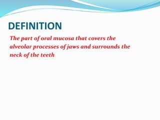
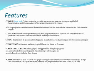
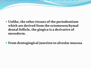
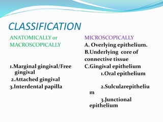
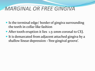
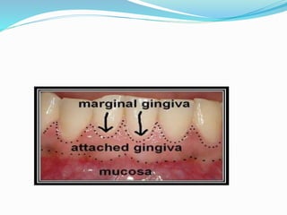
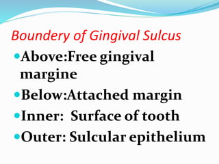
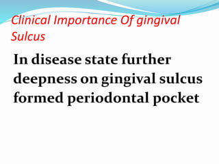
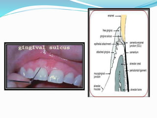
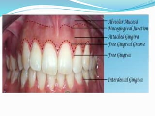
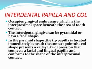
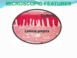
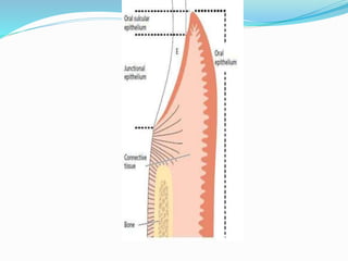
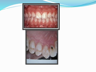
DEFINITION
The part of oral mucosa that covers the
alveolar processes of jaws and surrounds the
neck of the teeth
Features
COLOUR:Coral pink.Colour varies due to racial pigmentation, vascularity degree, epithelial
keratinization and fibrous nature of the underlying connective tissue
SIZE:Corresponds with the sum total of the bulk of cellular and intercellular elements and their vascular
supply
CONTOUR:Depends on shape of the tooth ,their alignment in arch, location and size of the area of
proximal contact and dimensions of facial and lingual embrasure
SHAPE: In anteriors its pyramidal in shape and more flattened in buccolingual direction in molar region
CONSISTENCY:Is firm and resilient.gingival fibres contribute in firmness
SURFACE TEXTURE : Attached gingiva is stippled and marginal gingiva is
not.Centralpart of interdentalpapilla has stippling while margins are
smooth.Prominent in facial than lingual
POSITION:Refers to level at which the gingival margin is attached to tooth.When tooth erupts margin
and sulcus are at the tip of the crown; as eruption progresses they are seen closer to the root
DEVELOPMENT and EXTENSION
 Unlike, the other tissues of the periodontium
which are derived from the ectomesenchymal
dental follicle, the gingiva is a derivative of
mesoderm.
 From dentogingival junction to alveolar mucosa
CLASSIFICATION
ANATOMICALLY or
MACROSCOPICALLY
1.Marginal gingival/Free
gingival
2.Attached gingival
3.Interdental papilla
MICROSCOPICALLY
A. Overlying epithelium.
B.Underlying core of
connective tissue
C.Gingival epithelium
1.Oral epithelium
2.Sulcularepitheliu
m
3.Junctional
epithelium
MARGINAL OR FREE GINGIVA
 Is the terminal edge/ border of gingiva surrounding
the teeth in collar like fashion
 After tooth eruption it lies 1.5-2mm coronal to CEJ.
 It is demarcated from adjacent attached gingiva by a
shallow linear depression –’free gingival groove’.
A.Free gingival groove- it’s a indentation that is
parallel to the vestibular surface of gingival
margin at a level corresponding at CEJ. It lies
opposite to the coronal margin of junctional
epithelium.
B. Gingival sulcus- it’s a shallow crevice or a space
around the tooth bounded by surface of the tooth on
one side and epithelium lining the free margin of
gingiva on another side. ‘
 V’ shaped , barely permits periodontal probe.
 Depth: 1-1.5 mm
 Probing depth of normal gingiva clinically – 2 to
3mm.
 Lining:Keratinied stratified squamous epithelium
 Content:Gingival fluid
Boundery of Gingival Sulcus
Above:Free gingival
margine
Below:Attached margin
Inner: Surface of tooth
Outer: Sulcular epithelium
Clinical Importance Of gingival
Sulcus
In disease state further
deepness on gingival sulcus
formed periodontal pocket
1.The attached gingiva is continuous with the marginal gingiva.
2.It is firm,resilient and tightly bound to the underlying
periosof alveolar bone.
3.The facial aspect of the attached gingiva extends to the
relatively loose and movable alveolar mucosa,from which it is
demarcated by the mucogingival junction
Function of Attached Gingiva
1.Allows proper deflection of
food.
2.Braces marginal gingiva.
3.Allows proper placement of
tooth.Esthetic value.
4.Maintenance of gingival
health
INTERDENTAL PAPILLA AND COL
 Occupies gingival embrassure,which is the
interproximal space beneath the area of tooth
contact.
 The interdental gingiva can be pyramidal or
hava a “col” shape.
 In the pyramid shape ,the tip papilla is located
immediately beneath the contact point;the col
shape presents a valley like depression that
connects a facial and lingual papilla and
conforms to the shape of the interproximal
contact.
The shape of the gingiva in a given interdental space depends
on the :
• Contact point between the two adjoining teeth
• The presence or absence of some degree of recession
If a diastema is present,the gingiva is
firmly bound over the interdental bone
and forms a smooth,rounded surface
without interdental papillae
The gingiva sulcus contains a
fluid GINGIVAL FLUID /
SULCULAR FLUID (GCF) that
seeps into it from the gingival
connective tissue through the
thin sulcular epithelium
Constituents Of GCF
Bacterial enymes
Bacterial degradation products
CT degradation products
Host mediated enymes
Extracellular matrix protein
Inflammatory chemical mediators such as
Histamin,Serotonin,Leukotriens,PG etc
Functions of GCF
 Active phase of periodontal disease process can
be measured or assesed by the constituents of
gingival fluid
 Cleanses material from the sulcus.
• Contains plasma proteins that may improve adhesion
of the epitheIt also possesses antimicrobial properties.
•It exerts antibody activity in defense of the gingival
lium to the tooth
MICROSCOPIC FEATURES
1.Overlying epithelium: Stratified
squamous epithelium
predominantly cellular in nature
2.Underlying core of CT: less cellular and
composed primarily of collagen fibers and
ground substance
3.Gingival epithelium:
The gingival epithelium is a
keratinized,stratified,squamous epithelium
1.Principal cell
•Keratinocytes
2.Clear cells/Nonkeratinocytes
•Langerhans cells
•Merkels cells
•Melanocyte
•Inflammatory cells
ORAL EPITHELIUM
 The epithelium forms the primary barrier between the
oral enviroment and deeper tissue.
 It consist of two cell populations:
Progenitor population-the function is to divide and
provide new cells
Maturing population-the cells continually
 undergo a process of differentiation or maturation
with the formation of a protective surface layer
SULCULAR EPITHELIUM
 Lines the gingival sulcus.
 Extends from coronal limit of JE to crest of gingival margin.
 Thin, non keratinized, stratified squamous squamous
epithelium.
 Without rete pegs and extends from the coronol limit of
junctional epithelium to the crest of gingival margin.
 Shows many cells with hydropic degeneratio
 It is extremely important because acts as a semipermeable
membrane through which bacterial products pass into gingival
fluide
•Provides connection between gingiva and tooth.
•Consist of collar like band of stratified squamous non keratinized
epithelium
•3-4 layers thick in early life, increases with age.
• Tapers from coronal end which may have10-20 cells wide to 1-2
cell wide at apical termination at CEJ in healthy tissue.
5.Cells grouped in 2 strata.
Basal layer: facing connective tissue
Supra basal layer: extending to tooth surface.
6.Length of JE: 0.25- 1.35mm.
7.Formed by confluence of oral epithelium and REE during tooth
eruption
The junction between the tooth and the gingival is
called Dentogingival junction
Other name: Epithelial attachment
Epithelial cuff
Structure:
1.Reduced enamel epithelium
2.Oral epithelium
3.Joint-Desmosome and hemidesmosome
MUCOGINGIVAL JUNCTION
 The facial aspect of attached gingiva extends to
the relatively loose and movable alveolar
mucosa(red) and is demarcated by the
mucogingival junction.
 In the palatal aspect it is continuous with palatal
mucosa and on the lingual aspect of the
mandible.it terminates at the junction of the
lingual alveolar mucosa
 Mucogingival junction remains stationary
throughout life
Connective tissue of marginal gingiva is densely collagenous
containing a prominent system of collagen bundles called
gingival fibres.
Consist of type 1 collagen
1.Brace the marginal gingiva firmly against
the tooth.
2.Provides rigidity necessary to
withstand the forces of mastication without
being deflected away from the tooth
surface.
3.Unites the free marginal gingiva with
cementum and adjacent attached gingival
ARRANGEMENT
Principle group Secondery group
 1.Dentogingival
 2.Dentoperiosteal
 3.Alveogingival
 4.Circular
 5.Transeptal
 1.periostogingival
 2.Interpapillary
 3.Transgingival
 4.Intergingival and
semicircular
•Posterior superior alveolar artery
•Infra orbital artery
•Greater palatine artery
• Inferior alveolar artery
•Supraperiosteal arterioles
•Arterioles from crest of interdental septa
•Vessels of PDL
•Maxillary & Mandibular branches of Trigeminal
nerve
•Superior labial branches from infraorbital nerve
•Posterior superior alveolar nerve
Palatal gingiva
•Greater palatine nerve
Area of incisors
• Long sphenopalatine nerve
Labial gingiva of mandibular incisors & canines
•Mental nerve
•Gingiva at buccal aspect of molars
•Buccal nerve
Age changes
 Thinning of epithelium
 Decreased keratinization
 ↑ cell density
 Morphology : Do not change
 Dental plaque : Etiological factor in development
of gingivitis
 Continuous presence of plaque & its quantity :
Development & course of periodontal disease

Gingiva

  • 2.
    DEFINITION The part oforal mucosa that covers the alveolar processes of jaws and surrounds the neck of the teeth
  • 3.
    Features COLOUR:Coral pink.Colour variesdue to racial pigmentation, vascularity degree, epithelial keratinization and fibrous nature of the underlying connective tissue SIZE:Corresponds with the sum total of the bulk of cellular and intercellular elements and their vascular supply CONTOUR:Depends on shape of the tooth ,their alignment in arch, location and size of the area of proximal contact and dimensions of facial and lingual embrasure SHAPE: In anteriors its pyramidal in shape and more flattened in buccolingual direction in molar region CONSISTENCY:Is firm and resilient.gingival fibres contribute in firmness SURFACE TEXTURE : Attached gingiva is stippled and marginal gingiva is not.Centralpart of interdentalpapilla has stippling while margins are smooth.Prominent in facial than lingual POSITION:Refers to level at which the gingival margin is attached to tooth.When tooth erupts margin and sulcus are at the tip of the crown; as eruption progresses they are seen closer to the root
  • 4.
    DEVELOPMENT and EXTENSION Unlike, the other tissues of the periodontium which are derived from the ectomesenchymal dental follicle, the gingiva is a derivative of mesoderm.  From dentogingival junction to alveolar mucosa
  • 5.
    CLASSIFICATION ANATOMICALLY or MACROSCOPICALLY 1.Marginal gingival/Free gingival 2.Attachedgingival 3.Interdental papilla MICROSCOPICALLY A. Overlying epithelium. B.Underlying core of connective tissue C.Gingival epithelium 1.Oral epithelium 2.Sulcularepitheliu m 3.Junctional epithelium
  • 7.
    MARGINAL OR FREEGINGIVA  Is the terminal edge/ border of gingiva surrounding the teeth in collar like fashion  After tooth eruption it lies 1.5-2mm coronal to CEJ.  It is demarcated from adjacent attached gingiva by a shallow linear depression –’free gingival groove’.
  • 9.
    A.Free gingival groove-it’s a indentation that is parallel to the vestibular surface of gingival margin at a level corresponding at CEJ. It lies opposite to the coronal margin of junctional epithelium.
  • 10.
    B. Gingival sulcus-it’s a shallow crevice or a space around the tooth bounded by surface of the tooth on one side and epithelium lining the free margin of gingiva on another side. ‘  V’ shaped , barely permits periodontal probe.  Depth: 1-1.5 mm  Probing depth of normal gingiva clinically – 2 to 3mm.  Lining:Keratinied stratified squamous epithelium  Content:Gingival fluid
  • 11.
    Boundery of GingivalSulcus Above:Free gingival margine Below:Attached margin Inner: Surface of tooth Outer: Sulcular epithelium
  • 12.
    Clinical Importance Ofgingival Sulcus In disease state further deepness on gingival sulcus formed periodontal pocket
  • 14.
    1.The attached gingivais continuous with the marginal gingiva. 2.It is firm,resilient and tightly bound to the underlying periosof alveolar bone. 3.The facial aspect of the attached gingiva extends to the relatively loose and movable alveolar mucosa,from which it is demarcated by the mucogingival junction
  • 16.
    Function of AttachedGingiva 1.Allows proper deflection of food. 2.Braces marginal gingiva. 3.Allows proper placement of tooth.Esthetic value. 4.Maintenance of gingival health
  • 17.
    INTERDENTAL PAPILLA ANDCOL  Occupies gingival embrassure,which is the interproximal space beneath the area of tooth contact.  The interdental gingiva can be pyramidal or hava a “col” shape.  In the pyramid shape ,the tip papilla is located immediately beneath the contact point;the col shape presents a valley like depression that connects a facial and lingual papilla and conforms to the shape of the interproximal contact.
  • 18.
    The shape ofthe gingiva in a given interdental space depends on the : • Contact point between the two adjoining teeth • The presence or absence of some degree of recession
  • 19.
    If a diastemais present,the gingiva is firmly bound over the interdental bone and forms a smooth,rounded surface without interdental papillae
  • 20.
    The gingiva sulcuscontains a fluid GINGIVAL FLUID / SULCULAR FLUID (GCF) that seeps into it from the gingival connective tissue through the thin sulcular epithelium
  • 21.
    Constituents Of GCF Bacterialenymes Bacterial degradation products CT degradation products Host mediated enymes Extracellular matrix protein Inflammatory chemical mediators such as Histamin,Serotonin,Leukotriens,PG etc
  • 22.
    Functions of GCF Active phase of periodontal disease process can be measured or assesed by the constituents of gingival fluid  Cleanses material from the sulcus. • Contains plasma proteins that may improve adhesion of the epitheIt also possesses antimicrobial properties. •It exerts antibody activity in defense of the gingival lium to the tooth
  • 23.
  • 24.
    1.Overlying epithelium: Stratified squamousepithelium predominantly cellular in nature 2.Underlying core of CT: less cellular and composed primarily of collagen fibers and ground substance 3.Gingival epithelium: The gingival epithelium is a keratinized,stratified,squamous epithelium
  • 26.
    1.Principal cell •Keratinocytes 2.Clear cells/Nonkeratinocytes •Langerhanscells •Merkels cells •Melanocyte •Inflammatory cells
  • 27.
    ORAL EPITHELIUM  Theepithelium forms the primary barrier between the oral enviroment and deeper tissue.  It consist of two cell populations: Progenitor population-the function is to divide and provide new cells Maturing population-the cells continually  undergo a process of differentiation or maturation with the formation of a protective surface layer
  • 28.
    SULCULAR EPITHELIUM  Linesthe gingival sulcus.  Extends from coronal limit of JE to crest of gingival margin.  Thin, non keratinized, stratified squamous squamous epithelium.  Without rete pegs and extends from the coronol limit of junctional epithelium to the crest of gingival margin.  Shows many cells with hydropic degeneratio  It is extremely important because acts as a semipermeable membrane through which bacterial products pass into gingival fluide
  • 30.
    •Provides connection betweengingiva and tooth. •Consist of collar like band of stratified squamous non keratinized epithelium •3-4 layers thick in early life, increases with age. • Tapers from coronal end which may have10-20 cells wide to 1-2 cell wide at apical termination at CEJ in healthy tissue. 5.Cells grouped in 2 strata. Basal layer: facing connective tissue Supra basal layer: extending to tooth surface. 6.Length of JE: 0.25- 1.35mm. 7.Formed by confluence of oral epithelium and REE during tooth eruption
  • 32.
    The junction betweenthe tooth and the gingival is called Dentogingival junction Other name: Epithelial attachment Epithelial cuff Structure: 1.Reduced enamel epithelium 2.Oral epithelium 3.Joint-Desmosome and hemidesmosome
  • 33.
    MUCOGINGIVAL JUNCTION  Thefacial aspect of attached gingiva extends to the relatively loose and movable alveolar mucosa(red) and is demarcated by the mucogingival junction.  In the palatal aspect it is continuous with palatal mucosa and on the lingual aspect of the mandible.it terminates at the junction of the lingual alveolar mucosa  Mucogingival junction remains stationary throughout life
  • 35.
    Connective tissue ofmarginal gingiva is densely collagenous containing a prominent system of collagen bundles called gingival fibres. Consist of type 1 collagen
  • 36.
    1.Brace the marginalgingiva firmly against the tooth. 2.Provides rigidity necessary to withstand the forces of mastication without being deflected away from the tooth surface. 3.Unites the free marginal gingiva with cementum and adjacent attached gingival
  • 37.
    ARRANGEMENT Principle group Seconderygroup  1.Dentogingival  2.Dentoperiosteal  3.Alveogingival  4.Circular  5.Transeptal  1.periostogingival  2.Interpapillary  3.Transgingival  4.Intergingival and semicircular
  • 38.
    •Posterior superior alveolarartery •Infra orbital artery •Greater palatine artery • Inferior alveolar artery •Supraperiosteal arterioles •Arterioles from crest of interdental septa •Vessels of PDL
  • 40.
    •Maxillary & Mandibularbranches of Trigeminal nerve •Superior labial branches from infraorbital nerve •Posterior superior alveolar nerve Palatal gingiva •Greater palatine nerve Area of incisors • Long sphenopalatine nerve Labial gingiva of mandibular incisors & canines •Mental nerve •Gingiva at buccal aspect of molars •Buccal nerve
  • 41.
    Age changes  Thinningof epithelium  Decreased keratinization  ↑ cell density  Morphology : Do not change  Dental plaque : Etiological factor in development of gingivitis  Continuous presence of plaque & its quantity : Development & course of periodontal disease