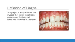
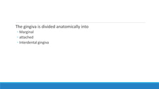
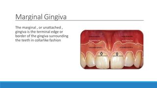
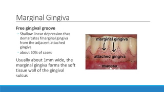
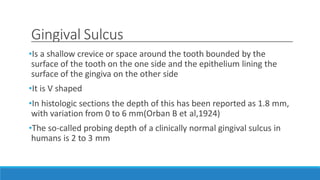
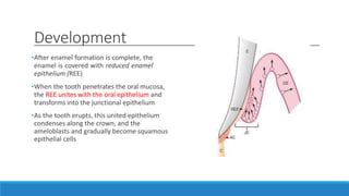
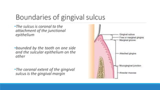
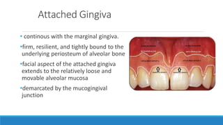
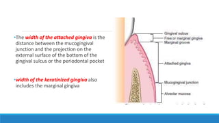
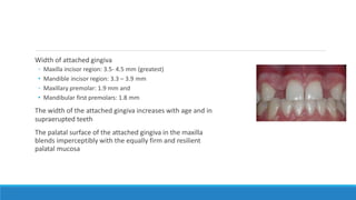
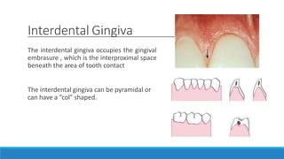
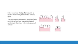
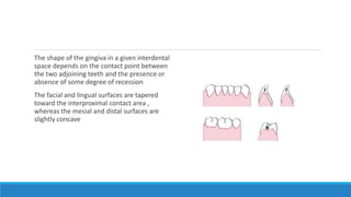
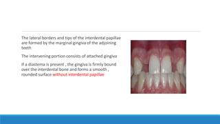
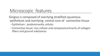
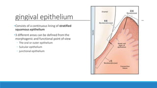
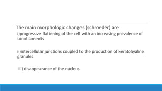
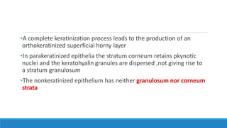
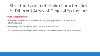
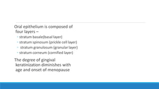
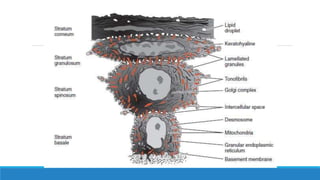
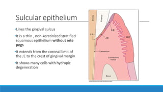
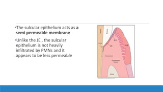
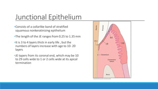
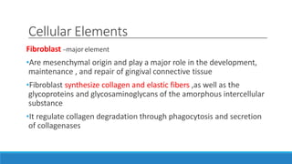
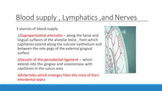
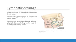
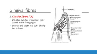
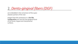
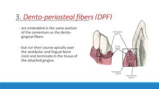
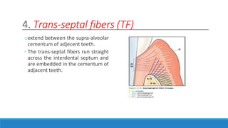
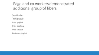
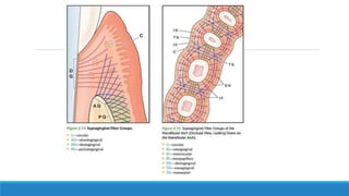
SlideShare a Scribd company logo
1 of 68
Gingiva: Anatomy,
Hisology & Biology
PRESENTED BY
DR ASIF K
DEPARTMENT OF PERIODONTICS
Content
Introduction
Development
Anatomic classification
Clinical features
Microscopic features
 Gingival fibers
Definition ofGingiva:
The gingiva is the part of the oral
mucosa that covers the alveolar
processes of the jaws and
surrounds the necks of the teeth
The gingiva is divided anatomically into
◦ Marginal
◦ attached
◦ Interdental gingiva
Marginal Gingiva
The marginal , or unattached ,
gingiva is the terminal edge or
border of the gingiva surrounding
the teeth in collarlike fashion
Marginal Gingiva
Free gingival groove
◦ Shallow linear depression that
demarcates fmarginal gingiva
from the adjacent attached
gingiva
◦ about 50% of cases
Usually about 1mm wide, the
marginal gingiva forms the soft
tissue wall of the gingival
sulcus
Marginal Gingiva
•The most apical point of the
marginal gingival scallop is called
the gingival zenith
•
Gingival Sulcus
•Is a shallow crevice or space around the tooth bounded by the
surface of the tooth on the one side and the epithelium lining the
surface of the gingiva on the other side
•It is V shaped
•In histologic sections the depth of this has been reported as 1.8 mm,
with variation from 0 to 6 mm(Orban B et al,1924)
•The so-called probing depth of a clinically normal gingival sulcus in
humans is 2 to 3 mm
Development
•After enamel formation is complete, the
enamel is covered with reduced enamel
epithelium (REE)
•When the tooth penetrates the oral mucosa,
the REE unites with the oral epithelium and
transforms into the junctional epithelium
•As the tooth erupts, this united epithelium
condenses along the crown, and the
ameloblasts and gradually become squamous
epithelial cells
Boundaries of gingival sulcus
•The sulcus is coronal to the
attachment of the junctional
epithelium
•bounded by the tooth on one side
and the sulcular epithelium on the
other
•The coronal extent of the gingival
sulcus is the gingival margin
Attached Gingiva
• continous with the marginal gingiva.
•firm, resilient, and tightly bound to the
underlying periosteum of alveolar bone
•facial aspect of the attached gingiva
extends to the relatively loose and
movable alveolar mucosa
•demarcated by the mucogingival
junction
•The width of the attached gingiva is the
distance between the mucogingival
junction and the projection on the
external surface of the bottom of the
gingival sulcus or the periodontal pocket
•width of the keratinized gingiva also
includes the marginal gingiva
Width of attached gingiva
◦ Maxilla incisor region: 3.5- 4.5 mm (greatest)
• Mandible incisor region: 3.3 – 3.9 mm
◦ Maxillary premolar: 1.9 mm and
• Mandibular first premolars: 1.8 mm
The width of the attached gingiva increases with age and in
supraerupted teeth
The palatal surface of the attached gingiva in the maxilla
blends imperceptibly with the equally firm and resilient
palatal mucosa
Interdental Gingiva
The interdental gingiva occupies the gingival
embrasure , which is the interproximal space
beneath the area of tooth contact
The interdental gingiva can be pyramidal or
can have a “colˮ shaped.
In the pyramidal the tip of one papilla is
located immediately beneath the contact
point
the Col presents a valley like depression that
connects a facial and lingual papilla and
conforms to the shape of the interproximal
contact
The shape of the gingiva in a given interdental
space depends on the contact point between
the two adjoining teeth and the presence or
absence of some degree of recession
The facial and lingual surfaces are tapered
toward the interproximal contact area ,
whereas the mesial and distal surfaces are
slightly concave
The lateral borders and tips of the interdental papillae
are formed by the marginal gingiva of the adjoining
teeth
The intervening portion consists of attached gingiva
If a diastema is present , the gingiva is firmly bound
over the interdental bone and forms a smooth ,
rounded surface without interdental papillae
Microscopic features
Gingiva is composed of overlying stratified squamous
epithelium and overlying central core of connective tissue
• Epithelium : predominantly cellular
• Connective tissue: less cellular and composed primarily of collagen
fibers and ground substances
gingival epithelium
•Consists of a continuous lining of stratified
squamous epithelium
•3 different areas can be defined from the
morphogenic and functional point of view
◦ The oral or outer epithelium
◦ Sulcular epithelium
◦ junctional epithelium
•The principal cell type – keratinocytes
•Clear cells or nonkeratinocytes
1) Langerhans cells
2) Merkel cells
3) Melanocytes
Merkel cells
•located in the deeper layers of the epithelium
•they harbor nerve endings
•connected to adjacent cells by desmosomes
•They have been identified as tactile perceptors
Langerhans Cell
•Langerhans cells are dendritic cells
located among keratinocytes at all
suprabasal levels
•They belong to the mononuclear
phagocytes system (reticuloendothelial
system )
•They contain elongated granules and are
considered macrophages with possible
antigenic properties
Melanocytes
•dendritic cells
•located in the basal and spinous layers of the gingival epithelium
•They synthesize melanin in organelles -premelanosomes or
melanosomes which contain tyrosinase which hydroxylates tyrosine
to dihydroxyphenylalanine(DOPA), which in turn is progressively
converted to melanin
•Melanin granules are phagocytosed and contained within other cells
of the epithelium and connective tissue called melanophages or
melanophores
Proliferation of keratinocytes takes place by mitosis in the basal
layer less frequently in the suprabasal layer
Differentiation involves the process of keratinization which consists
of biochemical and morphologic events that occur in the cell as they
migrate from the basal layer
The main morphologic changes (schroeder) are
i)progressive flattening of the cell with an increasing prevalence of
tonofilaments
ii)intercellular junctions coupled to the production of keratohyaline
granules
iii) disappearance of the nucleus
•A complete keratinization process leads to the production of an
orthokeratinized superficial horny layer
•In parakeratinized epithelia the stratum corneum retains pkynotic
nuclei and the keratohyalin granules are dispersed ,not giving rise to
a stratum granulosum
•The nonkeratinized epithelium has neither granulosum nor corneum
strata
Structural and metabolic characteristics
of Different Areas of Gingival Epithelium
Outer (Oral) epithelium –
•Covers the crest and outer surface of the marginal gingiva and the surface of the
attached gingiva
•On average, the oral epithelium is 0.2 to 0.3 mm in thickness
•It is keratinized or parakeratinized or presents various combination of these conditions
Oral epithelium is composed of
four layers –
◦ stratum basale(basal layer)
◦ stratum spinosum (prickle cell layer)
◦ stratum granulosum (granular layer)
◦ stratum corneum (cornified layer)
The degree of gingival
keratinization diminishes with
age and onset of menopause
Sulcular epithelium
•Lines the gingival sulcus
•It is a thin , non-keratinized stratified
squamous epithelium without rete
pegs
•it extends from the coronal limit of
the JE to the crest of gingival margin
•It shows many cells with hydropic
degeneration
•The sulcular epithelium acts as a
semi permeable membrane
•Unlike the JE , the sulcular
epithelium is not heavily
infiltrated by PMNs and it
appears to be less permeable
Junctional Epithelium
•Consists of a collarlike band of stratified
squamous nonkeratinizing epithelium
•The length of the JE ranges from 0.25 to 1.35 mm
•It is 3 to 4 layers thick in early life , but the
numbers of layers increase with age to 10- 20
layers
•JE tapers from its coronal end, which may be 10
to 29 cells wide to 1 or 2 cells wide at its apical
termination
•The JE is formed by the confluence of the oral epithelium and the reduced
enamel epithelium during tooth eruption
•The JE is attached to the tooth surface (epithelial attachment) by means of an
internal basal lamina
•It is attached to the gingival connective tissue by an external basal lamina
•The attachment of JE to the
tooth is reinforced by the
gingival fibers , which brace the
marginal gingiva against the
tooth surface
•For this reason ,the JE and the
gingival fibers are considered a
functional units referred to as
the dentogingival unit
Functions of JE
1)JE firmly attached to the tooth surface forming an epithelial barrier
against plaque bacteria
2)It allows access of gingival fluid inflammatory cells, and
components of the immunologic host defense to the gingival margin
3)JE cells exhibit rapid turnover - contributes to the host- parasite
equilibrium and rapid repair of damaged tissue
Gingival Connective Tissue
The major components of the gingiva connective tissue are collagen
fibres – approx 60% by volume , fibroblasts – 5% , vessels , nerves ,
and matrix – about 35%
The connective tissue of the gingiva is known as the lamina propria
and consists of two layers :
◦ 1) a papillary layer subjacent to the epithelium , which consists of papillary
projections between the epithelial rete pegs
◦ 2) a reticular layer contiguous with the periosteum of the alveolar bone
Connective tissue has a cellular and an extracellular compartment
composed of fibers and ground substance
The ground substance fills the space between fibers and cells , is
amorphous , and has a high content of water
•Ground substance is composed of proteoglycans , mainly hyaluronic
acid and chondroitin sulfate , and glycoproteins , mainly fibronectin
•Fibronectin binds fibroblasts to the fibers and many other
components of the intercellular matrix , helping mediate cell
adhesion and migration
•Laminin , another glycoprotein found in the basal lamina serves to
attach it to epithelial cells
•3 types of connective tissue fibers are collagen reticular and elastic
•Collagen type I forms the bulk of the lamina propria and provides
the tensile strength to the gingival tissue
•Type IV collagen (argyrophillic reticulum fiber) branches between the
collagen type I bundle and is continuous with fibers of the basement
membrane and blood vessel walls
•The elastic fiber system is composed of oxytalan , elaunin, and
elastin fibers distributed among collagen fibers
•Densely packed collagen bundles that are anchored into the acellular
extrinsic fiber cementum just below the terminal point of the JE form
the connective tissue attachment
•The stability of this attachment is a key factor in limiting the
migration of JE
Cellular Elements
Fibroblast –majorelement
•Are mesenchymal origin and play a major role in the development,
maintenance , and repair of gingival connective tissue
•Fibroblast synthesize collagen and elastic fibers ,as well as the
glycoproteins and glycosaminoglycans of the amorphous intercellular
substance
•It regulate collagen degradation through phagocytosis and secretion
of collagenases
Other cells
•Mast cell are numerous in connective tissue
•Fixed macrophages and histiocytes are present in the gingival connective tissue as components
of the mononuclear phagocytes system
•Adipose cells and eosinophils
•plasma cells and lymphocytes
•Neutrophils
Blood supply , Lymphatics ,and Nerves
3 sources of blood supply
1)Supraperiosteal arterioles – along the facial and
lingual surfaces of the alveolar bone , from which
capillaries extend along the sulcular epithelium and
between the rete pegs of the external gingival
surface
2)Vessels of the periodontal ligament – which
extend into the gingiva and anastomose with
capillaries in the sulcus area
3)Arterioles which emerges from the crest of their
interdental septa
Lymphatic drainage
From mandibular incisor gingiva  submental
lymph node.
From maxillary palatal gingiva  deep cervical
lymph nodes
Buccal gingiva of maxilla and buccal & lingual
gingiva in the mandibular premolar-molar 
submandibular lymph nodes
Nerve supply
•Branches of the trigeminal nerve provide sensory and
proprioceptive functions.
•Within the gingival connective tissues, most nerve fibers are
myelinated
•Gingival innervation is derived from fibers arising from nerves in the
PD and from the labial , buccal , and palatal nerves
The following nerve structures are present in the connective
tissue :
1. A meshwork of terminal argrophillic fibres
2. Meissener –type tactile corpuscles
3. Krause – type end bulbs
4. Encapsulated spindles
Gingival fibers
•The connective tissue of the marginal
gingiva is densely collagenous ,
containing a prominent system of
collagen fibres bundles called the
gingival fibers
•They consists of type I collagen
Functions
1) to brace the marginal gingiva firmly against the tooth
2) to provide the rigidity necessary to withstand the forces of mastication without being
deflected away from the tooth surface
3) to unite the free marginal gingiva with the cementum of the root and the adjacent gingiva
Gingival fibres
1. Circular fibers (CF)
◦ are fiber bundles which run their
course in the free gingiva
◦ encircle the tooth in a cuff- or ring-
like fashion.
2. Dento-gingival fibers (DGF)
are embedded in the cementum of the supra-
alveolar portion of the root
project from the cementum in a fan-like
configuration out into the free gingival tissue
of the facial, lingual and interproximal
surfaces.
3. Dento-periosteal fibers (DPF)
◦ are embedded in the same portion
of the cementum as the dento-
gingival fibers
◦ but run their course apically over
the vestibular and lingual bone
crest and terminate in the tissue of
the attached gingiva
4. Trans-septal fibers (TF)
oextend between the supra-alveolar
cementum of adjecent teeth.
◦ The trans-septal fibers run straight
across the interdental septum and
are embedded in the cementum of
adjacent teeth.
Page and co workersdemonstrated
additional group of fibers
Semicircular
Trans gingival
Inter gingival
Inter papillary
Inter circular
Periosteo gingival
Correlation of clinical and
microscopic features
Colour
Attached and marginal gingiva is “coral pink ˮ
The colour depends upon:
i. the vascular supply
ii. the thickness/degree of keratinization of the epithelium
iii. the presence of pigment containing cell
Size
•The size of the gingiva corresponds with the sum total of the bulk of
cellular and intercellular elements and their vascular supply
•Alteration of size denotes gingival disease
Contour
•Varies considerably and depends on the
• shape of the teeth and their alignment in the arch
• the location and size of the area of proximal contact
• the dimensions of the facial and lingual gingival embrasures
•The marginal gingiva envelops the teeth in collarlike fashion and
follows a scalloped outline on the facial and lingual surfaces
Contour
•It forms a straight line along
teeth in labial version , the
normal arcuate contour is
accentuated and the gingiva is
located farther apically
•On teeth in lingual version - the
gingiva is horizontal and
thickened
Shape
•The shape of the interdental gingiva is governed by the contour of
the proximal tooth surfaces and the location and shape of gingival
embrasures
•When the proximal surfaces of the crowns are relatively flat
faciolingually the roots are close together , the interdental bone is
thin mesiodistally ,and the gingival embrassures and interdental
gingiva are narrow mesiodistally
The height of the interdental gingiva varies with the location of the
proximal contact
◦ In the anterior region of the dentition ,the interdental papilla is pyramidal in
form whereas the papilla is more flattened in a buccolingual direction in the
molar region
Consistency
•The gingiva is firm and resilient and , with the exception of the
movable free margin, tightly bound to the underlying bone
•The collagenous nature of the alveolar bone determine the firmness
of the attached gingiva
•The gingiva fibers contribute to the firmness of the gingival margin
Surface Texture
The attached gingiva is stippled ; the marginal
gingiva is not
The central portion of the interdental papilla is
usually stippled , but the marginal borders are
smooth
Stippling varies with age.
It is absent in infancy , appears in some
children at about 5 yrs of age , increases until
adulthood ,and frequently begins to disappear
in old age
•Microscopically ,stippling is
produced by alternate rounded
protuberances and depressions in
the gingival surface
•The papillary layer of the
connective tissue projects into the
elevations
•The elevated and depressed areas
are covered by stratified squamous
epithelium
Position
•The position of the gingiva refers to the level at which the gingival margin is attached to the
tooth
•When the tooth erupts into the oral cavity ,the margin and sulcus are at the tip of the crown; as
eruption progress ,they are seen closer to the root
•Active eruption – movement of the teeth in the direction of the occlusal plane
•Passive eruption – exposure of the teeth by apical migration of the gingiva
Stages of passive eruption (Gottlieb and
Orban)
Stage 1: Base of the gingival
sulcus and JE are on the enamel
Stage 2: Base of the gingival
sulcus is on the enamel and the
part of the JE is on the root
Stage 3: Base of the gingival
sulcus is at the cementoenamel
line
Stage 4: Base of the gingival
sulcus and the JE are on the root
Exposure of the tooth via the apical migration
of the gingiva is called gingival recession or
atrophy
THANK YOU

More Related Content

What's hot (20)

Anatomy of periodontium
Anatomy of periodontiumAnatomy of periodontium
Anatomy of periodontium
 
Periodontal plastic surgery
Periodontal plastic surgeryPeriodontal plastic surgery
Periodontal plastic surgery
 
Free gingival grafts
Free gingival graftsFree gingival grafts
Free gingival grafts
 
Alveolar bone
Alveolar boneAlveolar bone
Alveolar bone
 
Phase 1 periodontal therapy
Phase 1 periodontal therapyPhase 1 periodontal therapy
Phase 1 periodontal therapy
 
Cementum
CementumCementum
Cementum
 
ROOT COVERAGE PROCEDURES
ROOT COVERAGE PROCEDURESROOT COVERAGE PROCEDURES
ROOT COVERAGE PROCEDURES
 
ATTACHED GINGIVA
ATTACHED GINGIVAATTACHED GINGIVA
ATTACHED GINGIVA
 
Aging and the periodontium
Aging and the periodontiumAging and the periodontium
Aging and the periodontium
 
Ridge Augmentation Procedures
Ridge Augmentation Procedures Ridge Augmentation Procedures
Ridge Augmentation Procedures
 
GINGIVECTOMY AND GINGIVOPLASTY
GINGIVECTOMY AND GINGIVOPLASTYGINGIVECTOMY AND GINGIVOPLASTY
GINGIVECTOMY AND GINGIVOPLASTY
 
"GUIDED TISSUE REGENERATION"
"GUIDED TISSUE REGENERATION""GUIDED TISSUE REGENERATION"
"GUIDED TISSUE REGENERATION"
 
Biologic width
Biologic widthBiologic width
Biologic width
 
Gingiva
GingivaGingiva
Gingiva
 
General principles of Periodontal surgery
General principles of Periodontal surgeryGeneral principles of Periodontal surgery
General principles of Periodontal surgery
 
Periodontal plastic and esthetic surgery
Periodontal plastic and esthetic surgeryPeriodontal plastic and esthetic surgery
Periodontal plastic and esthetic surgery
 
gingival connective tissue
 gingival connective tissue gingival connective tissue
gingival connective tissue
 
Risk factors in Periodontal Disease
Risk factors in Periodontal DiseaseRisk factors in Periodontal Disease
Risk factors in Periodontal Disease
 
Periodontal Flap
Periodontal FlapPeriodontal Flap
Periodontal Flap
 
030.furcation involvement and its therapy
030.furcation involvement and its therapy030.furcation involvement and its therapy
030.furcation involvement and its therapy
 

Similar to gingiva.pptx

Similar to gingiva.pptx (20)

Gingiva
GingivaGingiva
Gingiva
 
GINGIVA-DR JEEVANAND DESHMUKH.pptx
GINGIVA-DR JEEVANAND DESHMUKH.pptxGINGIVA-DR JEEVANAND DESHMUKH.pptx
GINGIVA-DR JEEVANAND DESHMUKH.pptx
 
gingiva.pptx
gingiva.pptxgingiva.pptx
gingiva.pptx
 
Gingiva
Gingiva Gingiva
Gingiva
 
Gingiva
GingivaGingiva
Gingiva
 
Gingiva
GingivaGingiva
Gingiva
 
Gingiva copy
Gingiva copyGingiva copy
Gingiva copy
 
Gingiva
GingivaGingiva
Gingiva
 
GINGIVA.pptx
GINGIVA.pptxGINGIVA.pptx
GINGIVA.pptx
 
Clinical Periodontology - Introduction & Anatomy
Clinical Periodontology - Introduction & AnatomyClinical Periodontology - Introduction & Anatomy
Clinical Periodontology - Introduction & Anatomy
 
003.biology of periodontal tissues
003.biology of periodontal tissues003.biology of periodontal tissues
003.biology of periodontal tissues
 
gigiva2ndseminar- 2.pdf
gigiva2ndseminar- 2.pdfgigiva2ndseminar- 2.pdf
gigiva2ndseminar- 2.pdf
 
486 junctional epithelium
486 junctional epithelium486 junctional epithelium
486 junctional epithelium
 
Gingiva in health and disease
Gingiva in health and diseaseGingiva in health and disease
Gingiva in health and disease
 
Gingiva. Lilly & Bong.pptx
Gingiva. Lilly & Bong.pptxGingiva. Lilly & Bong.pptx
Gingiva. Lilly & Bong.pptx
 
Gingiva
GingivaGingiva
Gingiva
 
GINGIVA SEMINAR .pptx
GINGIVA SEMINAR .pptxGINGIVA SEMINAR .pptx
GINGIVA SEMINAR .pptx
 
Ultrastructure of gingiva
Ultrastructure of gingiva Ultrastructure of gingiva
Ultrastructure of gingiva
 
Gingiva (2)
Gingiva (2)Gingiva (2)
Gingiva (2)
 
Ultrastructure of gingival epithelium in health and disease
Ultrastructure of gingival epithelium in health and diseaseUltrastructure of gingival epithelium in health and disease
Ultrastructure of gingival epithelium in health and disease
 

More from DentalYoutube

Introduction to Dental Anatomy.ppt
Introduction to Dental Anatomy.pptIntroduction to Dental Anatomy.ppt
Introduction to Dental Anatomy.pptDentalYoutube
 
rationale of endodontic treatment.pptx
rationale of endodontic treatment.pptxrationale of endodontic treatment.pptx
rationale of endodontic treatment.pptxDentalYoutube
 
diseasesofpulpandperiapicaltissues.pptx
diseasesofpulpandperiapicaltissues.pptxdiseasesofpulpandperiapicaltissues.pptx
diseasesofpulpandperiapicaltissues.pptxDentalYoutube
 
cleaningandshaping.pptx
cleaningandshaping.pptxcleaningandshaping.pptx
cleaningandshaping.pptxDentalYoutube
 
castrestorations-170210145741-converted.pptx
castrestorations-170210145741-converted.pptxcastrestorations-170210145741-converted.pptx
castrestorations-170210145741-converted.pptxDentalYoutube
 
caseselectionfinal-170514163212.pptx
caseselectionfinal-170514163212.pptxcaseselectionfinal-170514163212.pptx
caseselectionfinal-170514163212.pptxDentalYoutube
 
MERCURY TOXICITY.pptx
MERCURY TOXICITY.pptxMERCURY TOXICITY.pptx
MERCURY TOXICITY.pptxDentalYoutube
 
contactsandcontours.pptx
contactsandcontours.pptxcontactsandcontours.pptx
contactsandcontours.pptxDentalYoutube
 
resorption-160714175024.pptx
resorption-160714175024.pptxresorption-160714175024.pptx
resorption-160714175024.pptxDentalYoutube
 
NON CARIOUS LESIONS AND MANAGEMENT.pptx
NON CARIOUS LESIONS AND MANAGEMENT.pptxNON CARIOUS LESIONS AND MANAGEMENT.pptx
NON CARIOUS LESIONS AND MANAGEMENT.pptxDentalYoutube
 
introduction-ENDODONTICS.pptx
introduction-ENDODONTICS.pptxintroduction-ENDODONTICS.pptx
introduction-ENDODONTICS.pptxDentalYoutube
 
Intracanal Medicaments.pptx
Intracanal Medicaments.pptxIntracanal Medicaments.pptx
Intracanal Medicaments.pptxDentalYoutube
 
PAIN CONTROL in operative dentistry.pptx
PAIN CONTROL in operative dentistry.pptxPAIN CONTROL in operative dentistry.pptx
PAIN CONTROL in operative dentistry.pptxDentalYoutube
 

More from DentalYoutube (20)

Maxillary sinus.ppt
Maxillary sinus.pptMaxillary sinus.ppt
Maxillary sinus.ppt
 
Introduction to Dental Anatomy.ppt
Introduction to Dental Anatomy.pptIntroduction to Dental Anatomy.ppt
Introduction to Dental Anatomy.ppt
 
rationale of endodontic treatment.pptx
rationale of endodontic treatment.pptxrationale of endodontic treatment.pptx
rationale of endodontic treatment.pptx
 
diseasesofpulpandperiapicaltissues.pptx
diseasesofpulpandperiapicaltissues.pptxdiseasesofpulpandperiapicaltissues.pptx
diseasesofpulpandperiapicaltissues.pptx
 
cleaningandshaping.pptx
cleaningandshaping.pptxcleaningandshaping.pptx
cleaningandshaping.pptx
 
castrestorations-170210145741-converted.pptx
castrestorations-170210145741-converted.pptxcastrestorations-170210145741-converted.pptx
castrestorations-170210145741-converted.pptx
 
caseselectionfinal-170514163212.pptx
caseselectionfinal-170514163212.pptxcaseselectionfinal-170514163212.pptx
caseselectionfinal-170514163212.pptx
 
sealers.pptx
sealers.pptxsealers.pptx
sealers.pptx
 
MERCURY TOXICITY.pptx
MERCURY TOXICITY.pptxMERCURY TOXICITY.pptx
MERCURY TOXICITY.pptx
 
DFG.pptx
DFG.pptxDFG.pptx
DFG.pptx
 
contactsandcontours.pptx
contactsandcontours.pptxcontactsandcontours.pptx
contactsandcontours.pptx
 
resorption-160714175024.pptx
resorption-160714175024.pptxresorption-160714175024.pptx
resorption-160714175024.pptx
 
pulpirritants.pptx
pulpirritants.pptxpulpirritants.pptx
pulpirritants.pptx
 
pulp responses.pptx
pulp responses.pptxpulp responses.pptx
pulp responses.pptx
 
NON CARIOUS LESIONS AND MANAGEMENT.pptx
NON CARIOUS LESIONS AND MANAGEMENT.pptxNON CARIOUS LESIONS AND MANAGEMENT.pptx
NON CARIOUS LESIONS AND MANAGEMENT.pptx
 
introduction-ENDODONTICS.pptx
introduction-ENDODONTICS.pptxintroduction-ENDODONTICS.pptx
introduction-ENDODONTICS.pptx
 
Intracanal Medicaments.pptx
Intracanal Medicaments.pptxIntracanal Medicaments.pptx
Intracanal Medicaments.pptx
 
cavity designs.pptx
cavity designs.pptxcavity designs.pptx
cavity designs.pptx
 
airabrasion.pptx
airabrasion.pptxairabrasion.pptx
airabrasion.pptx
 
PAIN CONTROL in operative dentistry.pptx
PAIN CONTROL in operative dentistry.pptxPAIN CONTROL in operative dentistry.pptx
PAIN CONTROL in operative dentistry.pptx
 

Recently uploaded

Call Girl Chennai Indira 9907093804 Independent Call Girls Service Chennai
Call Girl Chennai Indira 9907093804 Independent Call Girls Service ChennaiCall Girl Chennai Indira 9907093804 Independent Call Girls Service Chennai
Call Girl Chennai Indira 9907093804 Independent Call Girls Service ChennaiNehru place Escorts
 
Call Girls Service In Shyam Nagar Whatsapp 8445551418 Independent Escort Service
Call Girls Service In Shyam Nagar Whatsapp 8445551418 Independent Escort ServiceCall Girls Service In Shyam Nagar Whatsapp 8445551418 Independent Escort Service
Call Girls Service In Shyam Nagar Whatsapp 8445551418 Independent Escort Serviceparulsinha
 
College Call Girls Pune Mira 9907093804 Short 1500 Night 6000 Best call girls...
College Call Girls Pune Mira 9907093804 Short 1500 Night 6000 Best call girls...College Call Girls Pune Mira 9907093804 Short 1500 Night 6000 Best call girls...
College Call Girls Pune Mira 9907093804 Short 1500 Night 6000 Best call girls...Miss joya
 
Call Girls Chennai Megha 9907093804 Independent Call Girls Service Chennai
Call Girls Chennai Megha 9907093804 Independent Call Girls Service ChennaiCall Girls Chennai Megha 9907093804 Independent Call Girls Service Chennai
Call Girls Chennai Megha 9907093804 Independent Call Girls Service ChennaiNehru place Escorts
 
Sonagachi Call Girls Services 9907093804 @24x7 High Class Babes Here Call Now
Sonagachi Call Girls Services 9907093804 @24x7 High Class Babes Here Call NowSonagachi Call Girls Services 9907093804 @24x7 High Class Babes Here Call Now
Sonagachi Call Girls Services 9907093804 @24x7 High Class Babes Here Call NowRiya Pathan
 
Call Girls Service in Bommanahalli - 7001305949 with real photos and phone nu...
Call Girls Service in Bommanahalli - 7001305949 with real photos and phone nu...Call Girls Service in Bommanahalli - 7001305949 with real photos and phone nu...
Call Girls Service in Bommanahalli - 7001305949 with real photos and phone nu...narwatsonia7
 
Hi,Fi Call Girl In Mysore Road - 7001305949 | 24x7 Service Available Near Me
Hi,Fi Call Girl In Mysore Road - 7001305949 | 24x7 Service Available Near MeHi,Fi Call Girl In Mysore Road - 7001305949 | 24x7 Service Available Near Me
Hi,Fi Call Girl In Mysore Road - 7001305949 | 24x7 Service Available Near Menarwatsonia7
 
Call Girls Service Jaipur Grishma WhatsApp ❤8445551418 VIP Call Girls Jaipur
Call Girls Service Jaipur Grishma WhatsApp ❤8445551418 VIP Call Girls JaipurCall Girls Service Jaipur Grishma WhatsApp ❤8445551418 VIP Call Girls Jaipur
Call Girls Service Jaipur Grishma WhatsApp ❤8445551418 VIP Call Girls Jaipurparulsinha
 
Call Girls Service Bellary Road Just Call 7001305949 Enjoy College Girls Service
Call Girls Service Bellary Road Just Call 7001305949 Enjoy College Girls ServiceCall Girls Service Bellary Road Just Call 7001305949 Enjoy College Girls Service
Call Girls Service Bellary Road Just Call 7001305949 Enjoy College Girls Servicenarwatsonia7
 
Russian Call Girl Brookfield - 7001305949 Escorts Service 50% Off with Cash O...
Russian Call Girl Brookfield - 7001305949 Escorts Service 50% Off with Cash O...Russian Call Girl Brookfield - 7001305949 Escorts Service 50% Off with Cash O...
Russian Call Girl Brookfield - 7001305949 Escorts Service 50% Off with Cash O...narwatsonia7
 
Call Girl Coimbatore Prisha☎️ 8250192130 Independent Escort Service Coimbatore
Call Girl Coimbatore Prisha☎️  8250192130 Independent Escort Service CoimbatoreCall Girl Coimbatore Prisha☎️  8250192130 Independent Escort Service Coimbatore
Call Girl Coimbatore Prisha☎️ 8250192130 Independent Escort Service Coimbatorenarwatsonia7
 
Call Girls In Andheri East Call 9920874524 Book Hot And Sexy Girls
Call Girls In Andheri East Call 9920874524 Book Hot And Sexy GirlsCall Girls In Andheri East Call 9920874524 Book Hot And Sexy Girls
Call Girls In Andheri East Call 9920874524 Book Hot And Sexy Girlsnehamumbai
 
CALL ON ➥9907093804 🔝 Call Girls Baramati ( Pune) Girls Service
CALL ON ➥9907093804 🔝 Call Girls Baramati ( Pune)  Girls ServiceCALL ON ➥9907093804 🔝 Call Girls Baramati ( Pune)  Girls Service
CALL ON ➥9907093804 🔝 Call Girls Baramati ( Pune) Girls ServiceMiss joya
 
Russian Call Girls in Pune Tanvi 9907093804 Short 1500 Night 6000 Best call g...
Russian Call Girls in Pune Tanvi 9907093804 Short 1500 Night 6000 Best call g...Russian Call Girls in Pune Tanvi 9907093804 Short 1500 Night 6000 Best call g...
Russian Call Girls in Pune Tanvi 9907093804 Short 1500 Night 6000 Best call g...Miss joya
 
VIP Call Girls Pune Vani 9907093804 Short 1500 Night 6000 Best call girls Ser...
VIP Call Girls Pune Vani 9907093804 Short 1500 Night 6000 Best call girls Ser...VIP Call Girls Pune Vani 9907093804 Short 1500 Night 6000 Best call girls Ser...
VIP Call Girls Pune Vani 9907093804 Short 1500 Night 6000 Best call girls Ser...Miss joya
 
Housewife Call Girls Hoskote | 7001305949 At Low Cost Cash Payment Booking
Housewife Call Girls Hoskote | 7001305949 At Low Cost Cash Payment BookingHousewife Call Girls Hoskote | 7001305949 At Low Cost Cash Payment Booking
Housewife Call Girls Hoskote | 7001305949 At Low Cost Cash Payment Bookingnarwatsonia7
 
Call Girls Colaba Mumbai ❤️ 9920874524 👈 Cash on Delivery
Call Girls Colaba Mumbai ❤️ 9920874524 👈 Cash on DeliveryCall Girls Colaba Mumbai ❤️ 9920874524 👈 Cash on Delivery
Call Girls Colaba Mumbai ❤️ 9920874524 👈 Cash on Deliverynehamumbai
 
Call Girls Yelahanka Just Call 7001305949 Top Class Call Girl Service Available
Call Girls Yelahanka Just Call 7001305949 Top Class Call Girl Service AvailableCall Girls Yelahanka Just Call 7001305949 Top Class Call Girl Service Available
Call Girls Yelahanka Just Call 7001305949 Top Class Call Girl Service Availablenarwatsonia7
 

Recently uploaded (20)

Escort Service Call Girls In Sarita Vihar,, 99530°56974 Delhi NCR
Escort Service Call Girls In Sarita Vihar,, 99530°56974 Delhi NCREscort Service Call Girls In Sarita Vihar,, 99530°56974 Delhi NCR
Escort Service Call Girls In Sarita Vihar,, 99530°56974 Delhi NCR
 
Call Girl Chennai Indira 9907093804 Independent Call Girls Service Chennai
Call Girl Chennai Indira 9907093804 Independent Call Girls Service ChennaiCall Girl Chennai Indira 9907093804 Independent Call Girls Service Chennai
Call Girl Chennai Indira 9907093804 Independent Call Girls Service Chennai
 
Call Girls Service In Shyam Nagar Whatsapp 8445551418 Independent Escort Service
Call Girls Service In Shyam Nagar Whatsapp 8445551418 Independent Escort ServiceCall Girls Service In Shyam Nagar Whatsapp 8445551418 Independent Escort Service
Call Girls Service In Shyam Nagar Whatsapp 8445551418 Independent Escort Service
 
College Call Girls Pune Mira 9907093804 Short 1500 Night 6000 Best call girls...
College Call Girls Pune Mira 9907093804 Short 1500 Night 6000 Best call girls...College Call Girls Pune Mira 9907093804 Short 1500 Night 6000 Best call girls...
College Call Girls Pune Mira 9907093804 Short 1500 Night 6000 Best call girls...
 
Call Girls Chennai Megha 9907093804 Independent Call Girls Service Chennai
Call Girls Chennai Megha 9907093804 Independent Call Girls Service ChennaiCall Girls Chennai Megha 9907093804 Independent Call Girls Service Chennai
Call Girls Chennai Megha 9907093804 Independent Call Girls Service Chennai
 
Sonagachi Call Girls Services 9907093804 @24x7 High Class Babes Here Call Now
Sonagachi Call Girls Services 9907093804 @24x7 High Class Babes Here Call NowSonagachi Call Girls Services 9907093804 @24x7 High Class Babes Here Call Now
Sonagachi Call Girls Services 9907093804 @24x7 High Class Babes Here Call Now
 
Call Girls Service in Bommanahalli - 7001305949 with real photos and phone nu...
Call Girls Service in Bommanahalli - 7001305949 with real photos and phone nu...Call Girls Service in Bommanahalli - 7001305949 with real photos and phone nu...
Call Girls Service in Bommanahalli - 7001305949 with real photos and phone nu...
 
Hi,Fi Call Girl In Mysore Road - 7001305949 | 24x7 Service Available Near Me
Hi,Fi Call Girl In Mysore Road - 7001305949 | 24x7 Service Available Near MeHi,Fi Call Girl In Mysore Road - 7001305949 | 24x7 Service Available Near Me
Hi,Fi Call Girl In Mysore Road - 7001305949 | 24x7 Service Available Near Me
 
Call Girls Service Jaipur Grishma WhatsApp ❤8445551418 VIP Call Girls Jaipur
Call Girls Service Jaipur Grishma WhatsApp ❤8445551418 VIP Call Girls JaipurCall Girls Service Jaipur Grishma WhatsApp ❤8445551418 VIP Call Girls Jaipur
Call Girls Service Jaipur Grishma WhatsApp ❤8445551418 VIP Call Girls Jaipur
 
Call Girls Service Bellary Road Just Call 7001305949 Enjoy College Girls Service
Call Girls Service Bellary Road Just Call 7001305949 Enjoy College Girls ServiceCall Girls Service Bellary Road Just Call 7001305949 Enjoy College Girls Service
Call Girls Service Bellary Road Just Call 7001305949 Enjoy College Girls Service
 
Russian Call Girl Brookfield - 7001305949 Escorts Service 50% Off with Cash O...
Russian Call Girl Brookfield - 7001305949 Escorts Service 50% Off with Cash O...Russian Call Girl Brookfield - 7001305949 Escorts Service 50% Off with Cash O...
Russian Call Girl Brookfield - 7001305949 Escorts Service 50% Off with Cash O...
 
Call Girl Coimbatore Prisha☎️ 8250192130 Independent Escort Service Coimbatore
Call Girl Coimbatore Prisha☎️  8250192130 Independent Escort Service CoimbatoreCall Girl Coimbatore Prisha☎️  8250192130 Independent Escort Service Coimbatore
Call Girl Coimbatore Prisha☎️ 8250192130 Independent Escort Service Coimbatore
 
Russian Call Girls in Delhi Tanvi ➡️ 9711199012 💋📞 Independent Escort Service...
Russian Call Girls in Delhi Tanvi ➡️ 9711199012 💋📞 Independent Escort Service...Russian Call Girls in Delhi Tanvi ➡️ 9711199012 💋📞 Independent Escort Service...
Russian Call Girls in Delhi Tanvi ➡️ 9711199012 💋📞 Independent Escort Service...
 
Call Girls In Andheri East Call 9920874524 Book Hot And Sexy Girls
Call Girls In Andheri East Call 9920874524 Book Hot And Sexy GirlsCall Girls In Andheri East Call 9920874524 Book Hot And Sexy Girls
Call Girls In Andheri East Call 9920874524 Book Hot And Sexy Girls
 
CALL ON ➥9907093804 🔝 Call Girls Baramati ( Pune) Girls Service
CALL ON ➥9907093804 🔝 Call Girls Baramati ( Pune)  Girls ServiceCALL ON ➥9907093804 🔝 Call Girls Baramati ( Pune)  Girls Service
CALL ON ➥9907093804 🔝 Call Girls Baramati ( Pune) Girls Service
 
Russian Call Girls in Pune Tanvi 9907093804 Short 1500 Night 6000 Best call g...
Russian Call Girls in Pune Tanvi 9907093804 Short 1500 Night 6000 Best call g...Russian Call Girls in Pune Tanvi 9907093804 Short 1500 Night 6000 Best call g...
Russian Call Girls in Pune Tanvi 9907093804 Short 1500 Night 6000 Best call g...
 
VIP Call Girls Pune Vani 9907093804 Short 1500 Night 6000 Best call girls Ser...
VIP Call Girls Pune Vani 9907093804 Short 1500 Night 6000 Best call girls Ser...VIP Call Girls Pune Vani 9907093804 Short 1500 Night 6000 Best call girls Ser...
VIP Call Girls Pune Vani 9907093804 Short 1500 Night 6000 Best call girls Ser...
 
Housewife Call Girls Hoskote | 7001305949 At Low Cost Cash Payment Booking
Housewife Call Girls Hoskote | 7001305949 At Low Cost Cash Payment BookingHousewife Call Girls Hoskote | 7001305949 At Low Cost Cash Payment Booking
Housewife Call Girls Hoskote | 7001305949 At Low Cost Cash Payment Booking
 
Call Girls Colaba Mumbai ❤️ 9920874524 👈 Cash on Delivery
Call Girls Colaba Mumbai ❤️ 9920874524 👈 Cash on DeliveryCall Girls Colaba Mumbai ❤️ 9920874524 👈 Cash on Delivery
Call Girls Colaba Mumbai ❤️ 9920874524 👈 Cash on Delivery
 
Call Girls Yelahanka Just Call 7001305949 Top Class Call Girl Service Available
Call Girls Yelahanka Just Call 7001305949 Top Class Call Girl Service AvailableCall Girls Yelahanka Just Call 7001305949 Top Class Call Girl Service Available
Call Girls Yelahanka Just Call 7001305949 Top Class Call Girl Service Available
 

gingiva.pptx

  • 1. Gingiva: Anatomy, Hisology & Biology PRESENTED BY DR ASIF K DEPARTMENT OF PERIODONTICS
  • 3. Definition ofGingiva: The gingiva is the part of the oral mucosa that covers the alveolar processes of the jaws and surrounds the necks of the teeth
  • 4. The gingiva is divided anatomically into ◦ Marginal ◦ attached ◦ Interdental gingiva
  • 5. Marginal Gingiva The marginal , or unattached , gingiva is the terminal edge or border of the gingiva surrounding the teeth in collarlike fashion
  • 6. Marginal Gingiva Free gingival groove ◦ Shallow linear depression that demarcates fmarginal gingiva from the adjacent attached gingiva ◦ about 50% of cases Usually about 1mm wide, the marginal gingiva forms the soft tissue wall of the gingival sulcus
  • 7. Marginal Gingiva •The most apical point of the marginal gingival scallop is called the gingival zenith •
  • 8. Gingival Sulcus •Is a shallow crevice or space around the tooth bounded by the surface of the tooth on the one side and the epithelium lining the surface of the gingiva on the other side •It is V shaped •In histologic sections the depth of this has been reported as 1.8 mm, with variation from 0 to 6 mm(Orban B et al,1924) •The so-called probing depth of a clinically normal gingival sulcus in humans is 2 to 3 mm
  • 9. Development •After enamel formation is complete, the enamel is covered with reduced enamel epithelium (REE) •When the tooth penetrates the oral mucosa, the REE unites with the oral epithelium and transforms into the junctional epithelium •As the tooth erupts, this united epithelium condenses along the crown, and the ameloblasts and gradually become squamous epithelial cells
  • 10. Boundaries of gingival sulcus •The sulcus is coronal to the attachment of the junctional epithelium •bounded by the tooth on one side and the sulcular epithelium on the other •The coronal extent of the gingival sulcus is the gingival margin
  • 11. Attached Gingiva • continous with the marginal gingiva. •firm, resilient, and tightly bound to the underlying periosteum of alveolar bone •facial aspect of the attached gingiva extends to the relatively loose and movable alveolar mucosa •demarcated by the mucogingival junction
  • 12. •The width of the attached gingiva is the distance between the mucogingival junction and the projection on the external surface of the bottom of the gingival sulcus or the periodontal pocket •width of the keratinized gingiva also includes the marginal gingiva
  • 13. Width of attached gingiva ◦ Maxilla incisor region: 3.5- 4.5 mm (greatest) • Mandible incisor region: 3.3 – 3.9 mm ◦ Maxillary premolar: 1.9 mm and • Mandibular first premolars: 1.8 mm The width of the attached gingiva increases with age and in supraerupted teeth The palatal surface of the attached gingiva in the maxilla blends imperceptibly with the equally firm and resilient palatal mucosa
  • 14. Interdental Gingiva The interdental gingiva occupies the gingival embrasure , which is the interproximal space beneath the area of tooth contact The interdental gingiva can be pyramidal or can have a “colˮ shaped.
  • 15. In the pyramidal the tip of one papilla is located immediately beneath the contact point the Col presents a valley like depression that connects a facial and lingual papilla and conforms to the shape of the interproximal contact
  • 16. The shape of the gingiva in a given interdental space depends on the contact point between the two adjoining teeth and the presence or absence of some degree of recession The facial and lingual surfaces are tapered toward the interproximal contact area , whereas the mesial and distal surfaces are slightly concave
  • 17. The lateral borders and tips of the interdental papillae are formed by the marginal gingiva of the adjoining teeth The intervening portion consists of attached gingiva If a diastema is present , the gingiva is firmly bound over the interdental bone and forms a smooth , rounded surface without interdental papillae
  • 18. Microscopic features Gingiva is composed of overlying stratified squamous epithelium and overlying central core of connective tissue • Epithelium : predominantly cellular • Connective tissue: less cellular and composed primarily of collagen fibers and ground substances
  • 19. gingival epithelium •Consists of a continuous lining of stratified squamous epithelium •3 different areas can be defined from the morphogenic and functional point of view ◦ The oral or outer epithelium ◦ Sulcular epithelium ◦ junctional epithelium
  • 20. •The principal cell type – keratinocytes •Clear cells or nonkeratinocytes 1) Langerhans cells 2) Merkel cells 3) Melanocytes
  • 21. Merkel cells •located in the deeper layers of the epithelium •they harbor nerve endings •connected to adjacent cells by desmosomes •They have been identified as tactile perceptors
  • 22. Langerhans Cell •Langerhans cells are dendritic cells located among keratinocytes at all suprabasal levels •They belong to the mononuclear phagocytes system (reticuloendothelial system ) •They contain elongated granules and are considered macrophages with possible antigenic properties
  • 23. Melanocytes •dendritic cells •located in the basal and spinous layers of the gingival epithelium •They synthesize melanin in organelles -premelanosomes or melanosomes which contain tyrosinase which hydroxylates tyrosine to dihydroxyphenylalanine(DOPA), which in turn is progressively converted to melanin •Melanin granules are phagocytosed and contained within other cells of the epithelium and connective tissue called melanophages or melanophores
  • 24. Proliferation of keratinocytes takes place by mitosis in the basal layer less frequently in the suprabasal layer Differentiation involves the process of keratinization which consists of biochemical and morphologic events that occur in the cell as they migrate from the basal layer
  • 25. The main morphologic changes (schroeder) are i)progressive flattening of the cell with an increasing prevalence of tonofilaments ii)intercellular junctions coupled to the production of keratohyaline granules iii) disappearance of the nucleus
  • 26. •A complete keratinization process leads to the production of an orthokeratinized superficial horny layer •In parakeratinized epithelia the stratum corneum retains pkynotic nuclei and the keratohyalin granules are dispersed ,not giving rise to a stratum granulosum •The nonkeratinized epithelium has neither granulosum nor corneum strata
  • 27. Structural and metabolic characteristics of Different Areas of Gingival Epithelium Outer (Oral) epithelium – •Covers the crest and outer surface of the marginal gingiva and the surface of the attached gingiva •On average, the oral epithelium is 0.2 to 0.3 mm in thickness •It is keratinized or parakeratinized or presents various combination of these conditions
  • 28. Oral epithelium is composed of four layers – ◦ stratum basale(basal layer) ◦ stratum spinosum (prickle cell layer) ◦ stratum granulosum (granular layer) ◦ stratum corneum (cornified layer) The degree of gingival keratinization diminishes with age and onset of menopause
  • 29.
  • 30. Sulcular epithelium •Lines the gingival sulcus •It is a thin , non-keratinized stratified squamous epithelium without rete pegs •it extends from the coronal limit of the JE to the crest of gingival margin •It shows many cells with hydropic degeneration
  • 31. •The sulcular epithelium acts as a semi permeable membrane •Unlike the JE , the sulcular epithelium is not heavily infiltrated by PMNs and it appears to be less permeable
  • 32. Junctional Epithelium •Consists of a collarlike band of stratified squamous nonkeratinizing epithelium •The length of the JE ranges from 0.25 to 1.35 mm •It is 3 to 4 layers thick in early life , but the numbers of layers increase with age to 10- 20 layers •JE tapers from its coronal end, which may be 10 to 29 cells wide to 1 or 2 cells wide at its apical termination
  • 33. •The JE is formed by the confluence of the oral epithelium and the reduced enamel epithelium during tooth eruption •The JE is attached to the tooth surface (epithelial attachment) by means of an internal basal lamina •It is attached to the gingival connective tissue by an external basal lamina
  • 34. •The attachment of JE to the tooth is reinforced by the gingival fibers , which brace the marginal gingiva against the tooth surface •For this reason ,the JE and the gingival fibers are considered a functional units referred to as the dentogingival unit
  • 35. Functions of JE 1)JE firmly attached to the tooth surface forming an epithelial barrier against plaque bacteria 2)It allows access of gingival fluid inflammatory cells, and components of the immunologic host defense to the gingival margin 3)JE cells exhibit rapid turnover - contributes to the host- parasite equilibrium and rapid repair of damaged tissue
  • 36. Gingival Connective Tissue The major components of the gingiva connective tissue are collagen fibres – approx 60% by volume , fibroblasts – 5% , vessels , nerves , and matrix – about 35% The connective tissue of the gingiva is known as the lamina propria and consists of two layers : ◦ 1) a papillary layer subjacent to the epithelium , which consists of papillary projections between the epithelial rete pegs ◦ 2) a reticular layer contiguous with the periosteum of the alveolar bone
  • 37. Connective tissue has a cellular and an extracellular compartment composed of fibers and ground substance The ground substance fills the space between fibers and cells , is amorphous , and has a high content of water
  • 38. •Ground substance is composed of proteoglycans , mainly hyaluronic acid and chondroitin sulfate , and glycoproteins , mainly fibronectin •Fibronectin binds fibroblasts to the fibers and many other components of the intercellular matrix , helping mediate cell adhesion and migration •Laminin , another glycoprotein found in the basal lamina serves to attach it to epithelial cells
  • 39. •3 types of connective tissue fibers are collagen reticular and elastic •Collagen type I forms the bulk of the lamina propria and provides the tensile strength to the gingival tissue •Type IV collagen (argyrophillic reticulum fiber) branches between the collagen type I bundle and is continuous with fibers of the basement membrane and blood vessel walls
  • 40. •The elastic fiber system is composed of oxytalan , elaunin, and elastin fibers distributed among collagen fibers •Densely packed collagen bundles that are anchored into the acellular extrinsic fiber cementum just below the terminal point of the JE form the connective tissue attachment •The stability of this attachment is a key factor in limiting the migration of JE
  • 41. Cellular Elements Fibroblast –majorelement •Are mesenchymal origin and play a major role in the development, maintenance , and repair of gingival connective tissue •Fibroblast synthesize collagen and elastic fibers ,as well as the glycoproteins and glycosaminoglycans of the amorphous intercellular substance •It regulate collagen degradation through phagocytosis and secretion of collagenases
  • 42. Other cells •Mast cell are numerous in connective tissue •Fixed macrophages and histiocytes are present in the gingival connective tissue as components of the mononuclear phagocytes system •Adipose cells and eosinophils •plasma cells and lymphocytes •Neutrophils
  • 43. Blood supply , Lymphatics ,and Nerves 3 sources of blood supply 1)Supraperiosteal arterioles – along the facial and lingual surfaces of the alveolar bone , from which capillaries extend along the sulcular epithelium and between the rete pegs of the external gingival surface 2)Vessels of the periodontal ligament – which extend into the gingiva and anastomose with capillaries in the sulcus area 3)Arterioles which emerges from the crest of their interdental septa
  • 44. Lymphatic drainage From mandibular incisor gingiva  submental lymph node. From maxillary palatal gingiva  deep cervical lymph nodes Buccal gingiva of maxilla and buccal & lingual gingiva in the mandibular premolar-molar  submandibular lymph nodes
  • 45. Nerve supply •Branches of the trigeminal nerve provide sensory and proprioceptive functions. •Within the gingival connective tissues, most nerve fibers are myelinated •Gingival innervation is derived from fibers arising from nerves in the PD and from the labial , buccal , and palatal nerves
  • 46. The following nerve structures are present in the connective tissue : 1. A meshwork of terminal argrophillic fibres 2. Meissener –type tactile corpuscles 3. Krause – type end bulbs 4. Encapsulated spindles
  • 47. Gingival fibers •The connective tissue of the marginal gingiva is densely collagenous , containing a prominent system of collagen fibres bundles called the gingival fibers •They consists of type I collagen
  • 48. Functions 1) to brace the marginal gingiva firmly against the tooth 2) to provide the rigidity necessary to withstand the forces of mastication without being deflected away from the tooth surface 3) to unite the free marginal gingiva with the cementum of the root and the adjacent gingiva
  • 49. Gingival fibres 1. Circular fibers (CF) ◦ are fiber bundles which run their course in the free gingiva ◦ encircle the tooth in a cuff- or ring- like fashion.
  • 50. 2. Dento-gingival fibers (DGF) are embedded in the cementum of the supra- alveolar portion of the root project from the cementum in a fan-like configuration out into the free gingival tissue of the facial, lingual and interproximal surfaces.
  • 51. 3. Dento-periosteal fibers (DPF) ◦ are embedded in the same portion of the cementum as the dento- gingival fibers ◦ but run their course apically over the vestibular and lingual bone crest and terminate in the tissue of the attached gingiva
  • 52. 4. Trans-septal fibers (TF) oextend between the supra-alveolar cementum of adjecent teeth. ◦ The trans-septal fibers run straight across the interdental septum and are embedded in the cementum of adjacent teeth.
  • 53. Page and co workersdemonstrated additional group of fibers Semicircular Trans gingival Inter gingival Inter papillary Inter circular Periosteo gingival
  • 54.
  • 55. Correlation of clinical and microscopic features
  • 56. Colour Attached and marginal gingiva is “coral pink ˮ The colour depends upon: i. the vascular supply ii. the thickness/degree of keratinization of the epithelium iii. the presence of pigment containing cell
  • 57. Size •The size of the gingiva corresponds with the sum total of the bulk of cellular and intercellular elements and their vascular supply •Alteration of size denotes gingival disease
  • 58. Contour •Varies considerably and depends on the • shape of the teeth and their alignment in the arch • the location and size of the area of proximal contact • the dimensions of the facial and lingual gingival embrasures •The marginal gingiva envelops the teeth in collarlike fashion and follows a scalloped outline on the facial and lingual surfaces
  • 59. Contour •It forms a straight line along teeth in labial version , the normal arcuate contour is accentuated and the gingiva is located farther apically •On teeth in lingual version - the gingiva is horizontal and thickened
  • 60. Shape •The shape of the interdental gingiva is governed by the contour of the proximal tooth surfaces and the location and shape of gingival embrasures •When the proximal surfaces of the crowns are relatively flat faciolingually the roots are close together , the interdental bone is thin mesiodistally ,and the gingival embrassures and interdental gingiva are narrow mesiodistally
  • 61. The height of the interdental gingiva varies with the location of the proximal contact ◦ In the anterior region of the dentition ,the interdental papilla is pyramidal in form whereas the papilla is more flattened in a buccolingual direction in the molar region
  • 62. Consistency •The gingiva is firm and resilient and , with the exception of the movable free margin, tightly bound to the underlying bone •The collagenous nature of the alveolar bone determine the firmness of the attached gingiva •The gingiva fibers contribute to the firmness of the gingival margin
  • 63. Surface Texture The attached gingiva is stippled ; the marginal gingiva is not The central portion of the interdental papilla is usually stippled , but the marginal borders are smooth Stippling varies with age. It is absent in infancy , appears in some children at about 5 yrs of age , increases until adulthood ,and frequently begins to disappear in old age
  • 64. •Microscopically ,stippling is produced by alternate rounded protuberances and depressions in the gingival surface •The papillary layer of the connective tissue projects into the elevations •The elevated and depressed areas are covered by stratified squamous epithelium
  • 65. Position •The position of the gingiva refers to the level at which the gingival margin is attached to the tooth •When the tooth erupts into the oral cavity ,the margin and sulcus are at the tip of the crown; as eruption progress ,they are seen closer to the root •Active eruption – movement of the teeth in the direction of the occlusal plane •Passive eruption – exposure of the teeth by apical migration of the gingiva
  • 66. Stages of passive eruption (Gottlieb and Orban) Stage 1: Base of the gingival sulcus and JE are on the enamel Stage 2: Base of the gingival sulcus is on the enamel and the part of the JE is on the root Stage 3: Base of the gingival sulcus is at the cementoenamel line Stage 4: Base of the gingival sulcus and the JE are on the root
  • 67. Exposure of the tooth via the apical migration of the gingiva is called gingival recession or atrophy