PROTEIN BIOSYNTHESIS
INTRODUCTION
• Proteinbiosynthesis is the biochemical translation of the
genetic information, from the four-letter language of nucleic
acids into the twenty letter language of proteins. The process
of protein biosynthesis is called as translation.
• Erythrocytes lack the machinery for translation, and therefore
cannot synthesize proteins.
• The normal liver cells are very rich in protein biosynthetic
machinery, and thus the liver may be regarded as the protein
factory in the human body.
2.
Genetic Code
• Thegenetic code is regarded as a dictionary of
nucleotide bases (A, G, C and U) that
determines the sequence of amino acids in
proteins.
3.
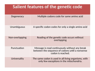
Salient features ofthe genetic code
Degeneracy Multiple codons code for same amino acid
Unambiguous A specific codon codes for only a single amino acid
Non-overlapping Reading of the genetic code occurs without
overlapping
Punctuation Message is read continuously without any break
between the sequence of codons until a nonsense
codon is reached.
Universality The same codon is used in all living organisms, with
only few exceptions in the mitochondria.
4.
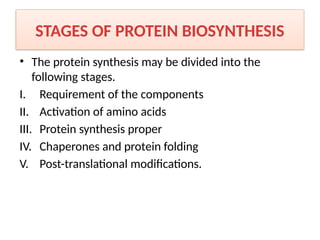
STAGES OF PROTEINBIOSYNTHESIS
• The protein synthesis may be divided into the
following stages.
I. Requirement of the components
II. Activation of amino acids
III. Protein synthesis proper
IV. Chaperones and protein folding
V. Post-translational modifications.
5.
REQUIREMENT OF THECOMPONENTS
1. Amino acids
• Of the 20 amino acids found in protein structure, half of
them (10) can be synthesized by man.
• Therefore, a regular dietary supply of essential amino acids,
should be maintained.
• In prokaryotes, all the 20 are synthesized from the
inorganic components.
2. Ribosomes
• The functionally active ribosomes are the centres or
factories for protein synthesis.
6.
3. Messenger RNA(mRNA)
• It is the carrier of information present in DNA.
4. Transfer RNAs (tRNAs)
• They carry the amino acids and hand over them to
the growing peptide chain.
5. Energy sources
• Both ATP and GTP are required as energy source.
6. Protein factors
• A number of protein factors are needed for initiation,
elongation and termination of protein synthesis.
7.
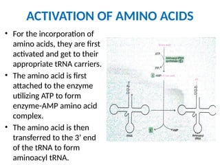
ACTIVATION OF AMINOACIDS
• For the incorporation of
amino acids, they are first
activated and get to their
appropriate tRNA carriers.
• The amino acid is first
attached to the enzyme
utilizing ATP to form
enzyme-AMP amino acid
complex.
• The amino acid is then
transferred to the 3’ end
of the tRNA to form
aminoacyl tRNA.
8.
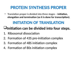
PROTEIN SYNTHESIS PROPER
•Translation proper is divided into three stages – initiation,
elongation and termination (as it is done for transcription).
INITIATION OF TRANSLATION
Initiation can be divided into four steps.
1. Ribosomal dissociation
2. Formation of 43S pre-initiation complex
3. Formation of 48S initiation complex
4. Formation of 80s initiation complex
9.
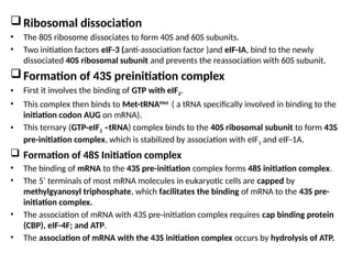
Ribosomal dissociation
• The80S ribosome dissociates to form 40S and 60S subunits.
• Two initiation factors eIF-3 (anti-association factor )and eIF-IA, bind to the newly
dissociated 40S ribosomal subunit and prevents the reassociation with 60S subunit.
Formation of 43S preinitiation complex
• First it involves the binding of GTP with eIF2.
• This complex then binds to Met-tRNAMet
( a tRNA specifically involved in binding to the
initiation codon AUG on mRNA).
• This ternary (GTP-eIF2 –tRNA) complex binds to the 40S ribosomal subunit to form 43S
pre-initiation complex, which is stabilized by association with eIF3 and eIF-1A.
Formation of 48S Initiation complex
• The binding of mRNA to the 43S pre-initiation complex forms 48S initiation complex.
• The 5’ terminals of most mRNA molecules in eukaryotic cells are capped by
methylgyanosyl triphosphate, which facilitates the binding of mRNA to the 43S pre-
initiation complex.
• The association of mRNA with 43S pre-initiation complex requires cap binding protein
(CBP), eIF-4F; and ATP.
• The association of mRNA with the 43S initiation complex occurs by hydrolysis of ATP.
10.
• The ribosomalinitiation complex scans the mRNA for the identification of
appropriate initiation codon.
• 5’-AUG is the initiation codon and its recognition is facilitated by a specific
sequence of nucleotides surrounding it.
• In case of prokaryotes the recognition sequence of initiation codon is referred
to as Shine-Dalgarno sequence.
Formation of 80S initiation complex
• Combinations of the 48S initiation complex with 60S ribosomal subunit forms
80S initiation complex.
• Binding of the 60S ribosomal subunit to the 48S initiation complex involves
the hydrolysis of the GTP bound to eIF2 by eIF5 with the release of the
initiation factors bound to the 48S initiation complex.
• At this stage, the Met-tRNAMet
is on the P site of the ribosome and is now
ready for the elongation process.
12.
ELONGATION OF TRANSLATION
•The amino acid sequence is determined by the
order of the codons in the specific mRNA.
• Elongation, a cyclic process involving certain
elongation factors (Efs) may be divided into three
steps.
1. Binding of aminoacyl t-RNA to A-site
2. Peptide bond formation
3. Translocation
13.
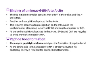
Binding of aminoacyl-tRNAto A-site
• The 80S initiation complex contains met-tRNAi
in the P-site, and the A-
site is free.
• Another aminoacyl-tRNA is placed in the A-site.
• This requires proper codon recognition on the mRNA and the
involvement of elongation factor 1a (EF-Ia) and supply of energy by GTP.
• As the aminoacyl-tRNA is placed in the A-site, EF-1α and GDP are recycled
to bring another aminoacyl-tRNA.
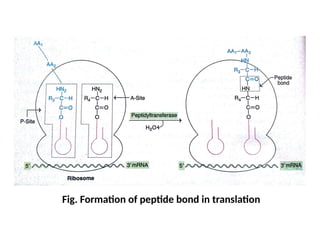
Peptide bond formation
• The enzyme peptidyltransferase catalyzes the formation of peptide bond.
• As the amino acid in the aminoacyl-tRNA is already activated, no
additional energy is required for peptide bond formation.
Translocation
• As thepeptide bond formation occurs, the ribosomes moves to the next
codon of the mRNA (towards 3’-end).
• This process called translocation, basically involves the movement of
growing peptide chain from A-site to P-site.
• Translocation requires EF-2 and GTP.
• GTP gets hydrolyzed and supplies energy to move mRNA.
Incorporation of amino acids
• It is estimated that about six amino acids per second are incorporated
during the course of elongation of translation in eukaryotes.
• In case of prokaryotes, as many as 20 amino acids can be incorporated
per second.
17.
TERMINATION OF TRANSLATION
•After several cycles of elongation, incorporating one of the
stop or termination signals (UAA, UAG and UGA) terminates
the growing polypeptide because termination codons do not
have specific tRNAs to bind.
• In this reaction, a water molecule, instead of an amino acid is
added.
• The hydrolysis releases the protein.
• The 80S ribosome dissociates to form 40S and 60S subunits
which are recycled.
• The mRNA is also releases.
18.
INHIBITORS OF PROTEINSYNTHESIS
• Majority of the antibiotics interfere with the bacterial protein
synthesis and are harmless to higher organisms.
1. Streptomycin
• It inhibits Initiation of protein synthesis.
2. Tetracycline
• Inhibits the binding of aminoacyl tRNA to the ribosomal
complex.
3. Puromycin
• It enters the A site and gets incorporated into the growing
peptide chain and causes its release.
19.
4. Chloramphenicol
• Itacts a competitive inhibitor of the enzyme
peptidyltransferase and thus interferes with elongation of
peptide chain.
5. Erythromycin
• It inhibits translocation by binding with 50s subunit of
bacterial ribosome.
6. Diphtheria toxin
• It prevents translocation in eukaryotic protein synthesis by
inactivating elongation factor eEF2.
20.
CHAPERONES AND PROTEINFOLDING
INTRODUCTION
• The three dimensional conformation of proteins is important for their
biological functions.
• Some of the proteins can spontaneously generate the correct functionally
active conformation e.g. denatured pancreatic ribonuclease.
• Majority of proteins can attain correct conformation, only through the
assistance of certain proteins referred to as chaperones (heat shock
proteins).
• Chaperones can reversibly bind to hydrophobic regions of unfolded proteins
and folding intermediates, prevent formation of incorrect intermediates,
and also prevent undesirable interactions with other proteins.
• All these activities of chaperones help the protein to attain compact and
biologically active conformation.
• The failure of a protein to fold properly generally leads to its rapid
degradation.
• Cystic fibrosis (CF) is a common autosomal recessive disease.
21.
POST-TRANSLATIONAL MODIFICATIONS OF
PROTEINS
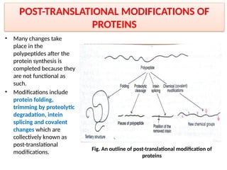
•Many changes take
place in the
polypeptides after the
protein synthesis is
completed because they
are not functional as
such.
• Modifications include
protein folding,
trimming by proteolytic
degradation, intein
splicing and covalent
changes which are
collectively known as
post-translational
modifications.
Fig. An outline of post-translational modification of
proteins