Download to read offline


![ Miller and Harley ..
Internet Kenhub,,
.Hodgkin AL, Huxley AF. A quantitative description of
membrane current and its application to conduction and
excitation in nerve. 1952. Bull. Math. Barnett MW,
Larkman PM. The action potential. Pract Neurol. 2007
Jun;7(3):192-7. [PubMed]
Rutecki PA. Neuronal excitability: voltage-dependent
currents and synaptic transmission. J Clin
Neurophysiol. 1992 Apr;9(2):195-211. [PubMed]
Outline:
Introduction](https://image.slidesharecdn.com/shahzaibkhurshid-210504052945/85/Generation-and-conduction-of-action-potential-2-320.jpg)
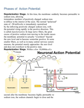
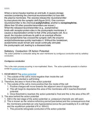
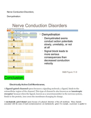

The document discusses the generation and conduction of action potentials in neurons. It covers several key topics: 1) An action potential is initiated when the membrane potential reaches threshold, opening voltage-gated sodium channels and causing rapid sodium influx. This depolarizes the membrane. 2) The membrane then repolarizes as sodium channels close and potassium channels open, allowing potassium efflux. 3) Action potentials propagate along axons via contiguous conduction, with adjacent segments of membrane depolarizing sequentially. Myelination allows faster saltatory conduction. 4) At synapses, neurotransmitters are released from presynaptic terminals and bind to receptors, sometimes depolarizing the postsynaptic cell and propagating the impulse.