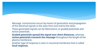
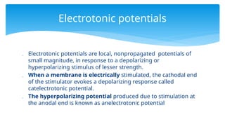
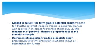
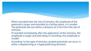
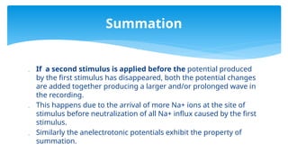
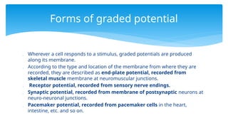
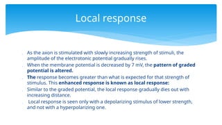
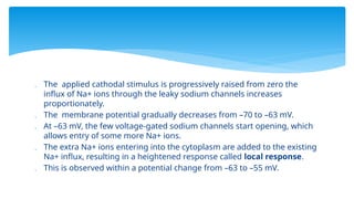
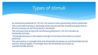
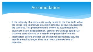
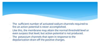
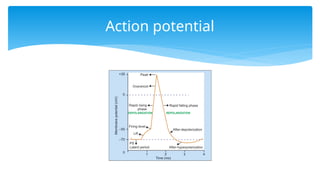
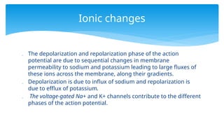
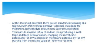
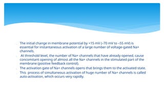
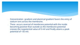
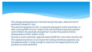
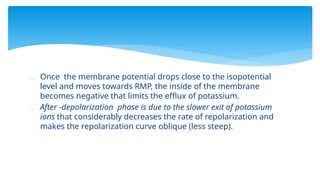
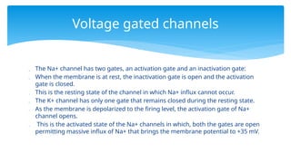
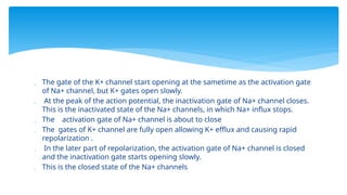
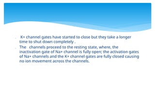
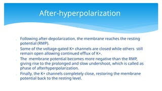
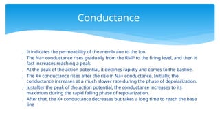
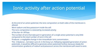
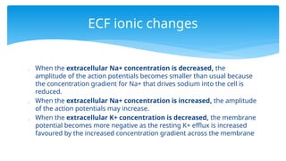
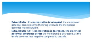
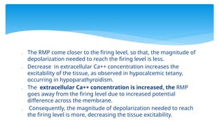
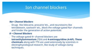
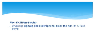
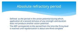
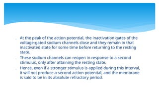
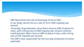
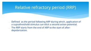
Nerve potential
26/1/2021
 Distribution and working of ion channels on the neuronal membrane.
 The importance of electrotonic potentials.
 Draw a labeled schematic diagram of nerve action potential (AP) and
describe the ionic basis of each phase of AP.
 Define refractory periods (ARP and RRP) of nerve AP and explain their
importance.
 Mechanism of propagation of action potential along the axon and the
importance of saltatory conduction.
 Differences between graded potential and action potential.
Objectives
 Neuronal membrane possesses numerous ion channels like Na+, K+, Ca++, Cl , etc.
−
 Categorized into three types:
i. Nongated or leaky channels
ii. Gated (voltage-gated, ligand-gated and mechanical- gated) channels.
iii. ATP-driven pumps:
 Nongated or leaky channels of Na+, K+, Cl are present throughout the neuronal
−
membrane.
 The voltage-gated Na+ channels are concentrated at the nodes of Ranvier.
 The voltage-gated Ca++ channels are mainly present at the axon terminals, where
they play important role in the secretion of neurotransmitters.
Neuronal ion channels
 The mechanical gated Na+ channels are involved in the genesis
of receptor potential in the somatic sensory nerve endings.
 Ligand-gated ion channels are present predominantly on
dendritic spines, dendrites and cell body of the neuron.
 They are important for receiving information from other neurons
at synaptic sites, in the form of released neurotransmitters.
In myelinated neurons, the number of Na+ channels per square micrometer of
membrane in different segments of the neuron is as follows:
 Cell body: 50–75 2.
 Initial segment: 350–500
 The surface of the myelin: 25 4.
 The nodes of Ranvier: 2000–12,000
 The axon terminals: 20–75
 Thus, the channels are concentrated in areas where the action potential is first
initiated (initial segment) and in regions where it is regenerated (nodes of
Ranvier) during its propagation.
Na+
channel distribution
 In unmyelinated neurons, about 110 Na+ channels are present
per square micrometer of the axonal membrane.
 Abnormalities of channels are called channelopathies
 The diseases caused due to the structural or functional changes
in the ion channels are known as channelopathies.
 Those affecting neurons include episodic and spinocerebellar
ataxias, some forms of epilepsy and familial hemiplegic migraine.
 When there is a voltage difference between the inside and outside
of the membrane, the membrane is said to be polarized.
 When a stimulus allows influx of positive charges or efflux of
negative charges, it decreases the membrane potential (i.e. the
membrane potential approaches towards zero) and the stimulus is
called a depolarizing stimulus. Thus, the membrane is said to
be depolarized
 When the membrane potential becomes positive or less negative
in relation to RMP.
Term definitions
 After the depolarization phase, return of the potential towards
the resting value is known as repolarization.
 As the interior of the cell becomes more negative in relation to
RMP, due to influx of negative charges or removal of positive
charges, the membrane is said to be hyperpolarized.
 In this state, the membrane potential is more negative in relation
to RMP.
 The ability of the cells to generate action potential in their
membrane is known as excitability.
 Nerve is a highly excitable tissue, which can be stimulated by
electrical, chemical and mechanical forms of energy.
 When a stimulus is applied, it induces ions to flow across the
membrane and alters the ionic balance on both sides of the
membrane, producing a voltage change.
 With application of a stronger stimulus, much larger disturbance
in ionic balance occurs.
Generation of nerve potential
 The ionic balance is promptly restored by 2 factors
(repolarizing forces):
1. Diffusion of ions across the cell membrane
2. Increased activity of Na+-K+ ATPase.
 The voltage changes across the membrane generate electrical signals, which on
recording show a wave like pattern.
 The transient and small voltage changes spread along the length of the nerve fiber
and die out after some time.
 When the stimulus is strong enough, the response does not die out fast, rather, it
travels along the whole length of the axon, being regenerated at regular intervals.
 This phenomenon is possible because the neuronal membrane is a biological
membrane studded with different ion channels, whose activation time is modifiable
with change in external environment.
 In the neuron, processing of information takes place chiefly in the cell body and to
some extent in the dendrites.
 Message transmission occurs by means of generation and propagation
of the electrical signals in the axon from one end to the other.
 These generated signals can be Electrotonic or graded potentials and
Action potentials.
 Graded potentials spread the signal over short distances, whereas
action potentials transmit the message throughout the length of the
plasma membrane.
 Another type of response is seen in neuronal membrane that is called
local response.
 Electrotonic potentials are local, nonpropagated potentials of
small magnitude, in response to a depolarizing or
hyperpolarizing stimulus of lesser strength.
 When a membrane is electrically stimulated, the cathodal end
of the stimulator evokes a depolarizing response called
catelectrotonic potential.
 The hyperpolarizing potential produced due to stimulation at
the anodal end is known as anelectrotonic potential
Electrotonic potentials
 Graded in nature: The term graded potential comes from the
fact that the potential change increases in a stepwise manner
with application of increasing strength of stimulus, i.e. the
magnitude of potential change is proportionate to the
stimulus strength.
 Decremental conduction: Graded potentials decay
progressively with time and distance, which is known as
decremental conduction
 When recorded near the site of stimulus, the amplitude of the
potential is larger and recorded at a farther place, it is smaller.
The potentials die out within a distance of 3 mm from the site of
stimulus.
 If recorded immediately after the application of the stimulus, the
amplitude is larger and with delay in recording, the amplitude is
smaller
 According to the type of stimulus, graded potentials can occur in
either a depolarizing or a hyperpolarizing direction.
 If a second stimulus is applied before the potential produced
by the first stimulus has disappeared, both the potential changes
are added together producing a larger and/or prolonged wave in
the recording.
 This happens due to the arrival of more Na+ ions at the site of
stimulus before neutralization of all Na+ influx caused by the first
stimulus.
 Similarly the anelectrotonic potentials exhibit the property of
summation.
Summation
 Wherever a cell responds to a stimulus, graded potentials are produced
along its membrane.
 According to the type and location of the membrane from where they are
recorded, they are described as end-plate potential, recorded from
skeletal muscle membrane at neuromuscular junctions.
 Receptor potential, recorded from sensory nerve endings.
 Synaptic potential, recorded from membrane of postsynaptic neurons at
neuro-neuronal junctions.
 Pacemaker potential, recorded from pacemaker cells in the heart,
intestine, etc. and so on.
Forms of graded potential
 As the axon is stimulated with slowly increasing strength of stimuli, the
amplitude of the electrotonic potential gradually rises.
 When the membrane potential is decreased by 7 mV, the pattern of graded
potential is altered.
 The response becomes greater than what is expected for that strength of
stimulus. This enhanced response is known as local response:
 Similar to the graded potential, the local response gradually dies out with
increasing distance.
 Local response is seen only with a depolarizing stimulus of lower strength,
and not with a hyperpolarizing one.
Local response
 The applied cathodal stimulus is progressively raised from zero the
influx of Na+ ions through the leaky sodium channels increases
proportionately.
 The membrane potential gradually decreases from –70 to –63 mV.
 At –63 mV, the few voltage-gated sodium channels start opening, which
allows entry of some more Na+ ions.
 The extra Na+ ions entering into the cytoplasm are added to the existing
Na+ influx, resulting in a heightened response called local response.
 This is observed within a potential change from –63 to –55 mV.
 At membrane potential of –55 mV, the neuron starts generating action potentials
 This is termed as firing or discharge of the neuron and the membrane potential at –
55 mV is known as the firing level or threshold.
 The stimulus that brings the membrane potential to –55 mV is known as
threshold stimulus.
 Threshold stimulus is the lowest strength of stimulus that elicits an action
potential.
 The stimuli less in strength than the threshold are known as sub threshold stimuli
and the stimuli higher in strength than the threshold are known as
suprathreshold stimuli.
Types of stimuli
 If the intensity of a stimulus is slowly raised to the threshold value,
the tissue fails to produce an action potential because it adapts to
the stimulus. This phenomenon is known as accommodation:
 During the slow depolarization, some of the voltage gated Na+
channels start opening at a membrane potential of –63 mV.
 Inactivated before another set of channel opens; because, the
membrane takes longer time to arrive at the next level of
potential.
Accomodation
 The sufficient number of activated sodium channels required to
fire an action potential is never accomplished.
 Like this, the membrane may attain the normal threshold level or
even surpass that level, but action potential is not produced.
 The potassium channels that open in response to the
depolarization drain off the positive charges.
Action potential
 Action potential is a transient change in membrane potential of
about 100 mV, which is conducted along the axon in an all-or-none
fashion.
 Characterized by a gradual depolarization to threshold, and a rapid
ascent in the membrane potential followed by a phase of
repolarization.
 Travel s along the axon with the same shape and amplitude being
regenerated at regular intervals.
 Also known as an impulse or spike potential.
 The duration of a single nerve action potential is about 1 msec,
during which the membrane potential sharply rises from –70 to
+35 mV, and then returns to its resting value.
Duration and amplitude
 Action potential is always preceded by a latent period, which is
the interval between the application of a stimulus and the
onset of action potential:
 Duration of latent period depends on the distance between
stimulating and recording electrodes and the type and diameter
of nerve fiber.
 The action potentials never summate and a definite interval
(refractory period) exists before the second action potential is
fired.
Latent period
 The phase of depolarization is recorded as a sharp upward wave during which the
membrane potential approaches zero and then attains a positive value.
 It consists of slow depolarization to threshold, rapid rising phase, overshoot and
peak. During overshoot, the membrane potential crosses the zero or isopotential
level and then at peak, it reaches a maximum potential of +35 mV.
 The phase of repolarization is recorded as downstroke during which the membrane
potential returns to the resting level.
 It includes a rapid falling phase and slower terminal part called after-depolarization.
 The phase of repolarization is followed by an afterhyperpolarization phase during
which the membrane potential undershoots (becomes more negative) and then
returns back to the resting level
Phases
 The depolarization and repolarization phase of the action
potential are due to sequential changes in membrane
permeability to sodium and potassium leading to large fluxes of
these ions across the membrane, along their gradients.
 Depolarization is due to influx of sodium and repolarization is
due to efflux of potassium.
 The voltage-gated Na+ and K+ channels contribute to the different
phases of the action potential.
Ionic changes
 Depolarization is due to opening of voltage-gated Na+ channels,
causing massive influx of sodium ions:
 When a threshold or suprathreshold stimulus is applied, the
influx of Na+ through leaky channels and later through opening
of few voltage-gated Na+ channels decreases the membrane
potential from –70 mV to –55 mV (threshold level).
 At this threshold potential, there occurs simultaneousopening of a
large number of the voltage-gatedNa+ channels, increasing the
membrane permeabilityto sodium ions several hundredfold.
 This leads to massive influx of sodium ions producing a swift,
large andsteep depolarization, changing the membrane
potentialto +35 mV (a change in membrane potential by 105 mV
starting from the resting value of –70 mV to +35 mV).
 The initial change in membrane potential by +15 mV (–70 mV to –55 mV) is
essential for instantaneous activation of a large number of voltage-gated Na+
channels.
 At threshold level, the number of Na+ channels that have already opened, cause
concomitant opening of almost all the Na+ channels in the stimulated part of the
membrane (positive feedback control).
 The activation gate of Na+ channels opens that brings them to the activated state.
 This process of simultaneous activation of huge number of Na+ channels is called
auto-activation, which occurs very rapidly.
 Concentration gradient and electrical gradient favors the entry of
sodium ions across the membrane.
 There occurs reversal of membrane potential with the inside
becoming positive than outside as the membrane potential
crosses the isopotential value of 0 mV and finally attains a peak
potential of +35 mV.
 During depolarization, the membrane potential approaches but
does not reach the equilibrium potential for sodium, which is +60
mV due to;
 At the peak of the action potential Sodium influx abruptly ceases
due to the closure of the inactivation gates of the Na+ channels.
 The Na+ channels open very fast, remain open for a very brief
period, and they close very fast.
 The process of speedy closure is called rapid autodeactivation.
MP below equilibrium
 The voltage-gated potassium channels being fully open, allow the exit of
positively charged K+ ions.
 The electrical gradient for Na+ is reversed subsequent to the overshoot, i.e.
after crossing RMP of 0 mV, inside of the cell membrane becomes positive
and it hinders the positively charged Na+ to enter the positive interior,
slowing down further sodium entry.
 During an action potential, approximately 20,000 Na+ ions enter into the cell.
 The repolarizing forces try to restore the resting membrane potential, but
the depolarization is large enough to overcome the opposing forces and
produce an action potential.
 Repolarization is due to opening of voltage-gated K+ channels,
causing efflux of K+.
 Actually, these K+ channels are sensitive to the same
depolarization that opens the voltage- gated Na+ channels but
they open more slowly:
 At the peak of the action potential, the voltage-gated Na+
channels enter a closed state whereas the voltage gated K+
channels are fully open.
Repolarization
 The membrane permeability to potassium ions increases several times
causing increased potassium efflux.
 The K+ concentration is much higher inside the cell and at the peak of the
action potential, outside of the membrane is negative in comparison to
inside, which is positive (+35 mV).
 These two factors favour the electrochemical gradient for potassium efflux.
 Rapid falling phase of repolarization is brought about by decline in sodium
influx together with increase in potassium efflux.
 The termination of action potential due to activation of voltage-gated
potassium channels is a negative feedback process.
 Once the membrane potential drops close to the isopotential
level and moves towards RMP, the inside of the membrane
becomes negative that limits the efflux of potassium.
 After -depolarization phase is due to the slower exit of potassium
ions that considerably decreases the rate of repolarization and
makes the repolarization curve oblique (less steep).
 The Na+ channel has two gates, an activation gate and an inactivation gate:
 When the membrane is at rest, the inactivation gate is open and the activation
gate is closed.
 This is the resting state of the channel in which Na+ influx cannot occur.
 The K+ channel has only one gate that remains closed during the resting state.
 As the membrane is depolarized to the firing level, the activation gate of Na+
channel opens.
 This is the activated state of the Na+ channels in which, both the gates are open
permitting massive influx of Na+ that brings the membrane potential to +35 mV.
Voltage gated channels
 The gate of the K+ channel start opening at the sametime as the activation gate
of Na+ channel, but K+ gates open slowly.
 At the peak of the action potential, the inactivation gate of Na+ channel closes.
This is the inactivated state of the Na+ channels, in which Na+ influx stops.
 The activation gate of Na+ channel is about to close
 The gates of K+ channel are fully open allowing K+ efflux and causing rapid
repolarization .
 In the later part of repolarization, the activation gate of Na+ channel is closed
and the inactivation gate starts opening slowly.
 This is the closed state of the Na+ channels
 K+ channel gates have started to close but they take a longer
time to shut down completely .
 The channels proceed to the resting state, where, the
inactivation gate of Na+ channel is fully open; the activation gates
of Na+ channels and the K+ channel gates are fully closed causing
no ion movement across the channels.
 Following after-depolarization, the membrane reaches the resting
potential (RMP).
 Some of the voltage-gated K+ channels are closed while others still
remain open allowing continued efflux of K+.
 The membrane potential becomes more negative than the RMP,
giving rise to the prolonged and slow undershoot, which is called as
phase of afterhyperpolarization.
 Finally, the K+ channels completely close, restoring the membrane
potential back to the resting level.
After-hyperpolarization
 It indicates the permeability of the membrane to the ion.
 The Na+ conductance rises gradually from the RMP to the firing level, and then it
fast increases reaching a peak.
 At the peak of the action potential, it declines rapidly and comes to the basline.
 The K+ conductance rises after the rise in Na+ conductance. Initially, the
conductance increases at a much slower rate during the phase of depolarization.
 Justafter the peak of the action potential, the conductance increases to its
maximum during the rapid falling phase of repolarization.
 After that, the K+ conductance decreases but takes a long time to reach the base
line
Conductance
 At the end of an action potential, the ionic composition on both sides of the membrane is
altered.
 More sodium and less potassium inside the cell:
 The ionic composition is restored by increased activity
 of the Na+-K+ ATPase.
 The number of ions that take part in generation of a single action potential is very little
compared to the total number of ions in the cell
 Produces a negligible change in the intracellular ionic concentration.
 This imbalance , not taken care of, in the long run, following repeated generation of action
potentials, it would be difficult to generate action potentials further, because slowly the
concentration gradients of sodium and potassium across the membrane will cease to exist.
Ionic activity after action potential
 When the extracellular Na+ concentration is decreased, the
amplitude of the action potentials becomes smaller than usual because
the concentration gradient for Na+ that drives sodium into the cell is
reduced.
 When the extracellular Na+ concentration is increased, the amplitude
of the action potentials may increase.
 When the extracellular K+ concentration is decreased, the membrane
potential becomes more negative as the resting K+ efflux is increased
favoured by the increased concentration gradient across the membrane
ECF ionic changes
 Extracellular K+ concentration is increased, the membrane
potential come closer to the firing level and the membrane
becomes more excitable.
 Extracellular Ca++ concentration is decreased, the electrical
potential difference across the membrane is decreased, as the
inside becomes less negative compared to outside.
 The RMP come closer to the firing level, so that, the magnitude of
depolarization needed to reach the firing level is less.
 Decrease in extracellular Ca++ concentration increases the
excitability of the tissue, as observed in hypocalcemic tetany,
occurring in hypoparathyroidism.
 The extracellular Ca++ concentration is increased, the RMP
goes away from the firing level due to increased potential
difference across the membrane.
 Consequently, the magnitude of depolarization needed to reach
the firing level is more, decreasing the tissue excitability.
Na+ Channel Blockers
 Drugs like lidocaine, procaine ‘etc., and neurotoxins like
tetrodotoxin, saxitoxin’ etc., block the voltage-gated Na+ channels
and hinder the generation of action potentials
K + Channel Blockers
 The voltage-gated K+ channel blockers are
tetraethylammonium (TEA) and 4-aminopyridine (4-AP). These
chemicals along with TTX are used extensively by scientists in
electrophysiological research, like study of voltage-clamp
techniques.
Ion channel blockers
Na+– K+ ATPase Blocker
 Drugs like digitalis and dinitrophenol block the Na+–K+ ATPase
pump.
 All or none states that the action potential occurs with a constant
amplitude and shape irrespective of magnitude of the stimulus.
 A subthreshold stimulus fails to excite the tissue.
 Only a stimulus of threshold magnitude elicits an action potential.
 If suprathreshold stimuli are applied, the action potentials resulting
from them have the same amplitude, duration and form as those
produced by threshold stimuli, provided the experimental conditions
like electrical potentials on both sides of the membrane,
concentration of ions in ICF and ECF, temperature’ etc. remain same.
All or None Law
 The activation gates of voltage-gated Na+ channels open as soon as 15
mV of depolarization is achieved.
 Any extra degree of depolarization is of no further use, as the membrane
automatically achieves another +90 mV of depolarization (–55 to +35 mV):
 Thus, after the threshold level is achieved, the amount of sodium influx
becomes independent of the stimulus factor.
 The number of voltage-gated Na+ channels over the axonal membrane of
unmyelinated axons as well as at the nodes of Ranvier in myelinated
axons remain fairly constant.
 Once the action potential is formed, it appears with its maximum
size and shape, otherwise it does not form at all.
 The action potential follows the all-or-none law; i.e. the action
potential occurs with a constant amplitude and shape whether
the stimulus is of threshold or suprathreshold magnitude.
 During the action potential, the stimulated area of the membrane
happens to be unresponsive to a second stimulus in most part,
and later it requires a stronger stimulus to get excited again.
 The length of time during which the membrane is
unresponsive to a second stimulus no matter how strong is the
stimulus, is known as refractory period.
 The periods of total and relative refractoriness are known as
absolute and relative refractory periods respectively.
Refractory period
 Defined as the period in the action potential during which,
application of a second stimulus of any strength and duration
does not produce another action potential.
 The ARP corresponds to the period from the time the firing level
is reached until repolarization is about one-third complete
Absolute refractory period
 At the peak of the action potential, the inactivation gates of the
voltage-gated sodium channels close and they remain in that
inactivated state for some time before returning to the resting
state.
 These sodium channels can reopen in response to a second
stimulus, only after attaining the resting state.
 Hence, even if a stronger stimulus is applied during this interval,
it will not produce a second action potential, and the membrane
is said to be in its absolute refractory period.
 ARP determines the rate of discharge of nerve fiber.
 In our body, nerves fire at a rate of 10 to 1000 impulses per
second.
 Generally, large diameter nerve fibers have an ARP of about 0.4
msec, with a firing rate of 2500 impulses per second, whereas
small diameter fibers have an ARP of about 4 msec, with a firing
rate of 250 impulses per second.
 The ARP is also responsible for the one-way conduction of action
potentials
 Defined as the period following ARP during which, application of
a suprathreshold stimulus can elicit a second action potential.
 The RRP starts from the end of ARP to the start of after-
depolarization.
Relative refractory period (RRP)
 All the sodium channels present at the site of stimulus do not
achieve the open state or inactivated state or resting state,
exactly at the same time.
 Few of them open when the membrane potential is –63 mV,
causing local response.
 By the time of relative refractory period, some of the channels
have returned to their initial resting state.
 These channels in resting state can open their activation gate and
allow the influx of Na+.
 A suprathreshold stimulus can spread to larger area over the membrane and
open extra voltage-gated sodium channel. only a suprathreshold stimulus
and not any threshold stimulus can open up sufficient number of sodium
channels to elicit an action potential during the RRP.
 The excitability of the membrane keeps on changing in different phases
of the action potential.
 The time the membrane potential is closer to the firing level, the membrane
is more excitable; and the excitability of the membrane decreases, when its
potential is away from the firing level.
 During the ARP, it is least excitable.
 Production of action potentials requires the presence of large number
of voltage-gated ion channels that are present mostly on the axons.
 Therefore, it is the axon, not the cell body or the dendrites that
generate and conduct the action potentials.
 The action potential is first initiated in the first node of Ranvier in
sensory neurons and initial segment-axon hillock area in motor
neurons.
 These areas are known as trigger zones that have a very high
concentration of voltage-gated sodium and potassium channels.
Initiation of action potential
 The synaptic potential generated at the dendrites and, or the cell
body is integrated by the cell body and transmitted to the axon
hillock.
 If this potential is sufficient to depolarize the membrane of the
axon hillock to firing level, the membrane easily fires an action
potential.
 Once formed, the action potential is regenerated at regular
intervals to be transmitted from the initial segment of the axon to
the axon terminal.
 This is known as the propagation of action potential.
 In myelinated axon, the speed and mode of propagation of
action potential is different from that in unmyelinated axon.
Propagation
The speed of conduction of the impulse depends on
 Myelination: Conduction velocity is more in myelinated axon and
is proportionate to the degree of myelination.
 Diameter of the axon: Conduction velocity is proportionate to the
diameter of the fiber.
 Fibers with larger diameter have faster rate of conduction. The
large diameter fibers have less cytoplasmic resistance.
 The flow of ions across the membrane is easier.
Unmyelinated fibres
Myelinated fibers
 In the motor neuron, the action potential is conducted from axon hillock
toward axon terminal.
 In the sensory neuron, it propagates from the first node of Ranvier toward
CNS.
 This is called anterograde conduction of impulse:
 The action potential does not travel from the axon back toward the trigger
zone. This is because; following depolarization, the area on the membrane
where action potential was produced becomes refractory.
 The action potential can be conducted only in the direction away from the
site of previous action potential.
Propagation of AP

Nerve potential 2021. it's transmissionn

  • 1.
  • 2.
     Distribution andworking of ion channels on the neuronal membrane.  The importance of electrotonic potentials.  Draw a labeled schematic diagram of nerve action potential (AP) and describe the ionic basis of each phase of AP.  Define refractory periods (ARP and RRP) of nerve AP and explain their importance.  Mechanism of propagation of action potential along the axon and the importance of saltatory conduction.  Differences between graded potential and action potential. Objectives
  • 3.
     Neuronal membranepossesses numerous ion channels like Na+, K+, Ca++, Cl , etc. −  Categorized into three types: i. Nongated or leaky channels ii. Gated (voltage-gated, ligand-gated and mechanical- gated) channels. iii. ATP-driven pumps:  Nongated or leaky channels of Na+, K+, Cl are present throughout the neuronal − membrane.  The voltage-gated Na+ channels are concentrated at the nodes of Ranvier.  The voltage-gated Ca++ channels are mainly present at the axon terminals, where they play important role in the secretion of neurotransmitters. Neuronal ion channels
  • 4.
     The mechanicalgated Na+ channels are involved in the genesis of receptor potential in the somatic sensory nerve endings.  Ligand-gated ion channels are present predominantly on dendritic spines, dendrites and cell body of the neuron.  They are important for receiving information from other neurons at synaptic sites, in the form of released neurotransmitters.
  • 5.
    In myelinated neurons,the number of Na+ channels per square micrometer of membrane in different segments of the neuron is as follows:  Cell body: 50–75 2.  Initial segment: 350–500  The surface of the myelin: 25 4.  The nodes of Ranvier: 2000–12,000  The axon terminals: 20–75  Thus, the channels are concentrated in areas where the action potential is first initiated (initial segment) and in regions where it is regenerated (nodes of Ranvier) during its propagation. Na+ channel distribution
  • 6.
     In unmyelinatedneurons, about 110 Na+ channels are present per square micrometer of the axonal membrane.  Abnormalities of channels are called channelopathies  The diseases caused due to the structural or functional changes in the ion channels are known as channelopathies.  Those affecting neurons include episodic and spinocerebellar ataxias, some forms of epilepsy and familial hemiplegic migraine.
  • 7.
     When thereis a voltage difference between the inside and outside of the membrane, the membrane is said to be polarized.  When a stimulus allows influx of positive charges or efflux of negative charges, it decreases the membrane potential (i.e. the membrane potential approaches towards zero) and the stimulus is called a depolarizing stimulus. Thus, the membrane is said to be depolarized  When the membrane potential becomes positive or less negative in relation to RMP. Term definitions
  • 8.
     After thedepolarization phase, return of the potential towards the resting value is known as repolarization.  As the interior of the cell becomes more negative in relation to RMP, due to influx of negative charges or removal of positive charges, the membrane is said to be hyperpolarized.  In this state, the membrane potential is more negative in relation to RMP.
  • 9.
     The abilityof the cells to generate action potential in their membrane is known as excitability.  Nerve is a highly excitable tissue, which can be stimulated by electrical, chemical and mechanical forms of energy.  When a stimulus is applied, it induces ions to flow across the membrane and alters the ionic balance on both sides of the membrane, producing a voltage change.  With application of a stronger stimulus, much larger disturbance in ionic balance occurs. Generation of nerve potential
  • 10.
     The ionicbalance is promptly restored by 2 factors (repolarizing forces): 1. Diffusion of ions across the cell membrane 2. Increased activity of Na+-K+ ATPase.
  • 11.
     The voltagechanges across the membrane generate electrical signals, which on recording show a wave like pattern.  The transient and small voltage changes spread along the length of the nerve fiber and die out after some time.  When the stimulus is strong enough, the response does not die out fast, rather, it travels along the whole length of the axon, being regenerated at regular intervals.  This phenomenon is possible because the neuronal membrane is a biological membrane studded with different ion channels, whose activation time is modifiable with change in external environment.  In the neuron, processing of information takes place chiefly in the cell body and to some extent in the dendrites.
  • 12.
     Message transmissionoccurs by means of generation and propagation of the electrical signals in the axon from one end to the other.  These generated signals can be Electrotonic or graded potentials and Action potentials.  Graded potentials spread the signal over short distances, whereas action potentials transmit the message throughout the length of the plasma membrane.  Another type of response is seen in neuronal membrane that is called local response.
  • 13.
     Electrotonic potentialsare local, nonpropagated potentials of small magnitude, in response to a depolarizing or hyperpolarizing stimulus of lesser strength.  When a membrane is electrically stimulated, the cathodal end of the stimulator evokes a depolarizing response called catelectrotonic potential.  The hyperpolarizing potential produced due to stimulation at the anodal end is known as anelectrotonic potential Electrotonic potentials
  • 15.
     Graded innature: The term graded potential comes from the fact that the potential change increases in a stepwise manner with application of increasing strength of stimulus, i.e. the magnitude of potential change is proportionate to the stimulus strength.  Decremental conduction: Graded potentials decay progressively with time and distance, which is known as decremental conduction
  • 16.
     When recordednear the site of stimulus, the amplitude of the potential is larger and recorded at a farther place, it is smaller. The potentials die out within a distance of 3 mm from the site of stimulus.  If recorded immediately after the application of the stimulus, the amplitude is larger and with delay in recording, the amplitude is smaller  According to the type of stimulus, graded potentials can occur in either a depolarizing or a hyperpolarizing direction.
  • 17.
     If asecond stimulus is applied before the potential produced by the first stimulus has disappeared, both the potential changes are added together producing a larger and/or prolonged wave in the recording.  This happens due to the arrival of more Na+ ions at the site of stimulus before neutralization of all Na+ influx caused by the first stimulus.  Similarly the anelectrotonic potentials exhibit the property of summation. Summation
  • 18.
     Wherever acell responds to a stimulus, graded potentials are produced along its membrane.  According to the type and location of the membrane from where they are recorded, they are described as end-plate potential, recorded from skeletal muscle membrane at neuromuscular junctions.  Receptor potential, recorded from sensory nerve endings.  Synaptic potential, recorded from membrane of postsynaptic neurons at neuro-neuronal junctions.  Pacemaker potential, recorded from pacemaker cells in the heart, intestine, etc. and so on. Forms of graded potential
  • 19.
     As theaxon is stimulated with slowly increasing strength of stimuli, the amplitude of the electrotonic potential gradually rises.  When the membrane potential is decreased by 7 mV, the pattern of graded potential is altered.  The response becomes greater than what is expected for that strength of stimulus. This enhanced response is known as local response:  Similar to the graded potential, the local response gradually dies out with increasing distance.  Local response is seen only with a depolarizing stimulus of lower strength, and not with a hyperpolarizing one. Local response
  • 20.
     The appliedcathodal stimulus is progressively raised from zero the influx of Na+ ions through the leaky sodium channels increases proportionately.  The membrane potential gradually decreases from –70 to –63 mV.  At –63 mV, the few voltage-gated sodium channels start opening, which allows entry of some more Na+ ions.  The extra Na+ ions entering into the cytoplasm are added to the existing Na+ influx, resulting in a heightened response called local response.  This is observed within a potential change from –63 to –55 mV.
  • 21.
     At membranepotential of –55 mV, the neuron starts generating action potentials  This is termed as firing or discharge of the neuron and the membrane potential at – 55 mV is known as the firing level or threshold.  The stimulus that brings the membrane potential to –55 mV is known as threshold stimulus.  Threshold stimulus is the lowest strength of stimulus that elicits an action potential.  The stimuli less in strength than the threshold are known as sub threshold stimuli and the stimuli higher in strength than the threshold are known as suprathreshold stimuli. Types of stimuli
  • 22.
     If theintensity of a stimulus is slowly raised to the threshold value, the tissue fails to produce an action potential because it adapts to the stimulus. This phenomenon is known as accommodation:  During the slow depolarization, some of the voltage gated Na+ channels start opening at a membrane potential of –63 mV.  Inactivated before another set of channel opens; because, the membrane takes longer time to arrive at the next level of potential. Accomodation
  • 23.
     The sufficientnumber of activated sodium channels required to fire an action potential is never accomplished.  Like this, the membrane may attain the normal threshold level or even surpass that level, but action potential is not produced.  The potassium channels that open in response to the depolarization drain off the positive charges.
  • 24.
  • 25.
     Action potentialis a transient change in membrane potential of about 100 mV, which is conducted along the axon in an all-or-none fashion.  Characterized by a gradual depolarization to threshold, and a rapid ascent in the membrane potential followed by a phase of repolarization.  Travel s along the axon with the same shape and amplitude being regenerated at regular intervals.  Also known as an impulse or spike potential.
  • 26.
     The durationof a single nerve action potential is about 1 msec, during which the membrane potential sharply rises from –70 to +35 mV, and then returns to its resting value. Duration and amplitude
  • 27.
     Action potentialis always preceded by a latent period, which is the interval between the application of a stimulus and the onset of action potential:  Duration of latent period depends on the distance between stimulating and recording electrodes and the type and diameter of nerve fiber.  The action potentials never summate and a definite interval (refractory period) exists before the second action potential is fired. Latent period
  • 28.
     The phaseof depolarization is recorded as a sharp upward wave during which the membrane potential approaches zero and then attains a positive value.  It consists of slow depolarization to threshold, rapid rising phase, overshoot and peak. During overshoot, the membrane potential crosses the zero or isopotential level and then at peak, it reaches a maximum potential of +35 mV.  The phase of repolarization is recorded as downstroke during which the membrane potential returns to the resting level.  It includes a rapid falling phase and slower terminal part called after-depolarization.  The phase of repolarization is followed by an afterhyperpolarization phase during which the membrane potential undershoots (becomes more negative) and then returns back to the resting level Phases
  • 29.
     The depolarizationand repolarization phase of the action potential are due to sequential changes in membrane permeability to sodium and potassium leading to large fluxes of these ions across the membrane, along their gradients.  Depolarization is due to influx of sodium and repolarization is due to efflux of potassium.  The voltage-gated Na+ and K+ channels contribute to the different phases of the action potential. Ionic changes
  • 30.
     Depolarization isdue to opening of voltage-gated Na+ channels, causing massive influx of sodium ions:  When a threshold or suprathreshold stimulus is applied, the influx of Na+ through leaky channels and later through opening of few voltage-gated Na+ channels decreases the membrane potential from –70 mV to –55 mV (threshold level).
  • 31.
     At thisthreshold potential, there occurs simultaneousopening of a large number of the voltage-gatedNa+ channels, increasing the membrane permeabilityto sodium ions several hundredfold.  This leads to massive influx of sodium ions producing a swift, large andsteep depolarization, changing the membrane potentialto +35 mV (a change in membrane potential by 105 mV starting from the resting value of –70 mV to +35 mV).
  • 32.
     The initialchange in membrane potential by +15 mV (–70 mV to –55 mV) is essential for instantaneous activation of a large number of voltage-gated Na+ channels.  At threshold level, the number of Na+ channels that have already opened, cause concomitant opening of almost all the Na+ channels in the stimulated part of the membrane (positive feedback control).  The activation gate of Na+ channels opens that brings them to the activated state.  This process of simultaneous activation of huge number of Na+ channels is called auto-activation, which occurs very rapidly.
  • 33.
     Concentration gradientand electrical gradient favors the entry of sodium ions across the membrane.  There occurs reversal of membrane potential with the inside becoming positive than outside as the membrane potential crosses the isopotential value of 0 mV and finally attains a peak potential of +35 mV.
  • 34.
     During depolarization,the membrane potential approaches but does not reach the equilibrium potential for sodium, which is +60 mV due to;  At the peak of the action potential Sodium influx abruptly ceases due to the closure of the inactivation gates of the Na+ channels.  The Na+ channels open very fast, remain open for a very brief period, and they close very fast.  The process of speedy closure is called rapid autodeactivation. MP below equilibrium
  • 35.
     The voltage-gatedpotassium channels being fully open, allow the exit of positively charged K+ ions.  The electrical gradient for Na+ is reversed subsequent to the overshoot, i.e. after crossing RMP of 0 mV, inside of the cell membrane becomes positive and it hinders the positively charged Na+ to enter the positive interior, slowing down further sodium entry.  During an action potential, approximately 20,000 Na+ ions enter into the cell.  The repolarizing forces try to restore the resting membrane potential, but the depolarization is large enough to overcome the opposing forces and produce an action potential.
  • 36.
     Repolarization isdue to opening of voltage-gated K+ channels, causing efflux of K+.  Actually, these K+ channels are sensitive to the same depolarization that opens the voltage- gated Na+ channels but they open more slowly:  At the peak of the action potential, the voltage-gated Na+ channels enter a closed state whereas the voltage gated K+ channels are fully open. Repolarization
  • 37.
     The membranepermeability to potassium ions increases several times causing increased potassium efflux.  The K+ concentration is much higher inside the cell and at the peak of the action potential, outside of the membrane is negative in comparison to inside, which is positive (+35 mV).  These two factors favour the electrochemical gradient for potassium efflux.  Rapid falling phase of repolarization is brought about by decline in sodium influx together with increase in potassium efflux.  The termination of action potential due to activation of voltage-gated potassium channels is a negative feedback process.
  • 38.
     Once themembrane potential drops close to the isopotential level and moves towards RMP, the inside of the membrane becomes negative that limits the efflux of potassium.  After -depolarization phase is due to the slower exit of potassium ions that considerably decreases the rate of repolarization and makes the repolarization curve oblique (less steep).
  • 39.
     The Na+channel has two gates, an activation gate and an inactivation gate:  When the membrane is at rest, the inactivation gate is open and the activation gate is closed.  This is the resting state of the channel in which Na+ influx cannot occur.  The K+ channel has only one gate that remains closed during the resting state.  As the membrane is depolarized to the firing level, the activation gate of Na+ channel opens.  This is the activated state of the Na+ channels in which, both the gates are open permitting massive influx of Na+ that brings the membrane potential to +35 mV. Voltage gated channels
  • 40.
     The gateof the K+ channel start opening at the sametime as the activation gate of Na+ channel, but K+ gates open slowly.  At the peak of the action potential, the inactivation gate of Na+ channel closes. This is the inactivated state of the Na+ channels, in which Na+ influx stops.  The activation gate of Na+ channel is about to close  The gates of K+ channel are fully open allowing K+ efflux and causing rapid repolarization .  In the later part of repolarization, the activation gate of Na+ channel is closed and the inactivation gate starts opening slowly.  This is the closed state of the Na+ channels
  • 42.
     K+ channelgates have started to close but they take a longer time to shut down completely .  The channels proceed to the resting state, where, the inactivation gate of Na+ channel is fully open; the activation gates of Na+ channels and the K+ channel gates are fully closed causing no ion movement across the channels.
  • 43.
     Following after-depolarization,the membrane reaches the resting potential (RMP).  Some of the voltage-gated K+ channels are closed while others still remain open allowing continued efflux of K+.  The membrane potential becomes more negative than the RMP, giving rise to the prolonged and slow undershoot, which is called as phase of afterhyperpolarization.  Finally, the K+ channels completely close, restoring the membrane potential back to the resting level. After-hyperpolarization
  • 44.
     It indicatesthe permeability of the membrane to the ion.  The Na+ conductance rises gradually from the RMP to the firing level, and then it fast increases reaching a peak.  At the peak of the action potential, it declines rapidly and comes to the basline.  The K+ conductance rises after the rise in Na+ conductance. Initially, the conductance increases at a much slower rate during the phase of depolarization.  Justafter the peak of the action potential, the conductance increases to its maximum during the rapid falling phase of repolarization.  After that, the K+ conductance decreases but takes a long time to reach the base line Conductance
  • 46.
     At theend of an action potential, the ionic composition on both sides of the membrane is altered.  More sodium and less potassium inside the cell:  The ionic composition is restored by increased activity  of the Na+-K+ ATPase.  The number of ions that take part in generation of a single action potential is very little compared to the total number of ions in the cell  Produces a negligible change in the intracellular ionic concentration.  This imbalance , not taken care of, in the long run, following repeated generation of action potentials, it would be difficult to generate action potentials further, because slowly the concentration gradients of sodium and potassium across the membrane will cease to exist. Ionic activity after action potential
  • 47.
     When theextracellular Na+ concentration is decreased, the amplitude of the action potentials becomes smaller than usual because the concentration gradient for Na+ that drives sodium into the cell is reduced.  When the extracellular Na+ concentration is increased, the amplitude of the action potentials may increase.  When the extracellular K+ concentration is decreased, the membrane potential becomes more negative as the resting K+ efflux is increased favoured by the increased concentration gradient across the membrane ECF ionic changes
  • 48.
     Extracellular K+concentration is increased, the membrane potential come closer to the firing level and the membrane becomes more excitable.  Extracellular Ca++ concentration is decreased, the electrical potential difference across the membrane is decreased, as the inside becomes less negative compared to outside.
  • 49.
     The RMPcome closer to the firing level, so that, the magnitude of depolarization needed to reach the firing level is less.  Decrease in extracellular Ca++ concentration increases the excitability of the tissue, as observed in hypocalcemic tetany, occurring in hypoparathyroidism.  The extracellular Ca++ concentration is increased, the RMP goes away from the firing level due to increased potential difference across the membrane.  Consequently, the magnitude of depolarization needed to reach the firing level is more, decreasing the tissue excitability.
  • 50.
    Na+ Channel Blockers Drugs like lidocaine, procaine ‘etc., and neurotoxins like tetrodotoxin, saxitoxin’ etc., block the voltage-gated Na+ channels and hinder the generation of action potentials K + Channel Blockers  The voltage-gated K+ channel blockers are tetraethylammonium (TEA) and 4-aminopyridine (4-AP). These chemicals along with TTX are used extensively by scientists in electrophysiological research, like study of voltage-clamp techniques. Ion channel blockers
  • 51.
    Na+– K+ ATPaseBlocker  Drugs like digitalis and dinitrophenol block the Na+–K+ ATPase pump.
  • 52.
     All ornone states that the action potential occurs with a constant amplitude and shape irrespective of magnitude of the stimulus.  A subthreshold stimulus fails to excite the tissue.  Only a stimulus of threshold magnitude elicits an action potential.  If suprathreshold stimuli are applied, the action potentials resulting from them have the same amplitude, duration and form as those produced by threshold stimuli, provided the experimental conditions like electrical potentials on both sides of the membrane, concentration of ions in ICF and ECF, temperature’ etc. remain same. All or None Law
  • 54.
     The activationgates of voltage-gated Na+ channels open as soon as 15 mV of depolarization is achieved.  Any extra degree of depolarization is of no further use, as the membrane automatically achieves another +90 mV of depolarization (–55 to +35 mV):  Thus, after the threshold level is achieved, the amount of sodium influx becomes independent of the stimulus factor.  The number of voltage-gated Na+ channels over the axonal membrane of unmyelinated axons as well as at the nodes of Ranvier in myelinated axons remain fairly constant.
  • 55.
     Once theaction potential is formed, it appears with its maximum size and shape, otherwise it does not form at all.  The action potential follows the all-or-none law; i.e. the action potential occurs with a constant amplitude and shape whether the stimulus is of threshold or suprathreshold magnitude.
  • 56.
     During theaction potential, the stimulated area of the membrane happens to be unresponsive to a second stimulus in most part, and later it requires a stronger stimulus to get excited again.  The length of time during which the membrane is unresponsive to a second stimulus no matter how strong is the stimulus, is known as refractory period.  The periods of total and relative refractoriness are known as absolute and relative refractory periods respectively. Refractory period
  • 57.
     Defined asthe period in the action potential during which, application of a second stimulus of any strength and duration does not produce another action potential.  The ARP corresponds to the period from the time the firing level is reached until repolarization is about one-third complete Absolute refractory period
  • 58.
     At thepeak of the action potential, the inactivation gates of the voltage-gated sodium channels close and they remain in that inactivated state for some time before returning to the resting state.  These sodium channels can reopen in response to a second stimulus, only after attaining the resting state.  Hence, even if a stronger stimulus is applied during this interval, it will not produce a second action potential, and the membrane is said to be in its absolute refractory period.
  • 60.
     ARP determinesthe rate of discharge of nerve fiber.  In our body, nerves fire at a rate of 10 to 1000 impulses per second.  Generally, large diameter nerve fibers have an ARP of about 0.4 msec, with a firing rate of 2500 impulses per second, whereas small diameter fibers have an ARP of about 4 msec, with a firing rate of 250 impulses per second.  The ARP is also responsible for the one-way conduction of action potentials
  • 61.
     Defined asthe period following ARP during which, application of a suprathreshold stimulus can elicit a second action potential.  The RRP starts from the end of ARP to the start of after- depolarization. Relative refractory period (RRP)
  • 62.
     All thesodium channels present at the site of stimulus do not achieve the open state or inactivated state or resting state, exactly at the same time.  Few of them open when the membrane potential is –63 mV, causing local response.  By the time of relative refractory period, some of the channels have returned to their initial resting state.  These channels in resting state can open their activation gate and allow the influx of Na+.
  • 63.
     A suprathresholdstimulus can spread to larger area over the membrane and open extra voltage-gated sodium channel. only a suprathreshold stimulus and not any threshold stimulus can open up sufficient number of sodium channels to elicit an action potential during the RRP.  The excitability of the membrane keeps on changing in different phases of the action potential.  The time the membrane potential is closer to the firing level, the membrane is more excitable; and the excitability of the membrane decreases, when its potential is away from the firing level.  During the ARP, it is least excitable.
  • 64.
     Production ofaction potentials requires the presence of large number of voltage-gated ion channels that are present mostly on the axons.  Therefore, it is the axon, not the cell body or the dendrites that generate and conduct the action potentials.  The action potential is first initiated in the first node of Ranvier in sensory neurons and initial segment-axon hillock area in motor neurons.  These areas are known as trigger zones that have a very high concentration of voltage-gated sodium and potassium channels. Initiation of action potential
  • 65.
     The synapticpotential generated at the dendrites and, or the cell body is integrated by the cell body and transmitted to the axon hillock.  If this potential is sufficient to depolarize the membrane of the axon hillock to firing level, the membrane easily fires an action potential.
  • 66.
     Once formed,the action potential is regenerated at regular intervals to be transmitted from the initial segment of the axon to the axon terminal.  This is known as the propagation of action potential.  In myelinated axon, the speed and mode of propagation of action potential is different from that in unmyelinated axon. Propagation
  • 67.
    The speed ofconduction of the impulse depends on  Myelination: Conduction velocity is more in myelinated axon and is proportionate to the degree of myelination.  Diameter of the axon: Conduction velocity is proportionate to the diameter of the fiber.  Fibers with larger diameter have faster rate of conduction. The large diameter fibers have less cytoplasmic resistance.  The flow of ions across the membrane is easier.
  • 68.
  • 69.
  • 70.
     In themotor neuron, the action potential is conducted from axon hillock toward axon terminal.  In the sensory neuron, it propagates from the first node of Ranvier toward CNS.  This is called anterograde conduction of impulse:  The action potential does not travel from the axon back toward the trigger zone. This is because; following depolarization, the area on the membrane where action potential was produced becomes refractory.  The action potential can be conducted only in the direction away from the site of previous action potential. Propagation of AP