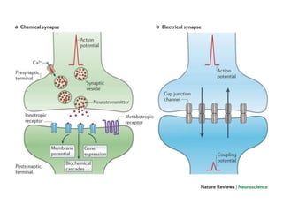
this ppt shares what synapses are and how information of one neuron is transmitted to other through the synapses. it also includes the properties and plasticity of synaptic transmission
Synapse – Greek word –synaptein. Syn –together; aptein –clasp.
Synapse – Clasping of hands (as in hand shaking between two friends).
Site of functional continuity (transneuronal junctional complex) between two neurons.
Why need of synapse?
these slides contain a brief introduction of neurons and its classification as well as details of generation of action potential, resting potential and eletrotonic potential.
Nervous system forms an interconnecting fibers of communication network.
In the ‘hard-wiring’ of the nerves, the signals travel in the form of a flow of electrical current called nerve impulses.
The stimulus-response reactions afford internal constancy in the face of environmental changes.
Receptor by Pandian M, Tutor, Dept of Physiology, DYPMCKOP, MH. This PPT for ...Pandian M
Introduction
SENSORY RECEPTORS
Structurally 3 types of receptors
Transducers
CLASSIFICATION OF RECEPTORS
A. Depending on the source of stimulus(Sherrington’s classification)
B. Depending upon type of stimulus
C. Clinical or anatomical classification of receptors
Production of receptor potential
Properties of receptors
Properties of receptor potential
Physiology of Neuromodulation and neuromodulators. Difference between neuromodulation and synapse. Recent advances in neuromodulation, clinical application of neuromodulation.
Classification and structure of synapsesAlaaAlchyad
Synapses can be classified by the type of cellular structures serving as the pre- and post-synaptic components. ... The axon can synapse onto a dendrite, onto a cell body, or onto another axon or axon terminal, as well as into the bloodstream or diffusely into the adjacent nervous tissue.
The majority of the content is physiology-based, but it's also built-in an anatomical perspective.i've also included disorder of synapses
Instagram @Rajamd_
This presentation contains the basic information about nerve cells and action potential. This work is done for academic purpose only so if you are using give proper reference.
Introduction:
RNA interference (RNAi) or Post-Transcriptional Gene Silencing (PTGS) is an important biological process for modulating eukaryotic gene expression.
It is highly conserved process of posttranscriptional gene silencing by which double stranded RNA (dsRNA) causes sequence-specific degradation of mRNA sequences.
dsRNA-induced gene silencing (RNAi) is reported in a wide range of eukaryotes ranging from worms, insects, mammals and plants.
This process mediates resistance to both endogenous parasitic and exogenous pathogenic nucleic acids, and regulates the expression of protein-coding genes.
What are small ncRNAs?
micro RNA (miRNA)
short interfering RNA (siRNA)
Properties of small non-coding RNA:
Involved in silencing mRNA transcripts.
Called “small” because they are usually only about 21-24 nucleotides long.
Synthesized by first cutting up longer precursor sequences (like the 61nt one that Lee discovered).
Silence an mRNA by base pairing with some sequence on the mRNA.
Discovery of siRNA?
The first small RNA:
In 1993 Rosalind Lee (Victor Ambros lab) was studying a non- coding gene in C. elegans, lin-4, that was involved in silencing of another gene, lin-14, at the appropriate time in the
development of the worm C. elegans.
Two small transcripts of lin-4 (22nt and 61nt) were found to be complementary to a sequence in the 3' UTR of lin-14.
Because lin-4 encoded no protein, she deduced that it must be these transcripts that are causing the silencing by RNA-RNA interactions.
Types of RNAi ( non coding RNA)
MiRNA
Length (23-25 nt)
Trans acting
Binds with target MRNA in mismatch
Translation inhibition
Si RNA
Length 21 nt.
Cis acting
Bind with target Mrna in perfect complementary sequence
Piwi-RNA
Length ; 25 to 36 nt.
Expressed in Germ Cells
Regulates trnasposomes activity
MECHANISM OF RNAI:
First the double-stranded RNA teams up with a protein complex named Dicer, which cuts the long RNA into short pieces.
Then another protein complex called RISC (RNA-induced silencing complex) discards one of the two RNA strands.
The RISC-docked, single-stranded RNA then pairs with the homologous mRNA and destroys it.
THE RISC COMPLEX:
RISC is large(>500kD) RNA multi- protein Binding complex which triggers MRNA degradation in response to MRNA
Unwinding of double stranded Si RNA by ATP independent Helicase
Active component of RISC is Ago proteins( ENDONUCLEASE) which cleave target MRNA.
DICER: endonuclease (RNase Family III)
Argonaute: Central Component of the RNA-Induced Silencing Complex (RISC)
One strand of the dsRNA produced by Dicer is retained in the RISC complex in association with Argonaute
ARGONAUTE PROTEIN :
1.PAZ(PIWI/Argonaute/ Zwille)- Recognition of target MRNA
2.PIWI (p-element induced wimpy Testis)- breaks Phosphodiester bond of mRNA.)RNAse H activity.
MiRNA:
The Double-stranded RNAs are naturally produced in eukaryotic cells during development, and they have a key role in regulating gene expression .
Deep Behavioral Phenotyping in Systems Neuroscience for Functional Atlasing a...Ana Luísa Pinho
Functional Magnetic Resonance Imaging (fMRI) provides means to characterize brain activations in response to behavior. However, cognitive neuroscience has been limited to group-level effects referring to the performance of specific tasks. To obtain the functional profile of elementary cognitive mechanisms, the combination of brain responses to many tasks is required. Yet, to date, both structural atlases and parcellation-based activations do not fully account for cognitive function and still present several limitations. Further, they do not adapt overall to individual characteristics. In this talk, I will give an account of deep-behavioral phenotyping strategies, namely data-driven methods in large task-fMRI datasets, to optimize functional brain-data collection and improve inference of effects-of-interest related to mental processes. Key to this approach is the employment of fast multi-functional paradigms rich on features that can be well parametrized and, consequently, facilitate the creation of psycho-physiological constructs to be modelled with imaging data. Particular emphasis will be given to music stimuli when studying high-order cognitive mechanisms, due to their ecological nature and quality to enable complex behavior compounded by discrete entities. I will also discuss how deep-behavioral phenotyping and individualized models applied to neuroimaging data can better account for the subject-specific organization of domain-general cognitive systems in the human brain. Finally, the accumulation of functional brain signatures brings the possibility to clarify relationships among tasks and create a univocal link between brain systems and mental functions through: (1) the development of ontologies proposing an organization of cognitive processes; and (2) brain-network taxonomies describing functional specialization. To this end, tools to improve commensurability in cognitive science are necessary, such as public repositories, ontology-based platforms and automated meta-analysis tools. I will thus discuss some brain-atlasing resources currently under development, and their applicability in cognitive as well as clinical neuroscience.
Seminar of U.V. Spectroscopy by SAMIR PANDASAMIR PANDA
Spectroscopy is a branch of science dealing the study of interaction of electromagnetic radiation with matter.
Ultraviolet-visible spectroscopy refers to absorption spectroscopy or reflect spectroscopy in the UV-VIS spectral region.
Ultraviolet-visible spectroscopy is an analytical method that can measure the amount of light received by the analyte.
A brief information about the SCOP protein database used in bioinformatics.
The Structural Classification of Proteins (SCOP) database is a comprehensive and authoritative resource for the structural and evolutionary relationships of proteins. It provides a detailed and curated classification of protein structures, grouping them into families, superfamilies, and folds based on their structural and sequence similarities.
What is greenhouse gasses and how many gasses are there to affect the Earth.moosaasad1975
What are greenhouse gasses how they affect the earth and its environment what is the future of the environment and earth how the weather and the climate effects.
Nutraceutical market, scope and growth: Herbal drug technologyLokesh Patil
As consumer awareness of health and wellness rises, the nutraceutical market—which includes goods like functional meals, drinks, and dietary supplements that provide health advantages beyond basic nutrition—is growing significantly. As healthcare expenses rise, the population ages, and people want natural and preventative health solutions more and more, this industry is increasing quickly. Further driving market expansion are product formulation innovations and the use of cutting-edge technology for customized nutrition. With its worldwide reach, the nutraceutical industry is expected to keep growing and provide significant chances for research and investment in a number of categories, including vitamins, minerals, probiotics, and herbal supplements.
Professional air quality monitoring systems provide immediate, on-site data for analysis, compliance, and decision-making.
Monitor common gases, weather parameters, particulates.
Earliest Galaxies in the JADES Origins Field: Luminosity Function and Cosmic ...Sérgio Sacani
We characterize the earliest galaxy population in the JADES Origins Field (JOF), the deepest
imaging field observed with JWST. We make use of the ancillary Hubble optical images (5 filters
spanning 0.4−0.9µm) and novel JWST images with 14 filters spanning 0.8−5µm, including 7 mediumband filters, and reaching total exposure times of up to 46 hours per filter. We combine all our data
at > 2.3µm to construct an ultradeep image, reaching as deep as ≈ 31.4 AB mag in the stack and
30.3-31.0 AB mag (5σ, r = 0.1” circular aperture) in individual filters. We measure photometric
redshifts and use robust selection criteria to identify a sample of eight galaxy candidates at redshifts
z = 11.5 − 15. These objects show compact half-light radii of R1/2 ∼ 50 − 200pc, stellar masses of
M⋆ ∼ 107−108M⊙, and star-formation rates of SFR ∼ 0.1−1 M⊙ yr−1
. Our search finds no candidates
at 15 < z < 20, placing upper limits at these redshifts. We develop a forward modeling approach to
infer the properties of the evolving luminosity function without binning in redshift or luminosity that
marginalizes over the photometric redshift uncertainty of our candidate galaxies and incorporates the
impact of non-detections. We find a z = 12 luminosity function in good agreement with prior results,
and that the luminosity function normalization and UV luminosity density decline by a factor of ∼ 2.5
from z = 12 to z = 14. We discuss the possible implications of our results in the context of theoretical
models for evolution of the dark matter halo mass function.
Slide 1: Title Slide
Extrachromosomal Inheritance
Slide 2: Introduction to Extrachromosomal Inheritance
Definition: Extrachromosomal inheritance refers to the transmission of genetic material that is not found within the nucleus.
Key Components: Involves genes located in mitochondria, chloroplasts, and plasmids.
Slide 3: Mitochondrial Inheritance
Mitochondria: Organelles responsible for energy production.
Mitochondrial DNA (mtDNA): Circular DNA molecule found in mitochondria.
Inheritance Pattern: Maternally inherited, meaning it is passed from mothers to all their offspring.
Diseases: Examples include Leber’s hereditary optic neuropathy (LHON) and mitochondrial myopathy.
Slide 4: Chloroplast Inheritance
Chloroplasts: Organelles responsible for photosynthesis in plants.
Chloroplast DNA (cpDNA): Circular DNA molecule found in chloroplasts.
Inheritance Pattern: Often maternally inherited in most plants, but can vary in some species.
Examples: Variegation in plants, where leaf color patterns are determined by chloroplast DNA.
Slide 5: Plasmid Inheritance
Plasmids: Small, circular DNA molecules found in bacteria and some eukaryotes.
Features: Can carry antibiotic resistance genes and can be transferred between cells through processes like conjugation.
Significance: Important in biotechnology for gene cloning and genetic engineering.
Slide 6: Mechanisms of Extrachromosomal Inheritance
Non-Mendelian Patterns: Do not follow Mendel’s laws of inheritance.
Cytoplasmic Segregation: During cell division, organelles like mitochondria and chloroplasts are randomly distributed to daughter cells.
Heteroplasmy: Presence of more than one type of organellar genome within a cell, leading to variation in expression.
Slide 7: Examples of Extrachromosomal Inheritance
Four O’clock Plant (Mirabilis jalapa): Shows variegated leaves due to different cpDNA in leaf cells.
Petite Mutants in Yeast: Result from mutations in mitochondrial DNA affecting respiration.
Slide 8: Importance of Extrachromosomal Inheritance
Evolution: Provides insight into the evolution of eukaryotic cells.
Medicine: Understanding mitochondrial inheritance helps in diagnosing and treating mitochondrial diseases.
Agriculture: Chloroplast inheritance can be used in plant breeding and genetic modification.
Slide 9: Recent Research and Advances
Gene Editing: Techniques like CRISPR-Cas9 are being used to edit mitochondrial and chloroplast DNA.
Therapies: Development of mitochondrial replacement therapy (MRT) for preventing mitochondrial diseases.
Slide 10: Conclusion
Summary: Extrachromosomal inheritance involves the transmission of genetic material outside the nucleus and plays a crucial role in genetics, medicine, and biotechnology.
Future Directions: Continued research and technological advancements hold promise for new treatments and applications.
Slide 11: Questions and Discussion
Invite Audience: Open the floor for any questions or further discussion on the topic.
2. SYNAPTIC INTEGRATION
• Neurons in the brain receive thousands of
synaptic inputs from other neurons.
• Synaptic integration is the term used to
describe how neurons ‘add up’ these inputs
before the generation of a nerve impulse, or
action potential.
• The ability of synaptic inputs to effect
neuronal output is determined by a number of
factors
3. Size, shape and relative timing of electrical
potentials generated by synaptic inputs
the geometric structure of the target neuron,
the physical location of synaptic inputs within
that structure
expression of voltage‐gated channels in
different regions of the neuronal membrane.
4. SYNAPTIC INTEGRATION AND ITS
MECHANISM
Neurons within a neural network receive information from,
and send information to, many other cells, at specialised
junctions called synapses.
Synaptic integration is the computational process by which an
individual neuron processes its synaptic inputs and converts
them into an output signal.
Neurons are specialised for electrical signalling, with the main
neuronal input signal (synaptic potentials) and the main
neuronal output signal (action potentials)
5. Synaptic potentials occur when neurotransmitter binds to and
opens ligand‐operated channels in the dendritic membrane,
allowing ions to move into or out of the cell according to their
electrochemical gradient.
Synaptic potentials can be either excitatory or inhibitory
depending on the direction and charge of ion movement.
Action potentials occur if the summed synaptic inputs to a
neuron reach a threshold level of depolarisation and trigger
regenerative opening of voltage‐gated ion channels.
Synaptic potentials are often brief and of small amplitude,
therefore summation of inputs in time (temporal summation)
or from multiple synaptic inputs (spatial summation) is usually
required to reach action potential firing threshold.
6.
7. TYPES OF SYNAPSES
• Types of synapses
• there are two types of synapses:
– electrical synapses
– chemical synapses
8. Electrical synapse
• electrical synapses are a direct electrical coupling between two
cells
– mediated by gap junctions, which are pores (as shown in the
electron micrograph) constructed of connexin proteins
– essentially result in the passing of a gradient potential (may be
depolarizing or hyperpolarizing) between two cells
• very rapid (no synaptic delay)
• passive process --> signal can degrade with distance-> may
not produce a large enough depolarization to initiate an
action potential in the postsynaptic cell
• bidirectional
– i.e., "post"synaptic cell can actually send messages to
the "pre"synaptic cell
9. Chemical synapse
• Chemical synapses coupling between two cells
through neuro-transmitters, ligand or voltage
gated channels, receptors.
• Influenced by the concentration and types of
ions on either side of the membrane.
• Glutamate, sodium, potassium, calcium are
positively charged.
• GABA, chloride are negatively charged.
10. Chemical synapse
• Ionotropic receptors are single protein
complexes that combine two functions.
• They have recognition sites on their surfaces
extending into the extracellular fluid that
allow them to interact with neurotransmitter
molecules.
• They also have the ability to open and close,
allowing ions to move across the neural
membrane.
• ionotropic receptors respond very quickly.
11. Chemical synapse
• Metabotropic receptors are made up of
multiple protein complexes embedded in the
neural membrane.
• One complex has the capacity to recognize
neurotransmitter molecules but cannot open
and close.
• Instead, the metabotropic receptor binds
molecules of neurotransmitter
• It releases a G protein, or "second
messenger," from its surface extending into
the intracellular fluid of the neuron
12. Chemical synapse
• This G protein travels away from the receptor
where it can interact with adjacent ion
channels, which can then open and close like
the ionotropic receptor.
• The eventual opening of ion channels is
slower than it is with ionotropic receptors.
13. • in contrast, chemical synapses are
• slow
• active (require ligand-gated channels)
• pseudo-unidirectional
14.
15. IPSP AND EPSP
• An electrical charge (hyperpolarisation) in the membrane of a
postsynaptic neuron caused by the binding of an inhibitory
neurotransmitter from a presynaptic cell to a postsynaptic
receptor; makes it more difficult for a postsynaptic neuron to
generate an action potential.
• An electrical change (depolarisation) in the membrane of a
postsynaptic neurone caused by the binding of an excitatory
neurotransmitter from a presynaptic cell to a postsynaptic
receptor; makes it more likely for a postsynaptic neurone to
generate an action potential
16.
17. EPSP
• Consider, for example, a neuronal synapse
that uses glutamate as receptor.
• Receptors open ion channels that are non-selectively
permeable to cations.
• When these glutamate receptors are
activated, both Na+ and K+ flow across
the postsynaptic membrane.
• The reversal potential (Erev) for the post -
synaptic current is approximately 0 mV.
18. EPSP
• The resting potential of neurons is
approximately -60 mV.
• The resulting EPSP will depolarize the post
synaptic membrane potential, bringing it
toward 0 mV.
19. IPSP
• As an example of inhibitory post synaptic
s action, consider a neuronal synapse that
uses GABA as its transmitter.
• At such synapses, the GABA receptors typically
open channels that are selectively permeable
to Cl-.
• When these channels open, negatively
charged chloride ions can flow across the
membrane.
20. IPSP
• Assume that the postsynaptic neuron has
a resting potential of -60 mV and an action
potential threshold of -40 mV.
• If ECl is -70 mV, transmitter release at this
synapse will inhibit the postsynaptic cell.
• Since ECl is more negative than the action
potential threshold.
• It reduces the probability that the
postsynaptic cell will fire an action potential.
21. • Some types of neurotransmitters, such as
glutamate, consistently result in EPSPs
• Others, such as GABA, consistently result in
IPSPs.
• The action potential lasts about one msec, or
1/1000th of a second.
• In contrast, the EPSPs and IPSPs can last as
long as 5 to 10 msec. This allows the effect of
one postsynaptic potential to build upon the
next and so on.