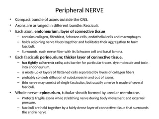
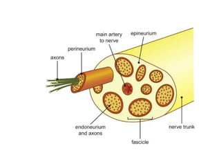
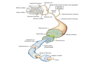
Peripheral NERVE
• Compactbundle of axons outside the CNS.
• Axons are arranged in different bundle: Fasciculi.
• Each axon: endoneurium; layer of connective tissue
– contains collagen, fibroblast, Schwann cells, endothelial cells and macrophages
– holds adjoining nerve fibers together and facilitates their aggregation to form
fasciculi.
– Surrounds each nerve fiber with its Schwann cell and basal lamina.
• Each fasciculi: perineurium; thicker layer of connective tissue.
– has tightly adherents cells; acts barrier for particular traces, dye molecule and toxin
into endoneurium.
– is made up of layers of flattened cells separated by layers of collagen fibers
– probably controls diffusion of substances in and out of axons.
– thin nerve may consist of single fasciculus, but usually a nerve is made of several
fasciculi.
• Whole nerve: epineurium, tubular sheath formed by areolar membrane.
– Protects fragile axons while stretching nerve during body movement and external
pressure.
– fasciculi are held together by a fairly dense layer of connective tissue that surrounds
the entire nerve
6.
Clinical correlation ofneuronal structure
• The epineurium contains nerve fibers. Loss of
this fat in bedridden patients can lead to
pressure on nerve fibers and paralysis.
• Blood vessels to a nerve travel through the
connective tissue that surrounds it. Severe
reduction in blood supply can lead to ischemic
neuritis and pain.
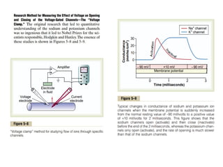
Roles of otherions during Action potential
• Impermeant Negatively Charged Ions (Anions) Inside the Nerve
Axon.
• anions of protein molecules and of many organic phosphate compounds,
sulfate compounds, and so forth.
• any deficit of positive ions inside the membrane responsible for the negative
charge inside the fiber
• Calcium ion
• voltage-gated calcium channels slightly permeable to sodium ions as well as
to calcium ions.
• 10-20 times slow for activation than sodium channel.
• increased Permeability of the Sodium Channels When there Is a Deficit of
Calcium Ions.
– spontaneous discharge occurs in some peripheral nerves, often causing muscle “tetany.
– tetanic contraction of the respiratory muscles can be lethal.
32.
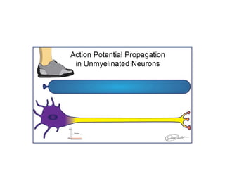
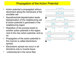
Propagation of theAction Potential
• Action potential is propagated without
decrement along the membrane of the
excitable cell
• Suprathreshold depolarization leads
depolarization of the neighbouring cell
& action potential is generated in the
neighbouring region
• Repetition of the process leads to
another action potential in the region
next to the new action potential, and so
on
• Propagation of the action potential in
this manner is called electrotonic
spread.
• Electrotonic spread can occur in all
directions only in muscle tissue
• Unidirectional in the nerve fiber
34.
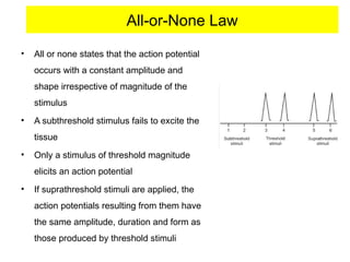
All-or-None Law
• Allor none states that the action potential
occurs with a constant amplitude and
shape irrespective of magnitude of the
stimulus
• A subthreshold stimulus fails to excite the
tissue
• Only a stimulus of threshold magnitude
elicits an action potential
• If suprathreshold stimuli are applied, the
action potentials resulting from them have
the same amplitude, duration and form as
those produced by threshold stimuli
35.
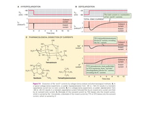
Inhibition of Excitability—“Stabilizers” and Local Anesthetics
• Calcium ion as stabilizer: For instance, a high extracellular fluid calcium ion
concentration decreases membrane permeability to sodium ions and simultaneously
reduces excitability
• Local Anesthetics:including procaine and tetracaine; act directly on the activation
gates of the sodium channels, making difficult for gates to open, thereby reducing
membrane excitability.
38.
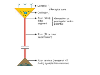
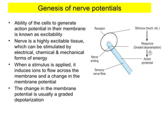
Genesis of nervepotentials
• Ability of the cells to generate
action potential in their membrane
is known as excitability
• Nerve is a highly excitable tissue,
which can be stimulated by
electrical, chemical & mechanical
forms of energy
• When a stimulus is applied, it
induces ions to flow across the
membrane and a change in the
membrane potential
• The change in the membrane
potential is usually a graded
depolarization
39.
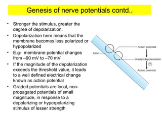
• Stronger thestimulus, greater the
degree of depolarization.
• Depolarization here means that the
membrane becomes less polarized or
hypopolarized
• E.g- membrane potential changes
from –90 mV to –70 mV
• If the magnitude of the depolarization
exceeds the threshold value, it leads
to a well defined electrical change
known as action potential
• Graded potentials are local, non-
propagated potentials of small
magnitude, in response to a
depolarizing or hyperpolarizing
stimulus of lesser strength
Genesis of nerve potentials contd..
40.
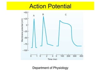
Action Potential
• Atransient change in membrane potential of about 100 mV,
which is conducted along the axon in an all-or-none fashion
• Features:
– characterized by a gradual depolarization to threshold,
and a rapid ascent in the membrane potential followed by
a phase of repolarization
– Travels along the axon with the same shape and
amplitude being regenerated at regular intervals.
– Also known as an impulse or spike potential
41.
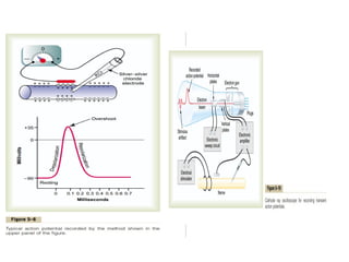
• Latent Period
–Action potential is always preceded by a latent period,
which is the interval between the application of a stimulus
and the onset of action potential
42.
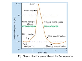
Phases of actionpotential
• Depolarization
– recorded as a sharp upward wave during which the
membrane potential approaches zero and then attains a
positive value
– It of slow depolarization to threshold, rapid rising phase,
overshoot and peak
• Repolarization
– Recorded as downstroke during which the membrane
potential returns to the resting level.
– includes a rapid falling phase & slower terminal part called
after-depolarization.
43.
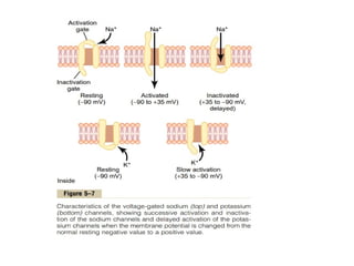
Ionic Basis ofAction Potential
• Action potential is generated by sequential changes in membrane
permeability to ions, principally Na & K ions
• Depolarization is due to an increase in permeability to Na+
which is
favoured by concentration gradient as well as the electrical gradient which
leads to a rapid entry of sodium ions
• Positively charged sodium ions leads to depolarization lasts only a short
while
• When the Na permeability starts declining, the membrane permeability to
potassium ions starts increasing. Since the concentration of K is higher
inside the cell, and the outside of the cell is negative at the peak of the
action potential, potassium ions diffuse outwards
• Decline in sodium permeability coupled with increase in potassium
permeability brings about repolarization
• At the end of an action potential, the excitable cell has more sodium ions
and less potassium ions than at the beginning
• Bringing the concentration back towards the original is the job of the
sodium pump, which it does slowly
44.
• Depolarization
– Whena threshold stimulus is applied, the influx of Na+
through leaky channels & later through opening of few
voltage-gated Na+
channels decreases the membrane
potential from –70 mV to –55 mV (threshold level)
– At this threshold potential, there occurs opening of a large
n.o of the voltage-gated Na+ channels, ↑ing the membrane
permeability to Na+
ions several 100x fold leads to massive
influx of sodium ions producing a swift, large & steep
depolarization, changing the membrane potential to +35
mV
– concentration gradient as well as electrical gradient favors
the entry of sodium ions across the membrane
45.
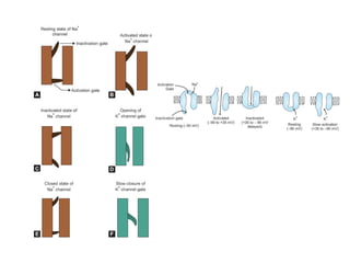
• Repolarization
– Dueto opening of voltage-gated K+ channels, causing
membrane permeability to potassium ions increases
several times causing increased potassium efflux
– At the peak of the action potential, voltage-gated Na+
channels enter a closed state whereas the voltage gated
K+ channels are fully open
– Termination of action potential due to activation of voltage-
gated potassium channels is a negative feedback process
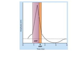
Refractory Period
• Duringthe action potential, the stimulated area of the
membrane happens to be unresponsive to a second stimulus
in most part, and later it requires a stronger stimulus to get
excited again.
• The length of time during which the membrane is
unresponsive to a second stimulus no matter how strong is
the stimulus, is known as refractory period
• 2 types
– Absolute refractory periods
– Relative refractory periods
50.
Absolute Refractory Period
•Period in the action potential during which, application of a second
stimulus of any strength and duration does not produce another
action potential
• ARP corresponds to the period from the time the firing level is
reached until repolarization is about one-third complete
• Mechanism- inactivation gates of the voltage-gated sodium
channels close and they remain in that inactivated state for some
time before returning to the resting state & can reopen in response
to a second stimulus, only after attaining the resting state
51.
Relative Refractory Period
•Period following ARP during which, application of a
suprathreshold stimulus can elicit a second action potential
• RRP starts from the end of ARP to the start of after-
depolarization
• Mechanism- all the sodium channels present at the site of
stimulus do not achieve the open state or inactivated state or
resting state, exactly at the same time