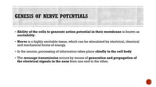
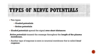
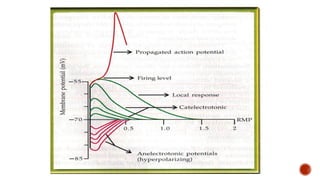
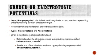
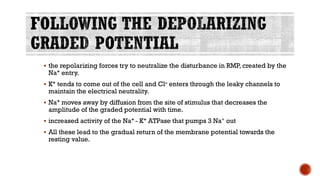
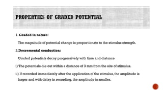
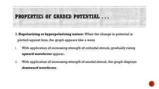
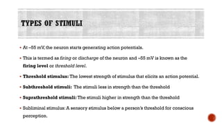
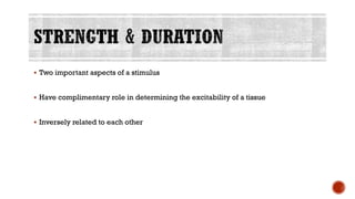
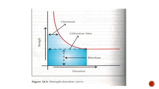
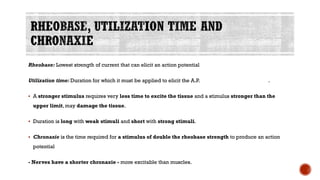
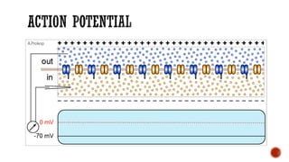
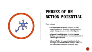
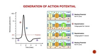
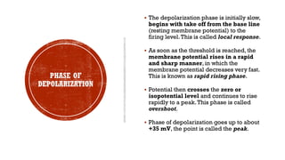
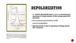
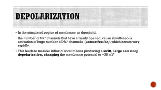
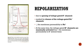
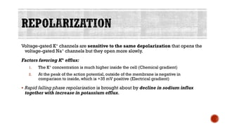
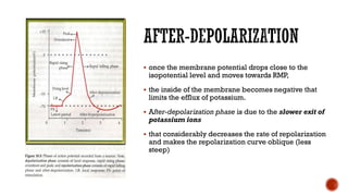
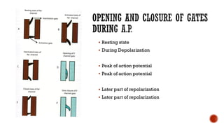
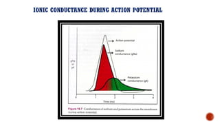
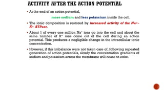
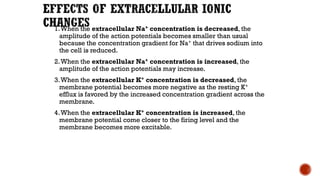
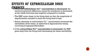
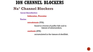
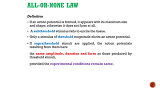
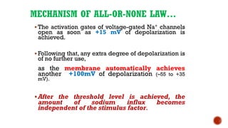
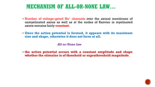
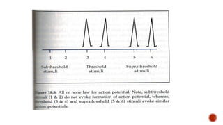
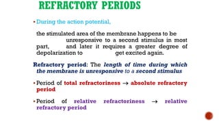
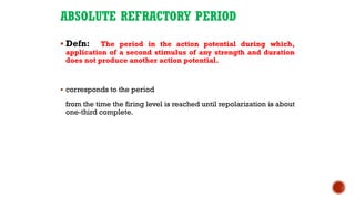
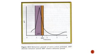
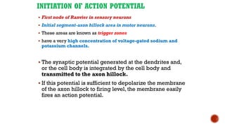
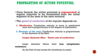
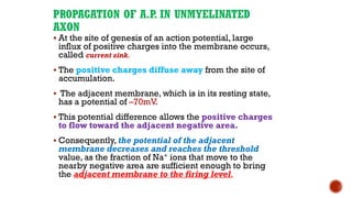
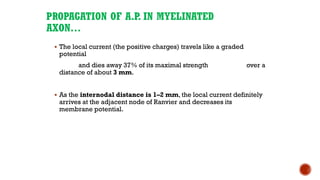
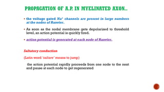
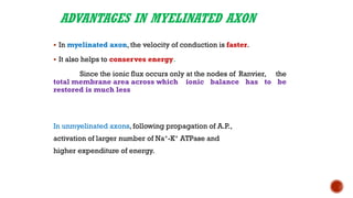
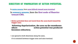
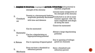
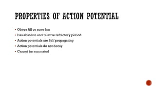
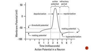
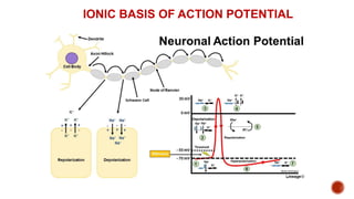
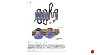
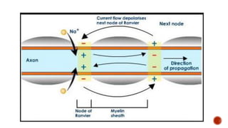
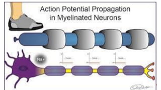
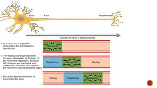
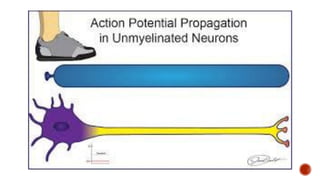
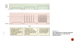
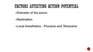
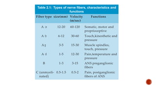
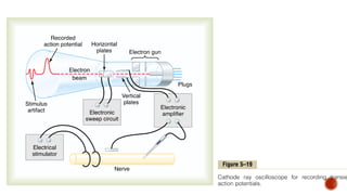
Dr. Saraswati JYadav
Associate Prof
Physiology
โ–ช Ability of the cells to generate action potential in their membrane is known as
excitability.
โ–ช Nerve is a highly excitable tissue, which can be stimulated by electrical, chemical
and mechanical forms of energy.
โ–ช In the neuron, processing of information takes place chiefly in the cell body
โ–ช The message transmission occurs by means of generation and propagation of
the electrical signals in the axon from one end to the other.
โ–ช Two types:
โ–ช Graded potentials
โ–ช Action potentials
โ–ช Graded potentials spread the signal over short distances
Action potentials transmit the message throughout the length of the plasma
membrane.
โ–ช Another type of response is seen in neuronal membrane that is called local
response.
โ–ช Local, Non-propagated potentials of small magnitude, in response to a depolarizing
or hyperpolarizing stimulus of lesser strength.
โ–ช Recorded from the membranes of dendrites and cell body.
โ–ช Types: Catelectrotonic and Anelectrotonic
โ–ช When a membrane is electrically stimulated,
โ–ช Cathodal end of the stimulator evokes a depolarizing response called
catelectrotonic potential.
โ–ช Anodal end of the stimulator evokes a hyperpolarizing response called
anelectrotonic potential.
โ–ช the repolarizing forces try to neutralize the disturbance in RMP, created by the
Na+ entry.
โ–ช K+ tends to come out of the cell and Clโ€“ enters through the leaky channels to
maintain the electrical neutrality.
โ–ช Na+ moves away by diffusion from the site of stimulus that decreases the
amplitude of the graded potential with time.
โ–ช increased activity of the Na+ - K+ ATPase that pumps 3 Na+ out
โ–ช All these lead to the gradual return of the membrane potential towards the
resting value.
1. Graded in nature:
The magnitude of potential change is proportionate to the stimulus strength.
2.Decremental conduction:
Graded potentials decay progressively with time and distance
i) The potentials die out within a distance of 3 mm from the site of stimulus.
ii) If recorded immediately after the application of the stimulus, the amplitude is
larger and with delay in recording, the amplitude is smaller.
3. Depolarizing or hyperpolarizing nature: When the change in potential is
plotted against time, the graph appears like a wave
I. With application of increasing strength of cathodal stimuli, gradually rising
upward waveforms appear.
II. With application of increasing strength of anodal stimuli, the graph displays
downward waveforms.
4. Summation:
โ–ช If a second stimulus is applied before the potential produced by the first stimulus
has disappeared, both the potential changes are added together producing a
larger and/or prolonged wave in the recording.
โ–ช This happens due to the arrival of more Na+ at the site of stimulus before
neutralization of all Na+ influx caused by the first stimulus.
โ–ช Similarly, the anelectrotonic potentials exhibit the property of summation.
They are named according to the membrane from where they are recorded.
โ–ช End-plate potential, recorded from end plate area of skeletal muscle membrane.
โ–ช Receptor potential, recorded from sensory nerve endings.
โ–ช Synaptic potential, recorded from membrane of post-synaptic neurons.
โ–ช Pacemaker potential, recorded from membrane of pacemaker cells in the heart,
intestine etc. and so on.
โ–ช If the axon is stimulated with slowly increasing strength of stimuli, when the
membrane potential is decreased by 7 mV, the pattern of graded potential is
altered.
โ–ช The response becomes greater than what is expected for that strength of
stimulus.This enhanced response is known as local response.
โ–ช Similar to the graded potential, the local response gradually dies out with
increasing distance.
โ–ช Local response is seen only with a depolarizing stimulus of lower strength, and
not with a hyperpolarizing one.
โ–ช As the applied cathodal stimulus is progressively raised from zero the influx of Na+
through the leaky sodium channels increases proportionately. Consequently, the
membrane potential gradually decreases from โ€“70 mV.
โ–ช As the membrane potential reaches โ€“63mV, few of the voltage-gated sodium channels
start opening, which allows entry of some more Na+.
โ–ช Thus, Na+ influx though the leaky channels added to Na+ influx through voltage gated
channels results in a heightened response called local response.
โ–ช This is observed within a potential change from โ€“63mV to โ€“55mV.
โ–ช At โ€“55 mV, the neuron starts generating action potentials.
โ–ช This is termed as firing or discharge of the neuron and โ€“55 mV is known as the
firing level or threshold level.
โ–ช Threshold stimulus: The lowest strength of stimulus that elicits an action potential.
โ–ช Subthreshold stimuli: The stimuli less in strength than the threshold
โ–ช Suprathreshold stimuli: The stimuli higher in strength than the threshold
โ–ช Subliminal stimulus: A sensory stimulus below a personโ€™s threshold for conscious
perception.
โ–ช Two important aspects of a stimulus
โ–ช Have complimentary role in determining the excitability of a tissue
โ–ช Inversely related to each other
Rheobase: Lowest strength of current that can elicit an action potential
Utilization time: Duration for which it must be applied to elicit the A.P. .
โ–ช A stronger stimulus requires very less time to excite the tissue and a stimulus stronger than the
upper limit, may damage the tissue.
โ–ช Duration is long with weak stimuli and short with strong stimuli.
โ–ช Chronaxie is the time required for a stimulus of double the rheobase strength to produce an action
potential
- Nerves have a shorter chronaxie - more excitable than muscles.
โ–ช If a slow depolarizing stimulus is applied, the tissue fails to produce an action
potential because it adapts to the stimulus.This phenomenon is known as
accommodation.
โ–ช Some of the voltage-gated Na+ channels start opening at a membrane potential of โ€“
63 mV. But they soon get inactivated before another set of channel opens; because,
the membrane takes longer time to arrive at the next level of potential.
โ–ช Besides, the slow voltage-gated K+ channels open allowing compensatory efflux of
K+ ions which opposes depolarization to critical firing potential.
Defn:
โ–ช A transient change in membrane potential of about 100
mV
โ–ช characterized by a gradual depolarization to threshold,
and
โ–ช a rapid ascent in the membrane potential followed by a
phase of repolarization,
โ–ช which is conducted along the axon in an all-or-none fashion
โ–ช with the same shape and amplitude.
It is also known as an impulse or spike potential.
ACTION
POTENTIAL..
Duration: 1 msec
Amplitude: 100mV (โ€“70 to +35
mV)
The action potentials never
summate
A definite interval (refractory
period) exists before the second
action potential is fired.
LATENT
PERIOD
โ–ช Interval between the application of
the stimulus and the onset of the
action potential.
โ–ช Action potential is always preceded by a
latent period
โ–ช Duration depends on
- the distance between the stimulating
and recording electrode
- the type and diameter of nerve fiber.
Three phases:
โ–ช Phase of depolarization consists of slow
depolarization to threshold (local response),
rapid rising phase, overshoot and peak.
โ–ช Phase of repolarization includes a rapid
falling phase and slower terminal part called
after-depolarization.
โ–ช Phase of after-hyperpolarization includes a
brief period of undershoot and the return of
the membrane potential to resting level.
GENERATION OF ACTION POTENTIAL
PHASE OF
DEPOLARIZATION
โ–ช The depolarization phase is initially slow,
begins with take off from the base line
(resting membrane potential) to the
firing level.This is called local response.
โ–ช As soon as the threshold is reached, the
membrane potential rises in a rapid
and sharp manner, in which the
membrane potential decreases very fast.
This is known as rapid rising phase.
โ–ช Potential then crosses the zero or
isopotential level and continues to rise
rapidly to a peak.This phase is called
overshoot.
โ–ช Phase of depolarization goes up to about
+35 mV, the point is called the peak.
โ–ช The repolarization process starts as soon as the depolarization is over and is
recorded as down-stroke.
โ–ช In the major part of the down-stroke, action potential falls rapidly.This is called
rapid falling phase of repolarization.
โ–ช This is followed by a slower repolarization, which is the terminal part of
repolarization phase that brings the membrane potential to base line.This is
called after-depolarization.
โ–ช The phase of repolarization is followed by an after-hyperpolarization phase
โ–ช the membrane potential undershoots (goes below the base line) and
โ–ช then membrane potential returns to the resting level.
โ–ช Sequential changes in membrane permeability to sodium and potassium
โ–ช leading to large fluxes of these ions across the membrane, along their gradients.
โ–ช Depolarization is due to influx of sodium
โ–ช Repolarization is due to efflux of potassium.
โ–ช The voltage-gated Na+ and K + channels contribute to the different phases of the
action potential.
โ–ช In the axon at resting state, only leaky channels are
open.
โ–ช The early part of local response is due to entry of
sodium ions through the leaky channels.
โ–ช When the change in membrane potential is about +7
mV (-63mV), few of the voltage-gated Na+
channels begin to open.
At -55mV (threshold level ), there is simultaneous
opening of a large number of the voltage-gated Na+
channels,
โ†‘ the membrane permeability to Na+
several hundredfold.
โ–ช Depolarization is due to opening of voltage-gated
Na+ channels.
โ–ช In the stimulated region of membrane, at threshold,
the number of Na+ channels that have already opened, cause simultaneous
activation of huge number of Na+ channels (autoactivation), which occurs very
rapidly.
โ–ช This leads to massive influx of sodium ions producing a swift, large and steep
depolarization, changing the membrane potential to +35 mV
HODGKINโ€™S
CYCLE
โ–ช The opening of few Na+ channels
leading to further opening of other Na
+ channels
โ–ช This is an example of positive
feedback control in which a stimulus
triggering an event further facilitates the
process.
โ–ช due to opening of voltage-gated K+ channels
โ–ช marked by closure of the voltage-gated Na+
channels
โ–ช โ†“ the membrane permeability to Na+
โ–ช At the same time, the voltage-gated K+ channels are
maximally activated that โ†‘ the membrane
permeability to K+ several times
Voltage-gated K+ channels are sensitive to the same depolarization that opens the
voltage-gated Na+ channels but they open more slowly.
Factors favoring K+ efflux:
1. The K+ concentration is much higher inside the cell (Chemical gradient)
2. At the peak of the action potential, outside of the membrane is negative in
comparison to inside, which is +35 mV positive (Electrical gradient)
โ–ช Rapid falling phase repolarization is brought about by decline in sodium influx
together with increase in potassium efflux.
โ–ช once the membrane potential drops close to the
isopotential level and moves towards RMP,
โ–ช the inside of the membrane becomes negative that
limits the efflux of potassium.
โ–ช After-depolarization phase is due to the slower exit of
potassium ions
โ–ช that considerably decreases the rate of repolarization
and makes the repolarization curve oblique (less
steep)
โ–ช Following after-depolarization, membrane potential reaches the resting level.
โ–ช At this level, though most of the voltage-gated K+ channels are closed, some of
them still remain open allowing continued efflux of K+ ions.
โ–ช As a result, the membrane potential becomes more negative than the RMP, giving
rise to the prolonged and slow undershoot, which is called as phase of after-
hyperpolarization.
โ–ช Finally, the K+ channels completely close, restoring the membrane potential to
the resting level.
โ–ช Resting state
โ–ช During Depolarization
โ–ช Peak of action potential
โ–ช Later part of repolarization
IONIC CONDUCTANCE DURING ACTION POTENTIAL
โ–ช At the end of an action potential,
more sodium and less potassium inside the cell.
โ–ช The ionic composition is restored by increased activity of the Na+-
K+ ATPase.
โ–ช About 1 of every one million Na+ ions go into the cell and about the
same number of K+ ions come out of the cell during an action
potential. This produces a negligible change in the intracellular ionic
concentration.
โ–ช However, if this imbalance were not taken care of, following repeated
generation of action potentials, slowly the concentration gradients of
sodium and potassium across the membrane will cease to exist.
1.When the extracellular Na+ concentration is decreased, the
amplitude of the action potentials becomes smaller than usual
because the concentration gradient for Na+ that drives sodium into
the cell is reduced.
2.When the extracellular Na+ concentration is increased, the
amplitude of the action potentials may increase.
3.When the extracellular K+ concentration is decreased, the
membrane potential becomes more negative as the resting K+
efflux is favored by the increased concentration gradient across the
membrane.
4.When the extracellular K+ concentration is increased, the
membrane potential come closer to the firing level and the
membrane becomes more excitable.
5.When the extracellular Ca2+ concentration is decreased, the
electrical potential difference across the membrane is decreased,
as the inside becomes less negative compared to outside.
โ–ช The RMP come closer to the firing level, so that, the magnitude of
depolarization needed to reach the firing level is less.
โ–ช Hence, decrease in extracellular Ca++ concentration increases the
excitability of the tissue, as observed in hypocalcemic tetany,
occurring in hypoparathyroidism.
6. If the extracellular Ca2+ concentration is increased, the RMP
goes away from the firing level decreasing the tissue excitability.
Na+ Channel Blockers
Local Anesthetics
Lidocaine, Procaine
Toxins
tetrodotoxin (TTX)
found in ovaries of puffer fish and in
tissues of salamanders,
saxitoxin (STX)
accumulated in the tissues of shellfish.
K+ Channel Blockers
โ–ช Tetraethylammonium (TEA)
โ–ช 4-aminopyridine (4-AP)
Na+ - K+ ATPase Blocker
โ–ช Dinitrophenol
ALL-OR-NONE LAW
Definition
โ–ช If an action potential is formed, it appears with its maximum size
and shape, otherwise it does not form at all.
โ–ช A subthreshold stimulus fails to excite the tissue.
โ–ช Only a stimulus of threshold magnitude elicits an action potential.
โ–ช If suprathreshold stimuli are applied, the action potentials
resulting from them have
the same amplitude, duration and form as those produced by
threshold stimuli,
provided the experimental conditions remain same.
MECHANISM OF ALL-OR-NONE LAWโ€ฆ
โ–ช The activation gates of voltage-gated Na+ channels
open as soon as +15 mV of depolarization is
achieved.
โ–ช Following that, any extra degree of depolarization is
of no further use,
as the membrane automatically achieves
another +100mV of depolarization (โ€“55 to +35
mV).
โ–ชAfter the threshold level is achieved, the
amount of sodium influx becomes
independent of the stimulus factor.
MECHANISM OF ALL-OR-NONE LAWโ€ฆ
โ–ช Number of voltage-gated Na+ channels over the axonal membrane of
unmyelinated axons as well as at the nodes of Ranvier in myelinated
axons remains fairly constant.
โ–ช Once the action potential is formed, it appears with its maximum
size and shape, otherwise it does not form at all.
All-or-None law
โ–ช the action potential occurs with a constant amplitude and shape
whether the stimulus is of threshold or suprathreshold magnitude.
REFRACTORY PERIODS
โ–ช During the action potential,
the stimulated area of the membrane happens to be
unresponsive to a second stimulus in most
part, and later it requires a greater degree of
depolarization to get excited again.
Refractory period: The length of time during which
the membrane is unresponsive to a second stimulus
โ–ช Period of total refractoriness โ†’ absolute refractory
period
โ–ช Period of relative refractoriness โ†’ relative
refractory period
ABSOLUTE REFRACTORY PERIOD
โ–ช Defn: The period in the action potential during which,
application of a second stimulus of any strength and duration
does not produce another action potential.
โ–ช corresponds to the period
from the time the firing level is reached until repolarization is about
one-third complete.
MECHANISM OF ABSOLUTE REFRACTORY PERIOD
โ–ช At the peak of the action potential,
the inactivation gates of the voltage-gated sodium channels close
and
they remain in that inactivated state for some time before
returning to the resting state.
โ–ช These sodium channels can reopen in response to a second
stimulus, only after attaining the resting state.
IMPORTANCE OF ARP
limits the number of action potentials that the nerve can fire in a given
period of time.
Functions of ARP:
1. ARP determines the rate of discharge of nerve fiber.
(Usual firing rate of 10 to 1000 impulses per second)
โ–ช Large diameter nerve fibers have
ARP - 0.4 msec, Firing rate 2500 impulses per second,
โ–ช Small diameter fibers have
ARP - 4 msec, Firing rate of 250 impulses per second.
2. ARP is also responsible for the
one-way conduction of action potentials.
RELATIVE REFRACTORY PERIOD
โ–ช Defn: The period following ARP
during which, application of a
suprathreshold stimulus can elicit
a second action potential.
โ–ช The RRP starts from the end of ARP to the start of after-
depolarization.
MECHANISM OF RRP
1. All of the sodium channels present at the site of stimulus does not
achieve the activated state or inactivated state or resting state,
exactly at the same time.
โ–ช By the time of relative refractory period, some of the channels have
returned to their initial resting state, and they can open in response
to a second stimulus.
2. A suprathreshold stimulus can spread to larger area over the
membrane and open more number of voltage-gated sodium
channel.
INITIATION OF ACTION POTENTIAL
โ–ช First node of Ranvier in sensory neurons
โ–ช Initial segment-axon hillock area in motor neurons.
โ–ช These areas are known as trigger zones
โ–ช have a very high concentration of voltage-gated sodium and
potassium channels.
โ–ช The synaptic potential generated at the dendrites and,
or the cell body is integrated by the cell body and
transmitted to the axon hillock.
โ–ช If this potential is sufficient to depolarize the membrane
of the axon hillock to firing level, the membrane easily
fires an action potential.
PROPAGATION OF ACTION POTENTIAL
โ–ช Once formed, the action potential is regenerated at
regular intervals to be transmitted from the initial
segment of the axon to the axon terminal
โ–ช The speed of conduction of the impulse depends on:
1. Myelination: Conduction velocity is more in myelinated
axon and is proportionate to the degree of myelination.
2. Diameter of the axon: Conduction velocity is proportionate
to the diameter of the fiber.
Larger diameter fiber - Faster rate of conduction.
Large diameter fibers have less cytoplasmic
resistance.
So, the flow of ions across the membrane is easier.
PROPAGATION OF A.P
. IN UNMYELINATED
AXON
โ–ช At the site of genesis of an action potential, large
influx of positive charges into the membrane occurs,
called current sink.
โ–ช The positive charges diffuse away from the site of
accumulation.
โ–ช The adjacent membrane, which is in its resting state,
has a potential of โ€“70mV.
โ–ช This potential difference allows the positive charges
to flow toward the adjacent negative area.
โ–ช Consequently, the potential of the adjacent
membrane decreases and reaches the threshold
value, as the fraction of Na+ ions that move to the
nearby negative area are sufficient enough to bring
the adjacent membrane to the firing level.
PROPAGATION OF A.P
. IN UNMYELINATED
AXONโ€ฆ
โ–ช This results in opening of the voltage gated Na+ channels
present in that area, firing an action potential.
โ–ช Similarly, from the site of second action potential, positive
charges flow to the adjacent resting membrane and
decrease its potential to the threshold level.
โ–ช This activates the voltage gated Na+ channels present in that
part of the membrane resulting in another action potential.
โ–ช In this manner, each point of the membrane gets
depolarized to the firing level and produces an action
potential.
โ–ช As the depolarization and repolarization phases of the
ensuing action potentials go on, there is a sequential
opening and closing of sodium and potassium channels
along the axonal membrane
PROPAGATION OF A.P
. IN MYELINATED AXON
(Saltatory Conduction)
In the myelinated portion of the axon:
โ–ช There are few voltage gated Na+ channels
โ–ช Myelin sheath acts as an insulator and does not allow free flow of
ions across the membrane.
โ–ช The concentration of positive charges does not decrease fast
because of less โ€˜leakageโ€™.
This helps the charges to spread farther along the axon.
PROPAGATION OF A.P. IN MYELINATED
AXONโ€ฆ
โ–ช The local current (the positive charges) travels like a graded
potential
and dies away 37% of its maximal strength over a
distance of about 3 mm.
โ–ช As the internodal distance is 1โ€“2 mm, the local current definitely
arrives at the adjacent node of Ranvier and decreases its
membrane potential.
PROPAGATION OF A.P. IN MYELINATED AXON..
โ–ช the voltage gated Na+ channels are present in large numbers
at the nodes of Ranvier.
โ–ช As soon as the nodal membrane gets depolarized to threshold
level, an action potential is quickly fired.
โ–ช action potential is generated at each node of Ranvier.
Saltatory conduction
(Latin word โ€˜saltareโ€™ means to jump)
the action potential rapidly proceeds from one node to the next
and pause at each node to get regenerated
ADVANTAGES IN MYELINATED AXON
โ–ช In myelinated axon, the velocity of conduction is faster.
โ–ช It also helps to conserves energy.
Since the ionic flux occurs only at the nodes of Ranvier, the
total membrane area across which ionic balance has to be
restored is much less
In unmyelinated axons, following propagation of A.P.,
activation of larger number of Na+-K+ ATPase and
higher expenditure of energy.
DIRECTION OF PROPAGATION OF ACTION POTENTIAL
In motor neuron, from axon hillock toward axon terminal
In sensory neuron, from the first node of Ranvier
toward CNS.
โ–ช Action potential does not travel from the axon back toward the
trigger zone.
following depolarization, the area on the membrane
where action potential was produced
becomes refractory.
โ–ช can spread in both directions along the axon,
if it is initiated between trigger zone and axon terminal.
Amplitude is proportional to the
strength of the stimulus
Travels in a decremental fashion;
amplitude gradually decreases
with time and distance
Can be summated
Can be a depolarizing or
hyperpolarizing potential
Due to opening of ligand-gated
Does not have a threshold or
refractory period
Once threshold potential is
reached, amplitude
remains same irrespective of
the strength of the stimulus
Conducted in an all-or-none
manner; appears with the
same amplitude and shape
all along the axon
Cannot be summated
Always a large depolarizing
potential
Due to opening of voltage-
gated ion channels
Have a threshold and
refractory period
1. Amplitude
2.
Conducti
on
3.
Summatio
n
4. Nature
5.
Mechanis
โ–ช Obeys All or none law
โ–ช Has absolute and relative refractory period
โ–ช Action potentials are Self propagating
โ–ช Action potentials do not decay
โ–ช Cannot be summated
IONIC BASIS OF ACTION POTENTIAL
โ–ชDiameter of the axons
โ–ชMyelination
โ–ชLocal Anesthetics - Procaine and Tetracaine
Nerve 29
d Gasser (1937)
ixed nerve and
ing to the fiber
assification, there
nd C
to a
a
a
a
a, b
b
b
b
b, g
g
g
g
g , d
d
d
d
d
Fiber type size(mm) Velocity Functions
(m/sec)
A a 12-20 60-120 Somatic, motor and
proprioceptive
A b 6-12 30-60 Touch,kinesthetic and
pressure
A g 3-5 15-30 Muscle spindles,
touch, pressure
A d 1-5 12-30 Pain,temperature and
pressure
B 1-3 3-15 ANS preganglionic
fibers
C (unmyeli- 0.5-1.5 0.5-2 Pain, postganglionic
nated) fibers of ANS
Table 2.1: Types of nerve fibers, characteristics and
functions
Nerve Potentials

Nerve Potentials

  • 1.
  • 2.
    โ–ช Ability ofthe cells to generate action potential in their membrane is known as excitability. โ–ช Nerve is a highly excitable tissue, which can be stimulated by electrical, chemical and mechanical forms of energy. โ–ช In the neuron, processing of information takes place chiefly in the cell body โ–ช The message transmission occurs by means of generation and propagation of the electrical signals in the axon from one end to the other.
  • 3.
    โ–ช Two types: โ–ชGraded potentials โ–ช Action potentials โ–ช Graded potentials spread the signal over short distances Action potentials transmit the message throughout the length of the plasma membrane. โ–ช Another type of response is seen in neuronal membrane that is called local response.
  • 5.
    โ–ช Local, Non-propagatedpotentials of small magnitude, in response to a depolarizing or hyperpolarizing stimulus of lesser strength. โ–ช Recorded from the membranes of dendrites and cell body. โ–ช Types: Catelectrotonic and Anelectrotonic โ–ช When a membrane is electrically stimulated, โ–ช Cathodal end of the stimulator evokes a depolarizing response called catelectrotonic potential. โ–ช Anodal end of the stimulator evokes a hyperpolarizing response called anelectrotonic potential.
  • 6.
    โ–ช the repolarizingforces try to neutralize the disturbance in RMP, created by the Na+ entry. โ–ช K+ tends to come out of the cell and Clโ€“ enters through the leaky channels to maintain the electrical neutrality. โ–ช Na+ moves away by diffusion from the site of stimulus that decreases the amplitude of the graded potential with time. โ–ช increased activity of the Na+ - K+ ATPase that pumps 3 Na+ out โ–ช All these lead to the gradual return of the membrane potential towards the resting value.
  • 7.
    1. Graded innature: The magnitude of potential change is proportionate to the stimulus strength. 2.Decremental conduction: Graded potentials decay progressively with time and distance i) The potentials die out within a distance of 3 mm from the site of stimulus. ii) If recorded immediately after the application of the stimulus, the amplitude is larger and with delay in recording, the amplitude is smaller.
  • 8.
    3. Depolarizing orhyperpolarizing nature: When the change in potential is plotted against time, the graph appears like a wave I. With application of increasing strength of cathodal stimuli, gradually rising upward waveforms appear. II. With application of increasing strength of anodal stimuli, the graph displays downward waveforms.
  • 9.
    4. Summation: โ–ช Ifa second stimulus is applied before the potential produced by the first stimulus has disappeared, both the potential changes are added together producing a larger and/or prolonged wave in the recording. โ–ช This happens due to the arrival of more Na+ at the site of stimulus before neutralization of all Na+ influx caused by the first stimulus. โ–ช Similarly, the anelectrotonic potentials exhibit the property of summation.
  • 10.
    They are namedaccording to the membrane from where they are recorded. โ–ช End-plate potential, recorded from end plate area of skeletal muscle membrane. โ–ช Receptor potential, recorded from sensory nerve endings. โ–ช Synaptic potential, recorded from membrane of post-synaptic neurons. โ–ช Pacemaker potential, recorded from membrane of pacemaker cells in the heart, intestine etc. and so on.
  • 11.
    โ–ช If theaxon is stimulated with slowly increasing strength of stimuli, when the membrane potential is decreased by 7 mV, the pattern of graded potential is altered. โ–ช The response becomes greater than what is expected for that strength of stimulus.This enhanced response is known as local response. โ–ช Similar to the graded potential, the local response gradually dies out with increasing distance. โ–ช Local response is seen only with a depolarizing stimulus of lower strength, and not with a hyperpolarizing one.
  • 12.
    โ–ช As theapplied cathodal stimulus is progressively raised from zero the influx of Na+ through the leaky sodium channels increases proportionately. Consequently, the membrane potential gradually decreases from โ€“70 mV. โ–ช As the membrane potential reaches โ€“63mV, few of the voltage-gated sodium channels start opening, which allows entry of some more Na+. โ–ช Thus, Na+ influx though the leaky channels added to Na+ influx through voltage gated channels results in a heightened response called local response. โ–ช This is observed within a potential change from โ€“63mV to โ€“55mV.
  • 13.
    โ–ช At โ€“55mV, the neuron starts generating action potentials. โ–ช This is termed as firing or discharge of the neuron and โ€“55 mV is known as the firing level or threshold level. โ–ช Threshold stimulus: The lowest strength of stimulus that elicits an action potential. โ–ช Subthreshold stimuli: The stimuli less in strength than the threshold โ–ช Suprathreshold stimuli: The stimuli higher in strength than the threshold โ–ช Subliminal stimulus: A sensory stimulus below a personโ€™s threshold for conscious perception.
  • 14.
    โ–ช Two importantaspects of a stimulus โ–ช Have complimentary role in determining the excitability of a tissue โ–ช Inversely related to each other
  • 16.
    Rheobase: Lowest strengthof current that can elicit an action potential Utilization time: Duration for which it must be applied to elicit the A.P. . โ–ช A stronger stimulus requires very less time to excite the tissue and a stimulus stronger than the upper limit, may damage the tissue. โ–ช Duration is long with weak stimuli and short with strong stimuli. โ–ช Chronaxie is the time required for a stimulus of double the rheobase strength to produce an action potential - Nerves have a shorter chronaxie - more excitable than muscles.
  • 17.
    โ–ช If aslow depolarizing stimulus is applied, the tissue fails to produce an action potential because it adapts to the stimulus.This phenomenon is known as accommodation. โ–ช Some of the voltage-gated Na+ channels start opening at a membrane potential of โ€“ 63 mV. But they soon get inactivated before another set of channel opens; because, the membrane takes longer time to arrive at the next level of potential. โ–ช Besides, the slow voltage-gated K+ channels open allowing compensatory efflux of K+ ions which opposes depolarization to critical firing potential.
  • 18.
    Defn: โ–ช A transientchange in membrane potential of about 100 mV โ–ช characterized by a gradual depolarization to threshold, and โ–ช a rapid ascent in the membrane potential followed by a phase of repolarization, โ–ช which is conducted along the axon in an all-or-none fashion โ–ช with the same shape and amplitude. It is also known as an impulse or spike potential.
  • 20.
    ACTION POTENTIAL.. Duration: 1 msec Amplitude:100mV (โ€“70 to +35 mV) The action potentials never summate A definite interval (refractory period) exists before the second action potential is fired.
  • 21.
    LATENT PERIOD โ–ช Interval betweenthe application of the stimulus and the onset of the action potential. โ–ช Action potential is always preceded by a latent period โ–ช Duration depends on - the distance between the stimulating and recording electrode - the type and diameter of nerve fiber.
  • 23.
    Three phases: โ–ช Phaseof depolarization consists of slow depolarization to threshold (local response), rapid rising phase, overshoot and peak. โ–ช Phase of repolarization includes a rapid falling phase and slower terminal part called after-depolarization. โ–ช Phase of after-hyperpolarization includes a brief period of undershoot and the return of the membrane potential to resting level.
  • 24.
  • 26.
    PHASE OF DEPOLARIZATION โ–ช Thedepolarization phase is initially slow, begins with take off from the base line (resting membrane potential) to the firing level.This is called local response. โ–ช As soon as the threshold is reached, the membrane potential rises in a rapid and sharp manner, in which the membrane potential decreases very fast. This is known as rapid rising phase. โ–ช Potential then crosses the zero or isopotential level and continues to rise rapidly to a peak.This phase is called overshoot. โ–ช Phase of depolarization goes up to about +35 mV, the point is called the peak.
  • 27.
    โ–ช The repolarizationprocess starts as soon as the depolarization is over and is recorded as down-stroke. โ–ช In the major part of the down-stroke, action potential falls rapidly.This is called rapid falling phase of repolarization. โ–ช This is followed by a slower repolarization, which is the terminal part of repolarization phase that brings the membrane potential to base line.This is called after-depolarization.
  • 28.
    โ–ช The phaseof repolarization is followed by an after-hyperpolarization phase โ–ช the membrane potential undershoots (goes below the base line) and โ–ช then membrane potential returns to the resting level.
  • 29.
    โ–ช Sequential changesin membrane permeability to sodium and potassium โ–ช leading to large fluxes of these ions across the membrane, along their gradients. โ–ช Depolarization is due to influx of sodium โ–ช Repolarization is due to efflux of potassium. โ–ช The voltage-gated Na+ and K + channels contribute to the different phases of the action potential.
  • 30.
    โ–ช In theaxon at resting state, only leaky channels are open. โ–ช The early part of local response is due to entry of sodium ions through the leaky channels. โ–ช When the change in membrane potential is about +7 mV (-63mV), few of the voltage-gated Na+ channels begin to open.
  • 31.
    At -55mV (thresholdlevel ), there is simultaneous opening of a large number of the voltage-gated Na+ channels, โ†‘ the membrane permeability to Na+ several hundredfold. โ–ช Depolarization is due to opening of voltage-gated Na+ channels.
  • 32.
    โ–ช In thestimulated region of membrane, at threshold, the number of Na+ channels that have already opened, cause simultaneous activation of huge number of Na+ channels (autoactivation), which occurs very rapidly. โ–ช This leads to massive influx of sodium ions producing a swift, large and steep depolarization, changing the membrane potential to +35 mV
  • 33.
    HODGKINโ€™S CYCLE โ–ช The openingof few Na+ channels leading to further opening of other Na + channels โ–ช This is an example of positive feedback control in which a stimulus triggering an event further facilitates the process.
  • 34.
    โ–ช due toopening of voltage-gated K+ channels โ–ช marked by closure of the voltage-gated Na+ channels โ–ช โ†“ the membrane permeability to Na+ โ–ช At the same time, the voltage-gated K+ channels are maximally activated that โ†‘ the membrane permeability to K+ several times
  • 35.
    Voltage-gated K+ channelsare sensitive to the same depolarization that opens the voltage-gated Na+ channels but they open more slowly. Factors favoring K+ efflux: 1. The K+ concentration is much higher inside the cell (Chemical gradient) 2. At the peak of the action potential, outside of the membrane is negative in comparison to inside, which is +35 mV positive (Electrical gradient) โ–ช Rapid falling phase repolarization is brought about by decline in sodium influx together with increase in potassium efflux.
  • 36.
    โ–ช once themembrane potential drops close to the isopotential level and moves towards RMP, โ–ช the inside of the membrane becomes negative that limits the efflux of potassium. โ–ช After-depolarization phase is due to the slower exit of potassium ions โ–ช that considerably decreases the rate of repolarization and makes the repolarization curve oblique (less steep)
  • 37.
    โ–ช Following after-depolarization,membrane potential reaches the resting level. โ–ช At this level, though most of the voltage-gated K+ channels are closed, some of them still remain open allowing continued efflux of K+ ions. โ–ช As a result, the membrane potential becomes more negative than the RMP, giving rise to the prolonged and slow undershoot, which is called as phase of after- hyperpolarization. โ–ช Finally, the K+ channels completely close, restoring the membrane potential to the resting level.
  • 38.
    โ–ช Resting state โ–ชDuring Depolarization โ–ช Peak of action potential โ–ช Peak of action potential โ–ช Later part of repolarization โ–ช Later part of repolarization
  • 39.
    IONIC CONDUCTANCE DURINGACTION POTENTIAL
  • 40.
    โ–ช At theend of an action potential, more sodium and less potassium inside the cell. โ–ช The ionic composition is restored by increased activity of the Na+- K+ ATPase. โ–ช About 1 of every one million Na+ ions go into the cell and about the same number of K+ ions come out of the cell during an action potential. This produces a negligible change in the intracellular ionic concentration. โ–ช However, if this imbalance were not taken care of, following repeated generation of action potentials, slowly the concentration gradients of sodium and potassium across the membrane will cease to exist.
  • 41.
    1.When the extracellularNa+ concentration is decreased, the amplitude of the action potentials becomes smaller than usual because the concentration gradient for Na+ that drives sodium into the cell is reduced. 2.When the extracellular Na+ concentration is increased, the amplitude of the action potentials may increase. 3.When the extracellular K+ concentration is decreased, the membrane potential becomes more negative as the resting K+ efflux is favored by the increased concentration gradient across the membrane. 4.When the extracellular K+ concentration is increased, the membrane potential come closer to the firing level and the membrane becomes more excitable.
  • 42.
    5.When the extracellularCa2+ concentration is decreased, the electrical potential difference across the membrane is decreased, as the inside becomes less negative compared to outside. โ–ช The RMP come closer to the firing level, so that, the magnitude of depolarization needed to reach the firing level is less. โ–ช Hence, decrease in extracellular Ca++ concentration increases the excitability of the tissue, as observed in hypocalcemic tetany, occurring in hypoparathyroidism. 6. If the extracellular Ca2+ concentration is increased, the RMP goes away from the firing level decreasing the tissue excitability.
  • 43.
    Na+ Channel Blockers LocalAnesthetics Lidocaine, Procaine Toxins tetrodotoxin (TTX) found in ovaries of puffer fish and in tissues of salamanders, saxitoxin (STX) accumulated in the tissues of shellfish.
  • 44.
    K+ Channel Blockers โ–ชTetraethylammonium (TEA) โ–ช 4-aminopyridine (4-AP) Na+ - K+ ATPase Blocker โ–ช Dinitrophenol
  • 45.
    ALL-OR-NONE LAW Definition โ–ช Ifan action potential is formed, it appears with its maximum size and shape, otherwise it does not form at all. โ–ช A subthreshold stimulus fails to excite the tissue. โ–ช Only a stimulus of threshold magnitude elicits an action potential. โ–ช If suprathreshold stimuli are applied, the action potentials resulting from them have the same amplitude, duration and form as those produced by threshold stimuli, provided the experimental conditions remain same.
  • 46.
    MECHANISM OF ALL-OR-NONELAWโ€ฆ โ–ช The activation gates of voltage-gated Na+ channels open as soon as +15 mV of depolarization is achieved. โ–ช Following that, any extra degree of depolarization is of no further use, as the membrane automatically achieves another +100mV of depolarization (โ€“55 to +35 mV). โ–ชAfter the threshold level is achieved, the amount of sodium influx becomes independent of the stimulus factor.
  • 47.
    MECHANISM OF ALL-OR-NONELAWโ€ฆ โ–ช Number of voltage-gated Na+ channels over the axonal membrane of unmyelinated axons as well as at the nodes of Ranvier in myelinated axons remains fairly constant. โ–ช Once the action potential is formed, it appears with its maximum size and shape, otherwise it does not form at all. All-or-None law โ–ช the action potential occurs with a constant amplitude and shape whether the stimulus is of threshold or suprathreshold magnitude.
  • 49.
    REFRACTORY PERIODS โ–ช Duringthe action potential, the stimulated area of the membrane happens to be unresponsive to a second stimulus in most part, and later it requires a greater degree of depolarization to get excited again. Refractory period: The length of time during which the membrane is unresponsive to a second stimulus โ–ช Period of total refractoriness โ†’ absolute refractory period โ–ช Period of relative refractoriness โ†’ relative refractory period
  • 50.
    ABSOLUTE REFRACTORY PERIOD โ–ชDefn: The period in the action potential during which, application of a second stimulus of any strength and duration does not produce another action potential. โ–ช corresponds to the period from the time the firing level is reached until repolarization is about one-third complete.
  • 52.
    MECHANISM OF ABSOLUTEREFRACTORY PERIOD โ–ช At the peak of the action potential, the inactivation gates of the voltage-gated sodium channels close and they remain in that inactivated state for some time before returning to the resting state. โ–ช These sodium channels can reopen in response to a second stimulus, only after attaining the resting state.
  • 53.
    IMPORTANCE OF ARP limitsthe number of action potentials that the nerve can fire in a given period of time. Functions of ARP: 1. ARP determines the rate of discharge of nerve fiber. (Usual firing rate of 10 to 1000 impulses per second) โ–ช Large diameter nerve fibers have ARP - 0.4 msec, Firing rate 2500 impulses per second, โ–ช Small diameter fibers have ARP - 4 msec, Firing rate of 250 impulses per second. 2. ARP is also responsible for the one-way conduction of action potentials.
  • 54.
    RELATIVE REFRACTORY PERIOD โ–ชDefn: The period following ARP during which, application of a suprathreshold stimulus can elicit a second action potential. โ–ช The RRP starts from the end of ARP to the start of after- depolarization.
  • 55.
    MECHANISM OF RRP 1.All of the sodium channels present at the site of stimulus does not achieve the activated state or inactivated state or resting state, exactly at the same time. โ–ช By the time of relative refractory period, some of the channels have returned to their initial resting state, and they can open in response to a second stimulus. 2. A suprathreshold stimulus can spread to larger area over the membrane and open more number of voltage-gated sodium channel.
  • 57.
    INITIATION OF ACTIONPOTENTIAL โ–ช First node of Ranvier in sensory neurons โ–ช Initial segment-axon hillock area in motor neurons. โ–ช These areas are known as trigger zones โ–ช have a very high concentration of voltage-gated sodium and potassium channels. โ–ช The synaptic potential generated at the dendrites and, or the cell body is integrated by the cell body and transmitted to the axon hillock. โ–ช If this potential is sufficient to depolarize the membrane of the axon hillock to firing level, the membrane easily fires an action potential.
  • 58.
    PROPAGATION OF ACTIONPOTENTIAL โ–ช Once formed, the action potential is regenerated at regular intervals to be transmitted from the initial segment of the axon to the axon terminal โ–ช The speed of conduction of the impulse depends on: 1. Myelination: Conduction velocity is more in myelinated axon and is proportionate to the degree of myelination. 2. Diameter of the axon: Conduction velocity is proportionate to the diameter of the fiber. Larger diameter fiber - Faster rate of conduction. Large diameter fibers have less cytoplasmic resistance. So, the flow of ions across the membrane is easier.
  • 59.
    PROPAGATION OF A.P .IN UNMYELINATED AXON โ–ช At the site of genesis of an action potential, large influx of positive charges into the membrane occurs, called current sink. โ–ช The positive charges diffuse away from the site of accumulation. โ–ช The adjacent membrane, which is in its resting state, has a potential of โ€“70mV. โ–ช This potential difference allows the positive charges to flow toward the adjacent negative area. โ–ช Consequently, the potential of the adjacent membrane decreases and reaches the threshold value, as the fraction of Na+ ions that move to the nearby negative area are sufficient enough to bring the adjacent membrane to the firing level.
  • 61.
    PROPAGATION OF A.P .IN UNMYELINATED AXONโ€ฆ โ–ช This results in opening of the voltage gated Na+ channels present in that area, firing an action potential. โ–ช Similarly, from the site of second action potential, positive charges flow to the adjacent resting membrane and decrease its potential to the threshold level. โ–ช This activates the voltage gated Na+ channels present in that part of the membrane resulting in another action potential. โ–ช In this manner, each point of the membrane gets depolarized to the firing level and produces an action potential. โ–ช As the depolarization and repolarization phases of the ensuing action potentials go on, there is a sequential opening and closing of sodium and potassium channels along the axonal membrane
  • 62.
    PROPAGATION OF A.P .IN MYELINATED AXON (Saltatory Conduction) In the myelinated portion of the axon: โ–ช There are few voltage gated Na+ channels โ–ช Myelin sheath acts as an insulator and does not allow free flow of ions across the membrane. โ–ช The concentration of positive charges does not decrease fast because of less โ€˜leakageโ€™. This helps the charges to spread farther along the axon.
  • 64.
    PROPAGATION OF A.P.IN MYELINATED AXONโ€ฆ โ–ช The local current (the positive charges) travels like a graded potential and dies away 37% of its maximal strength over a distance of about 3 mm. โ–ช As the internodal distance is 1โ€“2 mm, the local current definitely arrives at the adjacent node of Ranvier and decreases its membrane potential.
  • 65.
    PROPAGATION OF A.P.IN MYELINATED AXON.. โ–ช the voltage gated Na+ channels are present in large numbers at the nodes of Ranvier. โ–ช As soon as the nodal membrane gets depolarized to threshold level, an action potential is quickly fired. โ–ช action potential is generated at each node of Ranvier. Saltatory conduction (Latin word โ€˜saltareโ€™ means to jump) the action potential rapidly proceeds from one node to the next and pause at each node to get regenerated
  • 66.
    ADVANTAGES IN MYELINATEDAXON โ–ช In myelinated axon, the velocity of conduction is faster. โ–ช It also helps to conserves energy. Since the ionic flux occurs only at the nodes of Ranvier, the total membrane area across which ionic balance has to be restored is much less In unmyelinated axons, following propagation of A.P., activation of larger number of Na+-K+ ATPase and higher expenditure of energy.
  • 67.
    DIRECTION OF PROPAGATIONOF ACTION POTENTIAL In motor neuron, from axon hillock toward axon terminal In sensory neuron, from the first node of Ranvier toward CNS. โ–ช Action potential does not travel from the axon back toward the trigger zone. following depolarization, the area on the membrane where action potential was produced becomes refractory. โ–ช can spread in both directions along the axon, if it is initiated between trigger zone and axon terminal.
  • 68.
    Amplitude is proportionalto the strength of the stimulus Travels in a decremental fashion; amplitude gradually decreases with time and distance Can be summated Can be a depolarizing or hyperpolarizing potential Due to opening of ligand-gated Does not have a threshold or refractory period Once threshold potential is reached, amplitude remains same irrespective of the strength of the stimulus Conducted in an all-or-none manner; appears with the same amplitude and shape all along the axon Cannot be summated Always a large depolarizing potential Due to opening of voltage- gated ion channels Have a threshold and refractory period 1. Amplitude 2. Conducti on 3. Summatio n 4. Nature 5. Mechanis
  • 69.
    โ–ช Obeys Allor none law โ–ช Has absolute and relative refractory period โ–ช Action potentials are Self propagating โ–ช Action potentials do not decay โ–ช Cannot be summated
  • 71.
    IONIC BASIS OFACTION POTENTIAL
  • 78.
    โ–ชDiameter of theaxons โ–ชMyelination โ–ชLocal Anesthetics - Procaine and Tetracaine
  • 79.
    Nerve 29 d Gasser(1937) ixed nerve and ing to the fiber assification, there nd C to a a a a a, b b b b b, g g g g g , d d d d d Fiber type size(mm) Velocity Functions (m/sec) A a 12-20 60-120 Somatic, motor and proprioceptive A b 6-12 30-60 Touch,kinesthetic and pressure A g 3-5 15-30 Muscle spindles, touch, pressure A d 1-5 12-30 Pain,temperature and pressure B 1-3 3-15 ANS preganglionic fibers C (unmyeli- 0.5-1.5 0.5-2 Pain, postganglionic nated) fibers of ANS Table 2.1: Types of nerve fibers, characteristics and functions