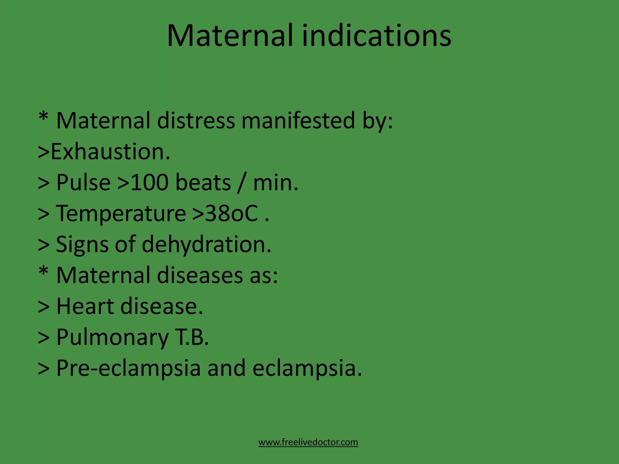
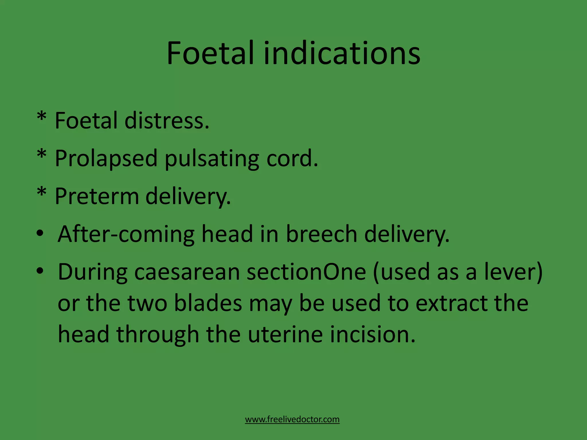
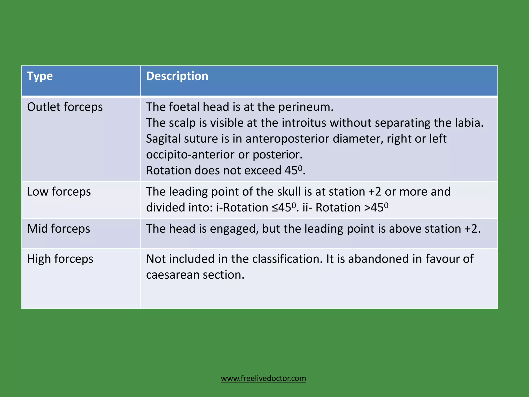
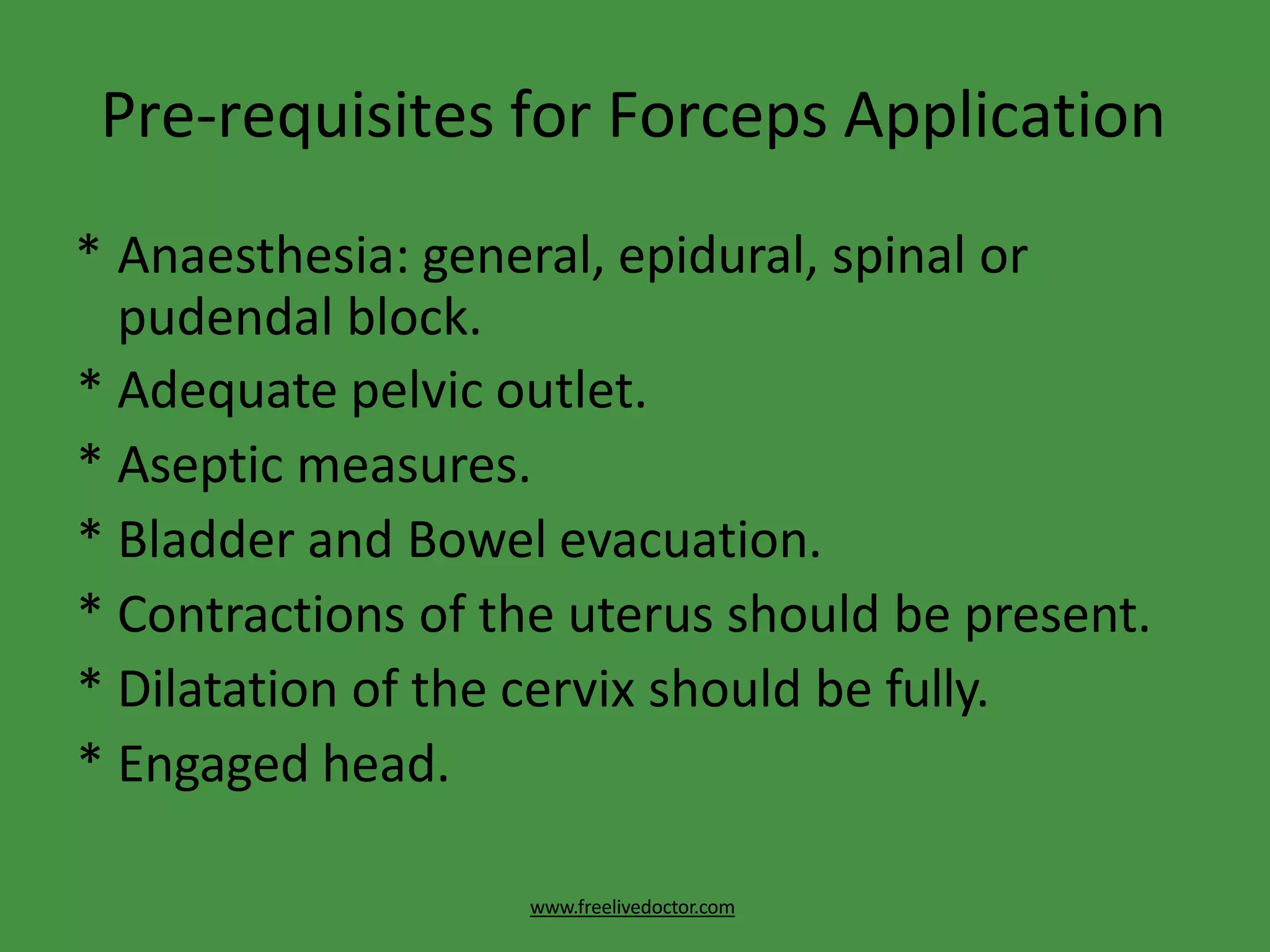
This document discusses forceps delivery, including definitions, types of forceps, indications for their use, techniques for application, and potential complications. Obstetric forceps are metal instruments used to extract the fetal head during delivery. They are indicated when delivery is prolonged or complicated by issues like fetal distress or maternal health conditions. Proper technique is important to minimize risks of injuries to the mother or baby. Complications can include lacerations, bone fractures, hemorrhage or problems for the infant like skull fractures or brain bleeding. Failed forceps require assessing the cause and may necessitate a C-section.
VACUUM DELIVERY - OBSTETRICS AND GYNAECOLOGY-
DEALS WITH THE DELIVERY OF HUMAN BABY BY VACUUM IN SPECIAL OBSTETRIC CONDITIONS.
VACUUM is an instrumental device designed to assist delivery by creating a vacuum between it and the fetal scalp.
Preterm labor is the labor that starts before the 37th completed week. In this presentation, we will discover causes, pathogenesis, diagnosis, clinical features, and management principles for preterm labor along with the most recent evidence.
This presentation contains :-
1.Introduction of normal labour
2. Definiation of normal labour
3.Criteria of normal labour
4. Physiology of normal labour
5. Pathophysiology of labor
6.Estrogen
7. Prostaglandin
8. Oxytocin
9. True labor and false labor difference
10. Uterine contraction in labor
11. Stages of labour
12. Management of 1 st stage
13. management of 2 nd stage
14. mamagement of 3 rd stage of labor
15. Cervix dilation
16. Friedman's curve
17. Fetal skull
18. Diameter of fetal skull
19. Sutures in fetal head
20. Moulding
21. Mechanism of labour
Obstetrical Surgeries - Operative vaginal deliveries are accomplished by appl...MariaDavis42
Operative vaginal deliveries are accomplished by applying direct traction on the fetal skull with forceps or by applying traction to the fetal scalp by means of a vacuum extractor
Introduction:
RNA interference (RNAi) or Post-Transcriptional Gene Silencing (PTGS) is an important biological process for modulating eukaryotic gene expression.
It is highly conserved process of posttranscriptional gene silencing by which double stranded RNA (dsRNA) causes sequence-specific degradation of mRNA sequences.
dsRNA-induced gene silencing (RNAi) is reported in a wide range of eukaryotes ranging from worms, insects, mammals and plants.
This process mediates resistance to both endogenous parasitic and exogenous pathogenic nucleic acids, and regulates the expression of protein-coding genes.
What are small ncRNAs?
micro RNA (miRNA)
short interfering RNA (siRNA)
Properties of small non-coding RNA:
Involved in silencing mRNA transcripts.
Called “small” because they are usually only about 21-24 nucleotides long.
Synthesized by first cutting up longer precursor sequences (like the 61nt one that Lee discovered).
Silence an mRNA by base pairing with some sequence on the mRNA.
Discovery of siRNA?
The first small RNA:
In 1993 Rosalind Lee (Victor Ambros lab) was studying a non- coding gene in C. elegans, lin-4, that was involved in silencing of another gene, lin-14, at the appropriate time in the
development of the worm C. elegans.
Two small transcripts of lin-4 (22nt and 61nt) were found to be complementary to a sequence in the 3' UTR of lin-14.
Because lin-4 encoded no protein, she deduced that it must be these transcripts that are causing the silencing by RNA-RNA interactions.
Types of RNAi ( non coding RNA)
MiRNA
Length (23-25 nt)
Trans acting
Binds with target MRNA in mismatch
Translation inhibition
Si RNA
Length 21 nt.
Cis acting
Bind with target Mrna in perfect complementary sequence
Piwi-RNA
Length ; 25 to 36 nt.
Expressed in Germ Cells
Regulates trnasposomes activity
MECHANISM OF RNAI:
First the double-stranded RNA teams up with a protein complex named Dicer, which cuts the long RNA into short pieces.
Then another protein complex called RISC (RNA-induced silencing complex) discards one of the two RNA strands.
The RISC-docked, single-stranded RNA then pairs with the homologous mRNA and destroys it.
THE RISC COMPLEX:
RISC is large(>500kD) RNA multi- protein Binding complex which triggers MRNA degradation in response to MRNA
Unwinding of double stranded Si RNA by ATP independent Helicase
Active component of RISC is Ago proteins( ENDONUCLEASE) which cleave target MRNA.
DICER: endonuclease (RNase Family III)
Argonaute: Central Component of the RNA-Induced Silencing Complex (RISC)
One strand of the dsRNA produced by Dicer is retained in the RISC complex in association with Argonaute
ARGONAUTE PROTEIN :
1.PAZ(PIWI/Argonaute/ Zwille)- Recognition of target MRNA
2.PIWI (p-element induced wimpy Testis)- breaks Phosphodiester bond of mRNA.)RNAse H activity.
MiRNA:
The Double-stranded RNAs are naturally produced in eukaryotic cells during development, and they have a key role in regulating gene expression .
Earliest Galaxies in the JADES Origins Field: Luminosity Function and Cosmic ...Sérgio Sacani
We characterize the earliest galaxy population in the JADES Origins Field (JOF), the deepest
imaging field observed with JWST. We make use of the ancillary Hubble optical images (5 filters
spanning 0.4−0.9µm) and novel JWST images with 14 filters spanning 0.8−5µm, including 7 mediumband filters, and reaching total exposure times of up to 46 hours per filter. We combine all our data
at > 2.3µm to construct an ultradeep image, reaching as deep as ≈ 31.4 AB mag in the stack and
30.3-31.0 AB mag (5σ, r = 0.1” circular aperture) in individual filters. We measure photometric
redshifts and use robust selection criteria to identify a sample of eight galaxy candidates at redshifts
z = 11.5 − 15. These objects show compact half-light radii of R1/2 ∼ 50 − 200pc, stellar masses of
M⋆ ∼ 107−108M⊙, and star-formation rates of SFR ∼ 0.1−1 M⊙ yr−1
. Our search finds no candidates
at 15 < z < 20, placing upper limits at these redshifts. We develop a forward modeling approach to
infer the properties of the evolving luminosity function without binning in redshift or luminosity that
marginalizes over the photometric redshift uncertainty of our candidate galaxies and incorporates the
impact of non-detections. We find a z = 12 luminosity function in good agreement with prior results,
and that the luminosity function normalization and UV luminosity density decline by a factor of ∼ 2.5
from z = 12 to z = 14. We discuss the possible implications of our results in the context of theoretical
models for evolution of the dark matter halo mass function.
Multi-source connectivity as the driver of solar wind variability in the heli...Sérgio Sacani
The ambient solar wind that flls the heliosphere originates from multiple
sources in the solar corona and is highly structured. It is often described
as high-speed, relatively homogeneous, plasma streams from coronal
holes and slow-speed, highly variable, streams whose source regions are
under debate. A key goal of ESA/NASA’s Solar Orbiter mission is to identify
solar wind sources and understand what drives the complexity seen in the
heliosphere. By combining magnetic feld modelling and spectroscopic
techniques with high-resolution observations and measurements, we show
that the solar wind variability detected in situ by Solar Orbiter in March
2022 is driven by spatio-temporal changes in the magnetic connectivity to
multiple sources in the solar atmosphere. The magnetic feld footpoints
connected to the spacecraft moved from the boundaries of a coronal hole
to one active region (12961) and then across to another region (12957). This
is refected in the in situ measurements, which show the transition from fast
to highly Alfvénic then to slow solar wind that is disrupted by the arrival of
a coronal mass ejection. Our results describe solar wind variability at 0.5 au
but are applicable to near-Earth observatories.
(May 29th, 2024) Advancements in Intravital Microscopy- Insights for Preclini...Scintica Instrumentation
Intravital microscopy (IVM) is a powerful tool utilized to study cellular behavior over time and space in vivo. Much of our understanding of cell biology has been accomplished using various in vitro and ex vivo methods; however, these studies do not necessarily reflect the natural dynamics of biological processes. Unlike traditional cell culture or fixed tissue imaging, IVM allows for the ultra-fast high-resolution imaging of cellular processes over time and space and were studied in its natural environment. Real-time visualization of biological processes in the context of an intact organism helps maintain physiological relevance and provide insights into the progression of disease, response to treatments or developmental processes.
In this webinar we give an overview of advanced applications of the IVM system in preclinical research. IVIM technology is a provider of all-in-one intravital microscopy systems and solutions optimized for in vivo imaging of live animal models at sub-micron resolution. The system’s unique features and user-friendly software enables researchers to probe fast dynamic biological processes such as immune cell tracking, cell-cell interaction as well as vascularization and tumor metastasis with exceptional detail. This webinar will also give an overview of IVM being utilized in drug development, offering a view into the intricate interaction between drugs/nanoparticles and tissues in vivo and allows for the evaluation of therapeutic intervention in a variety of tissues and organs. This interdisciplinary collaboration continues to drive the advancements of novel therapeutic strategies.
Observation of Io’s Resurfacing via Plume Deposition Using Ground-based Adapt...Sérgio Sacani
Since volcanic activity was first discovered on Io from Voyager images in 1979, changes
on Io’s surface have been monitored from both spacecraft and ground-based telescopes.
Here, we present the highest spatial resolution images of Io ever obtained from a groundbased telescope. These images, acquired by the SHARK-VIS instrument on the Large
Binocular Telescope, show evidence of a major resurfacing event on Io’s trailing hemisphere. When compared to the most recent spacecraft images, the SHARK-VIS images
show that a plume deposit from a powerful eruption at Pillan Patera has covered part
of the long-lived Pele plume deposit. Although this type of resurfacing event may be common on Io, few have been detected due to the rarity of spacecraft visits and the previously low spatial resolution available from Earth-based telescopes. The SHARK-VIS instrument ushers in a new era of high resolution imaging of Io’s surface using adaptive
optics at visible wavelengths.
This presentation explores a brief idea about the structural and functional attributes of nucleotides, the structure and function of genetic materials along with the impact of UV rays and pH upon them.
4. Action of the Forceps
www.freelivedoctor.com
• Traction: is the main action.
• Rotation: in deep transverse arrest, persistent
occipito-posterior and mento-posterior.
5. Indications of Forceps Delivery
www.freelivedoctor.com
• Prolonged 2nd stage
• It is prolongation for more than 1 hour in
primigravidae or 30 minutes in multiparae. This
may be due to:
* Inertia and poor voluntary bearing down.
* Large foetus.
* Rigid perineum.
* Malpositions: persistent occipito-posterior and
deep transverse arrest.
6. Maternal indications
www.freelivedoctor.com
* Maternal distress manifested by:
>Exhaustion.
> Pulse >100 beats / min.
> Temperature >38oC .
> Signs of dehydration.
* Maternal diseases as:
> Heart disease.
> Pulmonary T.B.
> Pre-eclampsia and eclampsia.
7. Foetal indications
www.freelivedoctor.com
* Foetal distress.
* Prolapsed pulsating cord.
* Preterm delivery.
• After-coming head in breech delivery.
• During caesarean sectionOne (used as a lever)
or the two blades may be used to extract the
head through the uterine incision.
8. Type Description
Outlet forceps The foetal head is at the perineum.
The scalp is visible at the introitus without separating the labia.
Sagital suture is in anteroposterior diameter, right or left
occipito-anterior or posterior.
Rotation does not exceed 450.
Low forceps The leading point of the skull is at station +2 or more and
divided into: i-Rotation ≤450. ii- Rotation >450
Mid forceps The head is engaged, but the leading point is above station +2.
High forceps Not included in the classification. It is abandoned in favour of
caesarean section.
www.freelivedoctor.com
9. Pre-requisites for Forceps Application
www.freelivedoctor.com
* Anaesthesia: general, epidural, spinal or
pudendal block.
* Adequate pelvic outlet.
* Aseptic measures.
* Bladder and Bowel evacuation.
* Contractions of the uterus should be present.
* Dilatation of the cervix should be fully.
* Engaged head.
10. Pre-requisites for Forceps Application
www.freelivedoctor.com
* Forewater rupture.
* Favourable position and presentation:
>Occipito-anterior.
> Occipito-posterior
> Face presentation.
>After-coming head in breech.
11. Types of Forceps Application
www.freelivedoctor.com
* Cephalic application: the forceps is applied on the sides
of the foetal head in the mento-vertical diameter so
injury of the foetal face, eyes and facial nerve is
avoided .
* Pelvic application: The forceps is applied along the
maternal pelvic wall irrespective to the position of the
head. It is easier for application but carries a great risk
of foetal injuries.
* Cephalo-pelvic application: It is the ideal application
and possible when the occiput is directly anterior or
posterior or in direct mento-anterior position.
12. How to know Right and Left Blades
www.freelivedoctor.com
• Putting in consideration that the mother is in
the lithotomy position, the blade will be
applied with the pelvic curve directed
anteriorly and the cephalic curve directed
medially. If the blade will be applied to the left
maternal side it is a left blade and vice versa.
13. Technique of Forceps Delivery
www.freelivedoctor.com
• In occipito- anterior position
* The left blade is applied first. It is held by its
handle between the thumb and fingers of the left
hand almost parallel with the right inguinal
ligament and passed along the left side of the
maternal pelvis between the guiding palm of the
right hand and foetal head.
* As the blade passes into the birth canal the
handle is carried backwards and towards the
midline. It is now the lower blade.
14. Technique of Forceps Delivery
www.freelivedoctor.com
* The fingers of the left hand are introduced
along the right side of the pelvis and the right
blade is held and passed in the same manner.
It is now the upper blade.
* The 2 blades should be locked easily, if not this
means that they were not correctly applied
and should be removed and re-assess the
position of the head.
15. Clinical checks for correct forceps
application:
www.freelivedoctor.com
* The sagittal suture lies in the midline of the
shanks.
* The operator cannot place more than a finger
tip between the fenestration of the blade and
the foetal head.
* The posterior fontanelle is not more than one
finger- breadth above the plane of the shanks.
16. Traction should be:
www.freelivedoctor.com
* gentle by the force of the arm only,
* intermittent with uterine contractions only,
* in correct direction i.e. downwards and
backwards till the occiput appears at the
vulva, then downwards and forwards.
* The 2 blades are unlocked between
contractions to minimise the period of head
compression.
17. Kielland forceps in deep transverse
arrest
www.freelivedoctor.com
• The forceps is locked outside with the knobs
towards the occiput to know the anterior
blade.
• The anterior blade is applied first by one of
the following methods:
18. • The wandering method: The anterior blade is guided
into the lateral side of the pelvis with the cephalic
curve facing the foetal head. It is then slid over the
forehead to fit against the anterior parietal eminence.
• The direct method: when the head is low down in the
pelvis, the anterior blade is slid between the head and
symphysis pubis with the cephalic curve facing the
foetal head.
• The old (classical) method: The anterior blade is
applied with the cephalic curve towards the symphysis
pubis then it is rotated 1800 to fit with the head. This
method is not recommended as the lower uterine
segment and bladder may be injured.
www.freelivedoctor.com
19. • The posterior blade is applied along the
concavity of the sacrum.
• The 2 blades are locked, head is rotated and
extracted as occipito-anterior.
www.freelivedoctor.com
22. Maternal complications
www.freelivedoctor.com
• Bone injuries: to pelvic joints, coccyx or
symphysis pubis.
• Pelvic nerve injuries.
• Postpartum haemorrhage: due to lacerations
or atony.
• Puerperal infections.
• Remote effects: genital prolapse, stress
incontinence, cervical incompetence and
genito-urinary fistulas.
23. Foetal complications
www.freelivedoctor.com
• Fracture of the skull.
• Cephalohaematoma.
• Intracranial haemorrhage.
• Facial nerve palsy.
• Trauma to the face, eyes or scalp.
• Asphyxia due to:
> intracranial haemorrhage or,
> cord compression between the head and the
forceps.
25. FAILED FORCEPS
www.freelivedoctor.com
• Causes
* Cephalo-pelvic disproportion.
* Contracted outlet.
* Incomplete cervical dilatation.
* Constriction ring.
* Head is not engaged.
* Malpositions as persistent occipito-posterior.
*Malpresentations as brow.
* Foetal congenital anomalies as hydrocephalus, ascitis
and conjoined twins.
26. Management
www.freelivedoctor.com
* Reassessment: The forceps is removed and the
patient is re-examined to detect the cause and
correct it if possible.
* Caesarean section: is indicated in
uncorrectable causes as cephalo-pelvic
disproportion, and contracted outlet.
* Exploration of the birth canal: for any injuries.