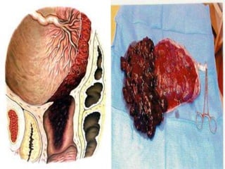
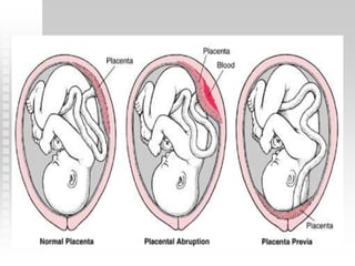
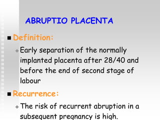
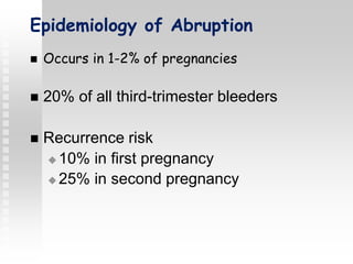
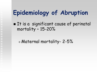
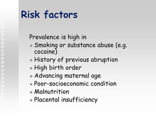
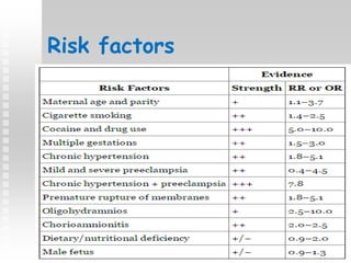
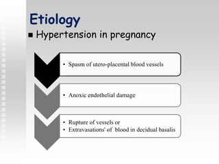
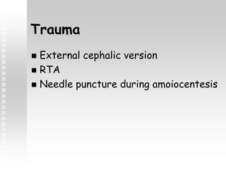
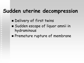
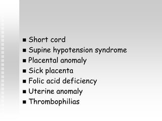
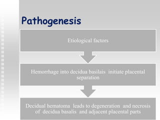
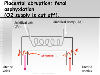
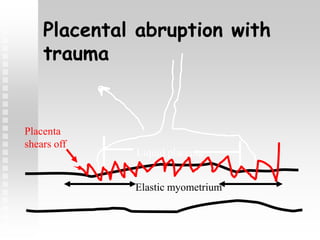
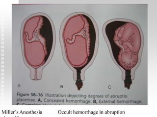
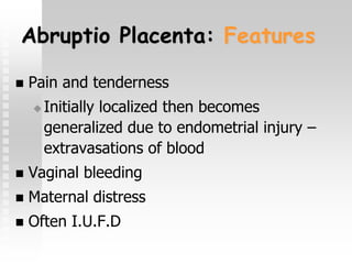
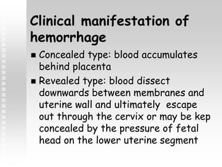
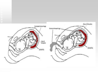
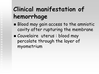
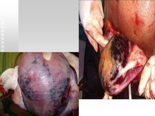
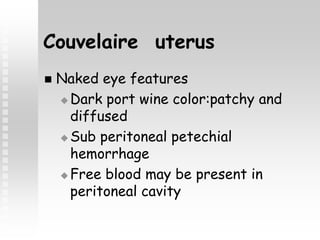
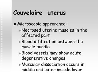
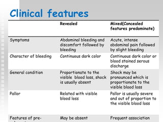
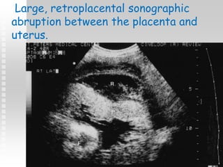
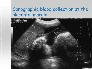
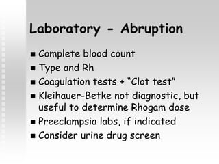
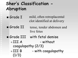
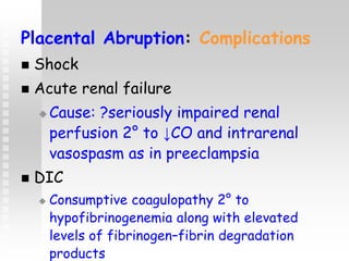
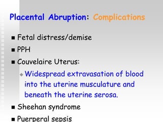
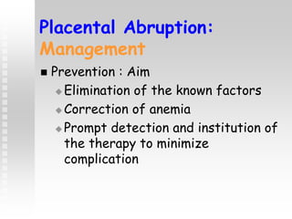
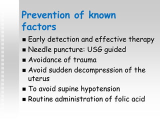
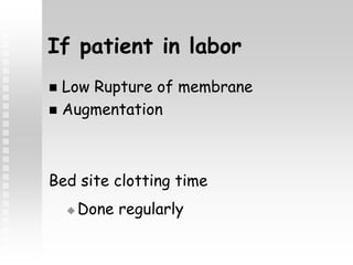
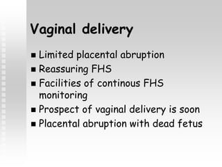
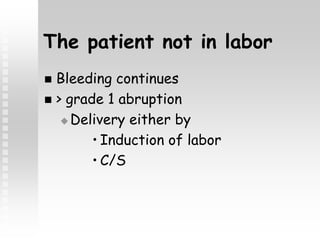
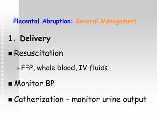
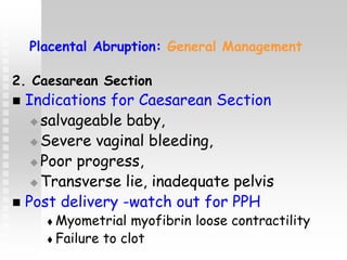
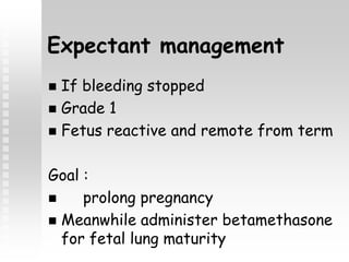
Vaginal Bleeding in
Late Pregnancy
Presented By
Nirsuba Gurung
Master in nursing
Women health and development
Obstetric Haemorrhage
 Ranks as the First cause of
maternal mortality accounting for
25 – 50 % of maternal deaths
APH: Epidemiology & Causes
 Magnitude: 4% of women may develop
APH.
 Causes:
 placenta previa (1/200)
 placental abruption (1/100)
 uterine rupture (<1% in scarred uterus)
 vasa previa (1/2000-3000)
 Local causes
 Cervical polyp
 Bloody show
 Cervicitis or cervical ectropion
 Cervical cancer
Vasa Previa
Velamentous Insertion of the
umbilical cord
ABRUPTIO PLACENTA
 Definition:
 Early separation of the normally
implanted placenta after 28/40 and
before the end of second stage of
labour
 Recurrence:
 The risk of recurrent abruption in a
subsequent pregnancy is high.
Epidemiology of Abruption
 Occurs in 1-2% of pregnancies
 20% of all third-trimester bleeders
 Recurrence risk
 10% in first pregnancy
 25% in second pregnancy
Epidemiology of Abruption
 It is a significant cause of perinatal
mortality – 15-20%
 Maternal mortality- 2-5%
Risk factors
Prevalence is high in
 Smoking or substance abuse (e.g.
cocaine)
 History of previous abruption
 High birth order
 Advancing maternal age
 Poor-socioeconomic condition
 Malnutrition
 Placental insufficiency
Risk factors
Etiology
 Hypertension in pregnancy
• Spasm of utero-placental blood vessels
• Anoxic endothelial damage
• Rupture of vessels or
• Extravasations' of blood in decidual basalis
Trauma
 External cephalic version
 RTA
 Needle puncture during amoiocentesis
Sudden uterine decompression
 Delivery of first twins
 Sudden escape of liquor amnii in
hydraminous
 Premature rupture of membrane
 Short cord
 Supine hypotension syndrome
 Placental anomaly
 Sick placenta
 Folic acid deficiency
 Uterine anomaly
 Thrombophilias
Pathogenesis
Decidual hematoma leads to degeneration and necrosis
of decidua basalis and adjacent placental parts
Hemorrhage into decidua basilais initiate placental
separation
Etiological factors
Umbilical artery (UA)Umbilical vein
(UV)
Uterine
arteries
Uterine
veins
Abruption
Archer TL 2006 unpublished
Placental abruption: fetal
asphyxiation
(O2 supply is cut off).
Placental abruption with
trauma
Elastic myometrium
Liquid placenta
Placenta
shears off
Miller’s Anesthesia Occult hemorrhage in abruption
Features of retroplacental
clots
 Depression found in maternal surface
of placenta
 Area of infraction with varying
degree of organization
Abruptio placenta:
Classifications
Are based on
1. Extent of separation: Partial vs complete
2. Location of separation: Marginal Vs
central
3. Clinical presentation: Revealed, concealed
and mixed
4. Clinical Severity: Mild, Moderate and
Severe
Grade 1 Mildest form:
approx 40 of all
cases.
• No vaginal bleeding to
mild vaginal bleeding
• Slightly tender uterus
• Normal maternal BP
and heart rate
• No coagulopathy
(clotting problems)
• No fetal distress
Clinical Severity
Grade 2: moderate -
approx
45% of all cases.
• No vaginal bleeding to moderate
vaginal bleeding
• Moderate-to-severe uterine
tenderness with possible tetanic
contractions
• Maternal tachycardia with
orthostatic changes in BP and
heart rate
• Fetal distress or even death
• Low fibrinogen levels present
(causing clotting problems)
Grade 0: no clinical features
•Diagnosis made after placental exmaninatio
Grade 3: Severe form: Approx 15% of all cases.
• No vaginal bleeding to heavy vaginal bleeding
• Very painful tetanic uterus
• Maternal shock
• Coagulopathy
• Fetal death
Clinical Severity
Abruptio Placenta: Features
 Pain and tenderness
 Initially localized then becomes
generalized due to endometrial injury –
extravasations of blood
 Vaginal bleeding
 Maternal distress
 Often I.U.F.D
Clinical manifestation of
hemorrhage
 Concealed type: blood accumulates
behind placenta
 Revealed type: blood dissect
downwards between membranes and
uterine wall and ultimately escape
out through the cervix or may be kep
concealed by the pressure of fetal
head on the lower uterine segment
Clinical manifestation of
hemorrhage
 Blood may gain access to the amniotic
cavity after rupturing the membrane
 Couvelaire uterus : blood may
percolate through the layer of
myometrium
Couvelaire uterus
 Naked eye features
 Dark port wine color:patchy and
diffused
 Sub peritoneal petechial
hemorrhage
 Free blood may be present in
peritoneal cavity
Couvelaire uterus
 Microscopic appearance:
 Necrosed uterine muscles in the
affected part
 Blood infiltration between the
muscle bundle
 Blood vessels may show acute
degenerative changes
 Muscular dissociation occurs in
middle and outer muscle layer
Clinical features
Revealed Mixed(Concealed
features predominate)
Symptoms Abdominal bleeding and
discomfort followed by
bleeding
Acute, intense
abdominal pain followed
by slight bleeding
Character of bleeding Continuous dark color Continuous dark color or
blood stained serous
discharge
General condition Proportionate to the
visible blood loss, shock
is usually absent
Shock may be
pronounced which is
proportionate to the
visible blood loss
Pallor Related with visible
blood loss
Pallor is usually severe
and out of proportion to
the visible blood loss
Features of pre- May be absent Frequent association
Revealed Mixed(Concealed
features predominate)
Uterine height Proportionate to the
POG
May be
disproportionately
enlarged and globular
Uterine feel Normal feel with
localized tenderness,
contractions frequent
and local amplitude
Uterus is tense ,tender
and rigid
Fetal parts Can be identified easily Difficult to make out
FHS Usually present Usually absent
Laboratory test
Revealed Mixed(Concealed features
predominate)
Hemoglobin Low value proportionate to
the blood loss
Markedly lower than vi
Coagulation profile Usually unchanged Variable changes
•Clotting time increased(>6min)
•Fibrinogen level
low(<150mg/dl)
•Low platelet count
•^ Partial thromboplastin time
•^ FDP and D-dimer
Urine for protein
Confusion in diagnosis
May be absent Usually present
Confusion in diagnosis Placenta previa Acute obstretrical-
gynaecological –surgical
complication
Ultrasound - Abruption
 Abruption is a clinical diagnosis!
 Placental location and appearance
 Retroplacental echolucency
 Abnormal thickening of placenta
 “Torn” edge of placenta
 Fetal lie
 Estimated fetal weight
Large, extensive sonographic
preplacental collection beneath the
chorionic plate
Large, retroplacental sonographic
abruption between the placenta and
uterus.
Sonographic blood collection at the
placental margin
Laboratory - Abruption
 Complete blood count
 Type and Rh
 Coagulation tests + “Clot test”
 Kleihauer-Betke not diagnostic, but
useful to determine Rhogam dose
 Preeclampsia labs, if indicated
 Consider urine drug screen
Sher’s Classification -
Abruption
 Grade I
 Grade II
 Grade III with fetal demise
 III A - without
coagulopathy (2/3)
 III B - with coagulopathy
(1/3)
mild, often retroplacental
clot identified at delivery
tense, tender abdomen and
live fetus
Placental Abruption: Complications
 Shock
 Acute renal failure
 Cause: ?seriously impaired renal
perfusion 2° to ↓CO and intrarenal
vasospasm as in preeclampsia
 DIC
 Consumptive coagulopathy 2° to
hypofibrinogenemia along with elevated
levels of fibrinogen–fibrin degradation
products
Placental Abruption: Complications
 Fetal distress/demise
 PPH
 Couvelaire Uterus:
 Widespread extravasation of blood
into the uterine musculature and
beneath the uterine serosa.
 Sheehan syndrome
 Puerperal sepsis
Placental Abruption:
Management
 Prevention : Aim
 Elimination of the known factors
 Correction of anemia
 Prompt detection and institution of
the therapy to minimize
complication
Prevention of known
factors
 Early detection and effective therapy
 Needle puncture: USG guided
 Avoidance of trauma
 Avoid sudden decompression of the
uterus
 To avoid supine hypotension
 Routine administration of folic acid
Placental Abruption:
Management
 Management depends on:
 fetal maturity,
 degree of severity,
 viability of the fetus/fetal distress
Assessment of case
 Blood loss
 Maturity of fetus
 Whether patient is in labor or not
 Presence of complication
 Types and grade of abruption
Emergency measures
 Sent blood for Hb and hematocrit,
coagulation profile, ABO and Rh
grouping
 Urine for detection of protein
 IV RL drip with wide bore cannula and
arrangement for BT
 Close monitoring of maternal and
fetal well being
Treatment modalities
 Expectant management of
pregnancy
 Definitive management
 Induction/augmentation of labor
 Caesarean section
If patient in labor
 Low Rupture of membrane
 Augmentation
Bed site clotting time
 Done regularly
Vaginal delivery
 Limited placental abruption
 Reassuring FHS
 Facilities of continous FHS
monitoring
 Prospect of vaginal delivery is soon
 Placental abruption with dead fetus
The patient not in labor
 Bleeding continues
 > grade 1 abruption
 Delivery either by
• Induction of labor
• C/S
Placental Abruption: General Management
1. Delivery
 Resuscitation
 FFP, whole blood, IV fluids
 Monitor BP
 Catherization - monitor urine output
Placental Abruption: General Management
2. Caesarean Section
 Indications for Caesarean Section
 salvageable baby,
 Severe vaginal bleeding,
 Poor progress,
 Transverse lie, inadequate pelvis
 Post delivery -watch out for PPH
 Myometrial myofibrin loose contractility
 Failure to clot
Expectant management
 If bleeding stopped
 Grade 1
 Fetus reactive and remote from term
Goal :
 prolong pregnancy
 Meanwhile administer betamethasone
for fetal lung maturity
Abruptio placenta
Abruptio placenta

Abruptio placenta

  • 2.
    Vaginal Bleeding in LatePregnancy Presented By Nirsuba Gurung Master in nursing Women health and development
  • 3.
    Obstetric Haemorrhage  Ranksas the First cause of maternal mortality accounting for 25 – 50 % of maternal deaths
  • 4.
    APH: Epidemiology &Causes  Magnitude: 4% of women may develop APH.  Causes:  placenta previa (1/200)  placental abruption (1/100)  uterine rupture (<1% in scarred uterus)  vasa previa (1/2000-3000)  Local causes  Cervical polyp  Bloody show  Cervicitis or cervical ectropion  Cervical cancer
  • 6.
  • 7.
    Velamentous Insertion ofthe umbilical cord
  • 8.
    ABRUPTIO PLACENTA  Definition: Early separation of the normally implanted placenta after 28/40 and before the end of second stage of labour  Recurrence:  The risk of recurrent abruption in a subsequent pregnancy is high.
  • 9.
    Epidemiology of Abruption Occurs in 1-2% of pregnancies  20% of all third-trimester bleeders  Recurrence risk  10% in first pregnancy  25% in second pregnancy
  • 10.
    Epidemiology of Abruption It is a significant cause of perinatal mortality – 15-20%  Maternal mortality- 2-5%
  • 11.
    Risk factors Prevalence ishigh in  Smoking or substance abuse (e.g. cocaine)  History of previous abruption  High birth order  Advancing maternal age  Poor-socioeconomic condition  Malnutrition  Placental insufficiency
  • 12.
  • 13.
    Etiology  Hypertension inpregnancy • Spasm of utero-placental blood vessels • Anoxic endothelial damage • Rupture of vessels or • Extravasations' of blood in decidual basalis
  • 14.
    Trauma  External cephalicversion  RTA  Needle puncture during amoiocentesis
  • 15.
    Sudden uterine decompression Delivery of first twins  Sudden escape of liquor amnii in hydraminous  Premature rupture of membrane
  • 16.
     Short cord Supine hypotension syndrome  Placental anomaly  Sick placenta  Folic acid deficiency  Uterine anomaly  Thrombophilias
  • 17.
    Pathogenesis Decidual hematoma leadsto degeneration and necrosis of decidua basalis and adjacent placental parts Hemorrhage into decidua basilais initiate placental separation Etiological factors
  • 18.
    Umbilical artery (UA)Umbilicalvein (UV) Uterine arteries Uterine veins Abruption Archer TL 2006 unpublished Placental abruption: fetal asphyxiation (O2 supply is cut off).
  • 19.
    Placental abruption with trauma Elasticmyometrium Liquid placenta Placenta shears off
  • 20.
    Miller’s Anesthesia Occulthemorrhage in abruption
  • 21.
    Features of retroplacental clots Depression found in maternal surface of placenta  Area of infraction with varying degree of organization
  • 22.
    Abruptio placenta: Classifications Are basedon 1. Extent of separation: Partial vs complete 2. Location of separation: Marginal Vs central 3. Clinical presentation: Revealed, concealed and mixed 4. Clinical Severity: Mild, Moderate and Severe
  • 23.
    Grade 1 Mildestform: approx 40 of all cases. • No vaginal bleeding to mild vaginal bleeding • Slightly tender uterus • Normal maternal BP and heart rate • No coagulopathy (clotting problems) • No fetal distress Clinical Severity Grade 2: moderate - approx 45% of all cases. • No vaginal bleeding to moderate vaginal bleeding • Moderate-to-severe uterine tenderness with possible tetanic contractions • Maternal tachycardia with orthostatic changes in BP and heart rate • Fetal distress or even death • Low fibrinogen levels present (causing clotting problems) Grade 0: no clinical features •Diagnosis made after placental exmaninatio
  • 24.
    Grade 3: Severeform: Approx 15% of all cases. • No vaginal bleeding to heavy vaginal bleeding • Very painful tetanic uterus • Maternal shock • Coagulopathy • Fetal death Clinical Severity
  • 25.
    Abruptio Placenta: Features Pain and tenderness  Initially localized then becomes generalized due to endometrial injury – extravasations of blood  Vaginal bleeding  Maternal distress  Often I.U.F.D
  • 26.
    Clinical manifestation of hemorrhage Concealed type: blood accumulates behind placenta  Revealed type: blood dissect downwards between membranes and uterine wall and ultimately escape out through the cervix or may be kep concealed by the pressure of fetal head on the lower uterine segment
  • 28.
    Clinical manifestation of hemorrhage Blood may gain access to the amniotic cavity after rupturing the membrane  Couvelaire uterus : blood may percolate through the layer of myometrium
  • 30.
    Couvelaire uterus  Nakedeye features  Dark port wine color:patchy and diffused  Sub peritoneal petechial hemorrhage  Free blood may be present in peritoneal cavity
  • 31.
    Couvelaire uterus  Microscopicappearance:  Necrosed uterine muscles in the affected part  Blood infiltration between the muscle bundle  Blood vessels may show acute degenerative changes  Muscular dissociation occurs in middle and outer muscle layer
  • 32.
    Clinical features Revealed Mixed(Concealed featurespredominate) Symptoms Abdominal bleeding and discomfort followed by bleeding Acute, intense abdominal pain followed by slight bleeding Character of bleeding Continuous dark color Continuous dark color or blood stained serous discharge General condition Proportionate to the visible blood loss, shock is usually absent Shock may be pronounced which is proportionate to the visible blood loss Pallor Related with visible blood loss Pallor is usually severe and out of proportion to the visible blood loss Features of pre- May be absent Frequent association
  • 33.
    Revealed Mixed(Concealed features predominate) Uterineheight Proportionate to the POG May be disproportionately enlarged and globular Uterine feel Normal feel with localized tenderness, contractions frequent and local amplitude Uterus is tense ,tender and rigid Fetal parts Can be identified easily Difficult to make out FHS Usually present Usually absent
  • 34.
    Laboratory test Revealed Mixed(Concealedfeatures predominate) Hemoglobin Low value proportionate to the blood loss Markedly lower than vi Coagulation profile Usually unchanged Variable changes •Clotting time increased(>6min) •Fibrinogen level low(<150mg/dl) •Low platelet count •^ Partial thromboplastin time •^ FDP and D-dimer Urine for protein Confusion in diagnosis May be absent Usually present Confusion in diagnosis Placenta previa Acute obstretrical- gynaecological –surgical complication
  • 35.
    Ultrasound - Abruption Abruption is a clinical diagnosis!  Placental location and appearance  Retroplacental echolucency  Abnormal thickening of placenta  “Torn” edge of placenta  Fetal lie  Estimated fetal weight
  • 36.
    Large, extensive sonographic preplacentalcollection beneath the chorionic plate
  • 37.
    Large, retroplacental sonographic abruptionbetween the placenta and uterus.
  • 38.
    Sonographic blood collectionat the placental margin
  • 39.
    Laboratory - Abruption Complete blood count  Type and Rh  Coagulation tests + “Clot test”  Kleihauer-Betke not diagnostic, but useful to determine Rhogam dose  Preeclampsia labs, if indicated  Consider urine drug screen
  • 40.
    Sher’s Classification - Abruption Grade I  Grade II  Grade III with fetal demise  III A - without coagulopathy (2/3)  III B - with coagulopathy (1/3) mild, often retroplacental clot identified at delivery tense, tender abdomen and live fetus
  • 41.
    Placental Abruption: Complications Shock  Acute renal failure  Cause: ?seriously impaired renal perfusion 2° to ↓CO and intrarenal vasospasm as in preeclampsia  DIC  Consumptive coagulopathy 2° to hypofibrinogenemia along with elevated levels of fibrinogen–fibrin degradation products
  • 42.
    Placental Abruption: Complications Fetal distress/demise  PPH  Couvelaire Uterus:  Widespread extravasation of blood into the uterine musculature and beneath the uterine serosa.  Sheehan syndrome  Puerperal sepsis
  • 43.
    Placental Abruption: Management  Prevention: Aim  Elimination of the known factors  Correction of anemia  Prompt detection and institution of the therapy to minimize complication
  • 44.
    Prevention of known factors Early detection and effective therapy  Needle puncture: USG guided  Avoidance of trauma  Avoid sudden decompression of the uterus  To avoid supine hypotension  Routine administration of folic acid
  • 45.
    Placental Abruption: Management  Managementdepends on:  fetal maturity,  degree of severity,  viability of the fetus/fetal distress
  • 46.
    Assessment of case Blood loss  Maturity of fetus  Whether patient is in labor or not  Presence of complication  Types and grade of abruption
  • 47.
    Emergency measures  Sentblood for Hb and hematocrit, coagulation profile, ABO and Rh grouping  Urine for detection of protein  IV RL drip with wide bore cannula and arrangement for BT  Close monitoring of maternal and fetal well being
  • 48.
    Treatment modalities  Expectantmanagement of pregnancy  Definitive management  Induction/augmentation of labor  Caesarean section
  • 49.
    If patient inlabor  Low Rupture of membrane  Augmentation Bed site clotting time  Done regularly
  • 50.
    Vaginal delivery  Limitedplacental abruption  Reassuring FHS  Facilities of continous FHS monitoring  Prospect of vaginal delivery is soon  Placental abruption with dead fetus
  • 51.
    The patient notin labor  Bleeding continues  > grade 1 abruption  Delivery either by • Induction of labor • C/S
  • 52.
    Placental Abruption: GeneralManagement 1. Delivery  Resuscitation  FFP, whole blood, IV fluids  Monitor BP  Catherization - monitor urine output
  • 53.
    Placental Abruption: GeneralManagement 2. Caesarean Section  Indications for Caesarean Section  salvageable baby,  Severe vaginal bleeding,  Poor progress,  Transverse lie, inadequate pelvis  Post delivery -watch out for PPH  Myometrial myofibrin loose contractility  Failure to clot
  • 54.
    Expectant management  Ifbleeding stopped  Grade 1  Fetus reactive and remote from term Goal :  prolong pregnancy  Meanwhile administer betamethasone for fetal lung maturity