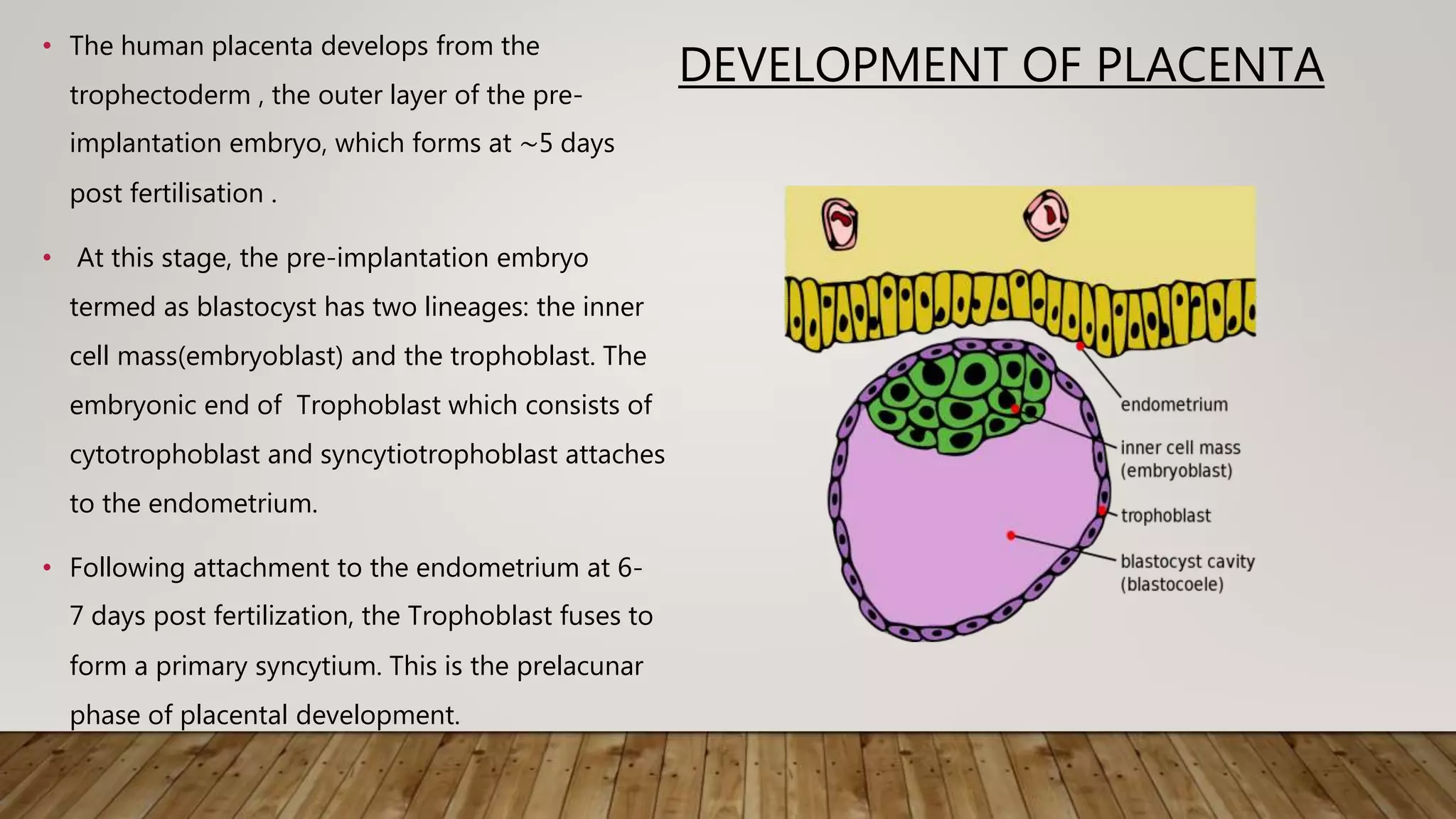
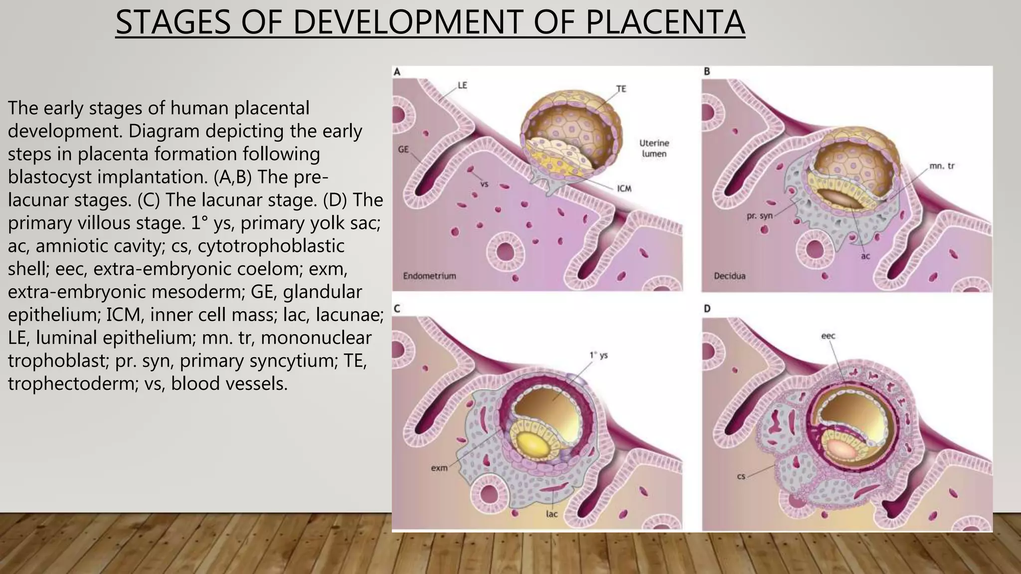
2. DEVELOPMENT OF PLACENTA
ā¢ The human placenta develops from the
trophectoderm , the outer layer of the pre-
implantation embryo, which forms at ā¼5 days
post fertilisation .
ā¢ At this stage, the pre-implantation embryo
termed as blastocyst has two lineages: the inner
cell mass(embryoblast) and the trophoblast. The
embryonic end of Trophoblast which consists of
cytotrophoblast and syncytiotrophoblast attaches
to the endometrium.
ā¢ Following attachment to the endometrium at 6-
7 days post fertilization, the Trophoblast fuses to
form a primary syncytium. This is the prelacunar
phase of placental development.
3.
4. PLACENTA DEVELOPMENT CONTDā¦
ā¢ Following implantation,
ā¢ The primary syncytium quickly invades through the surface epithelium into the underlying
endometrium, which is transformed during pregnancy into a specialised tissue known as
decidua.
ā¢ By the time of the first missed menstrual period (ā¼14 days post fertilization), the blastocyst
is completely embedded in the decidua and is covered by the surface epithelium.
ā¢ Fluid-filled spaces (lacunae) then appear within the syncytial mass that enlarge and merge,
partitioning it into a system of trabeculae. This is the lacunar stage. The syncytium also
erodes into decidual glands, allowing secretions to bathe the syncytial mass.
5.
6. PLACENTA DEVELOPMENT CONTD..
ā¢ The trophoblast cells beneath the syncytium (termed cytotrophoblast cells) are initially not in direct contact with
maternal tissue but rapidly proliferate to form projections that push through the primary syncytium to form
primary villi (a cytotrophoblast core with an outer layer of syncytiotrophoblast, ); this is the villous stage of
development (The villous trees are formed by further proliferation and branching, and the lacunae become the
intervillous space.
ā¢ Cytotrophoblast cells eventually penetrate through the primary syncytium and merge laterally to surround the
conceptus in a continuous cytotrophoblast shell between the villi and the decidua The blastocyst is now covered
by three layers: the inner chorionic plate in contact with the original cavity; the villi separated by the intervillous
space; and the cytotrophoblast shell in contact with the decidua.
7. PLACENTA DEVELOPMENT CONTD..
ā¢ Soon afterwards, around day 17-18, extraembryonic mesenchymal cells penetrate through the villous core
to form secondary villi.
ā¢ By day 18 dpf, fetal capillaries appear within the core, marking the development of tertiary villi. The villous
tree continues to rapidly enlarge by progressive branching from the chorionic plate to form a system of
villous trees.
ā¢ Where the cytotrophoblast shell is in contact with the decidua (the maternal-fetal interface), individual
cytotrophoblast cells leave the shell to invade into decidua as extravillous trophoblast in a process closely
resembling epithelial-mesenchymal transition. In this way, by the end of the first trimester, the blueprint
of the placenta is established.
8. STAGES OF DEVELOPMENT OF PLACENTA
The early stages of human placental
development. Diagram depicting the early
steps in placenta formation following
blastocyst implantation. (A,B) The pre-
lacunar stages. (C) The lacunar stage. (D) The
primary villous stage. 1Ā° ys, primary yolk sac;
ac, amniotic cavity; cs, cytotrophoblastic
shell; eec, extra-embryonic coelom; exm,
extra-embryonic mesoderm; GE, glandular
epithelium; ICM, inner cell mass; lac, lacunae;
LE, luminal epithelium; mn. tr, mononuclear
trophoblast; pr. syn, primary syncytium; TE,
trophectoderm; vs, blood vessels.
9. NORMAL ANATOMY OF PLACENTA
ā¢ At term: Weight: 470 grams, round to oval in shape
ā¢ Diameter: 22cm and central thickness of 2.5 cm.
ā¢ Composed of a Placental Disk, Extra-Placental membranes and a three vessel
umbilical cord.
ā¢ Maternal Surface is Basal Plate
ā¢ Fetal Surface is the chorionic plate into which umbilical cord inserts into the
center.
ā¢ Chorionic plate and its vessels are covered by amnion.
10. ULTRASOUND APPEARANCE
ā¢ In first and early second trimester placenta appears homogenous in echotexture and
mildly hyperechoic compared to underlying myometrium.
ā¢ It becomes more isoechoic with advancing gestation.
ā¢ After midpregnancy, it is common to identify small placental lucencies.
ā¢ In the third trimester, placenta appears more heterogenous with visible calcifications.
ā¢ Placental Thickness in mm roughly approximates to gestational age in weeks.
ā¢ Does not normally exceed 4 mm in second trimester or 6 cm in the third trimester.
ā¢ Retroplacental clear space normally measures less than 1 to 2 cm and is hypoechoic.
ā¢ Retroplacental clear space common location for hematoma development.
11. Normal placenta at 10 weeks gestation.
Transverse gray-scale Ultrasounfd image shows
the chorion laeve (right arrow) and chorion
frondosum (left arrows) of the placenta.
12. Normal placenta at 12 weeks gestation.
Transverse color Doppler image shows
intervillous flow (arrow). M = myometrium, P =
placenta.
13. Normal placenta at 18 weeks gestation. Longitudinal
gray-scale US image shows a homogeneous
placenta (P) with central placental cord
insertion (CI) and the hypoechoic retroplacental
complex (arrows) behind the placenta.
15. PLACENTAL GRADING (GRANNUM
CLASSIFICATION)
ā¢ The grading system is as follows:
ā¢ grade 0: <18 weeks
ā¢ uniform echogenicity
ā¢ Smooth chorionic plate
ā¢ grade I: 18-29 weeks
ā¢ occasional parenchymal calcification/hyperechoic areas
ā¢ subtle indentations of chorionic plate
ā¢ grade II: ā30-38 weeks
ā¢ occasional basal calcification/hyperechoic areas
ā¢ deeper indentations of the chorionic plate: seen as comma type densities at the chorionic plate.
ā¢ grade III: āāā„ 39 weeks
ā¢ significant basal plate calcification
ā¢ chorionic plate interrupted by indentations that reach up to the basal plate: cotyledons
ā¢ an early progression to a grade III placenta is concerning and is termed as hyper mature placenta and sometimes associated with
placental insufficiency.
ā¢ associated with smoking, chronic hypertension, SLE, diabetes
21. BILOBED PLACENTA
ā¢ Bilobed placenta is a placenta with two roughly
equal-sized lobes separated by a membrane.
ā¢ The umbilical cord may insert in either lobe, in
velamentous fashion, or in between the lobes.
ā¢ Bilobed placentas can be associated with first-
trimester bleeding, polyhydramnios, abruption and
retained placenta.
ā¢ A placenta with more than two lobes is rare and is
termed a multilobate placenta.
22. These are two views of a bilobate placenta, where each of the lobes (one on the anterior and one on
the posterior aspects of the uterine cavity) are marked with an *
23. US image shows a bilobed
placenta. The two lobes of the
placenta (P1 and P2) are separated
by a thin bridge of placental tissue
that covers the internal os. In this
case, the umbilical cord
(arrowhead) inserts into the bridge
of tissue.
24. CIRCUMVALLATE PLACENTA
ā¢ Circumvallate placenta is a placenta that is an annularly-shaped
placenta with raised edges composed of a double fold of chorion,
amnion, degenerated decidua, and fibrin deposits.
ā¢ In this condition, the chorionic plate is smaller than the basal plate,
resulting in hematoma retention in the placental margin.
ā¢ Within the ring, the fetal surface has the usual appearance except that
the large vessels terminate abruptly at the margin of the ring.
ā¢ Circumvallate placenta is associated with poor pregnancy outcomes
due to increased risk of vaginal bleeding beginning in the first
trimester, premature rupture of the membranes , preterm delivery,
placental insufficiency, and placental abruption.
25. CIRCUMVALLATE PLACENTA USG
Longitudinal gray-scale US image at 21 weeks
gestation shows the raised edge of the
placenta (P) as a linear band of tissue or shelf-like
structure (arrow) that may mimic a uterine
synechia.
26. US image shows a circumvallate placenta.
The chorionic plate (the fetal surface of
the placenta) (black arrowheads) is
smaller than the basal plate (the surface
interfacing with the uterus), with rolling
and shouldering of the placental margins
(white arrowheads). F = fetus.
27. SUCCENTURIATE PLACENTA
ā¢ Succenturiate placenta is a condition in which one or more accessory lobes
develop in the membranes apart from the main placental body to which vessels
of fetal origin usually connect them. It is a smaller variant of a bilobed placenta.
ā¢ The vessels are supported only by communicating membranes. If the
communicating membranes do not have vessels, it is called placenta supuria.
ā¢ .Advanced maternal age and in vitro fertilization are risk factors for the
succenturiate placenta.
ā¢ Other factors leading to succenturiate placentas include implantation over
leiomyomas, in areas of previous surgery, in the cornu or over the cervical os.
Ultrasound, particularly color Doppler, can be used to identify this condition
ā¢ . The risks of vasa previa and retained placenta are increased with this condition
like with bilobed and multilobate placentas.
28. US image shows a placenta (P) with a
succenturiate lobe (S). The main body of the
placenta is located along the posterior uterine
wall. A second soft-tissue structure of the same
echogenicity but located anteriorly is the
succenturiate lobe.
29. PLACENTA MEMBRANACEA
ā¢ Placenta membranacea or placenta diffusa is a rare
abnormality in which all or most fetal membranes remain
covered by chorionic villi, because the chorion has failed to
differentiate into chorion leave and chorion frondosum.
ā¢ Clinically the abnormality can present with vaginal
bleeding in the second or third trimester which is often
painless or during labor.
ā¢ Other placenta abnormalities, such as placenta previa and
placenta accreta, can be associated with this condition.
ā¢ Ultrasound has been reported as a diagnostic tool for this
condition, but due to the conditionās rarity, there is no data
on its sensitivity and specificity.
30.
31. RING SHAPED PLACENTA
ā¢ The ring-shaped placenta is an annularly-shaped placenta that is a variant of placenta
membranacea.
ā¢ It can sometimes be a complete ring of placental tissue, but more often tissue atrophy in a
portion of the ring results in a horseshoe shape.
ā¢ The ring-shaped placenta can cause antepartum and postpartum bleeding as well as fetal
growth restriction.
32. ABNORMAL PLACENTAL THICKNESS
ā¢ Placentomegally, if placental thickness exceeds
4cm in 2nd trimester and 6 cm in 3rd trimester.
ā¢ Causes: maternal diabetes
ā¢ Severe maternal anemia,
ā¢ Severe fetal growth restriction
ā¢ Aneuploidy
ā¢ Congenital Infections: Syphillis,cmv,parvovirus,
toxoplasmosis,herpesvirus,
rubella,schistosomiasis.
ā¢ Is a component of hydrops fetalis (Immune or
Non-Immune Hydrops)
ā¢ In rare cases it may be due to:
Collection of blood or fibrin within
placneta.
ā¢ Neoplasia
ā¢ GTD creates a thick cystic appearing
placenta , cystic vesicles are also seen
with placental mesenchymal dysplasia.
33. PLACENTAL THICKNESS ON USG
Longitudinal US image of a normal placenta at 18
weeks gestation shows the measurement calipers
appropriately positioned at the anterior and posterior
margins of the placenta (P), perpendicular to the long
axis of the placenta near the umbilical cord origin
34. ABNORMALITIES OF PLACENTAL LOCATION
ā¢ Placental location is determined according to the main placental body position from
the uterine equator. It can be anterior or posterior, fundal, or left or right.
ā¢ The term low-lying placenta is used when the placental edge is located in the lower
uterine segment within 2 cm or less of the internal cervical os.
ā¢ The term placenta previa is used when the placental edge covers the internal cervical
os
ā¢ Historically, terms such as complete, marginal, and partial were used to describe how
much of the placenta was covering the internal os.
ā¢ Transvaginal ultrasonography has improved sonographic description of the precise
location of the placenta, and these terms have been abandoned.
35. PLACENTA PREVIA USG
US is the gold standard for diagnosing placenta
previa
Complete placenta previa. Longitudinal gray-scale
US image at 28 weeks gestation shows the
placenta (P) completely covering the internal os (x)
36. PLACENTA PREVIA USG
Longitudinal gray-scale US image of another patient at
16 weeks gestation shows a low-lying
placenta (P) extending to, but not covering, the internal
os (x).