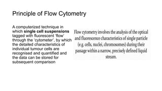
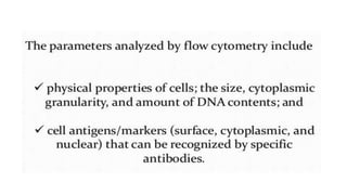
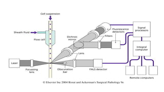
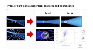
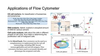
Flow Cytometry
SDL-11
Learning Objectives
Know the principle of flow cytometry
Identify the diagnostic value of FCM
Principle of Flow Cytometry
A computerized technique in
which single cell suspensions
tagged with fluorescent ‘flow’
through the ‘cytometer’, by which
the detailed characteristics of
individual tumour cells are
recognised and quantified and
the data can be stored for
subsequent comparison
Parts of a Flow Cytometer
A flow cytometer is made up of three main
systems
The fluidics system; transports particles in
a stream to the laser beam for interrogation
The optics system; consists of lasers to
illuminate the particles in the sample stream
and optical filters to direct the resulting light
signals to the appropriate detectors
The electronics system; converts the
detected light signals into electronic signals
that can be processed by the computer
How Flow Cytometer Works
To identify particular subpopulations of cells different fluorescent dyes are
conjugated to monocolonal antibodies directed towards the antigens on a
particular cell subset
When cells labeled with fluorescent molecules pass through the focused
laser beam, two types of light signals are generated scattered and
fluorescence which are picked up by photodetectors which produce
electronic signals proportional to the optical signals striking them
The electronic signals are then collected and stored in the computer and can
be analyzed to provide information about subpopulations of cells within the
sample and is plotted on histogram
Types of light signals generated; scattered and fluorescence
Specimen Collection for Flow Cytometry
Specimen required
Single cell suspension
Specimen preparation
Aspiration biopsies; peripheral blood, bone
marrow aspirate and FNA specimen have
cells in suspension form, to prevent them
from clotting they were kept in disodium
EDTA, sodium citrate or heparin
Solid biopsy tissue; should be collected in
a RPMI medium and the tissue must be
minced into small pieces and pass through
fine mesh to prepare a cell suspension
before analysis
Applications of Flow Cytometer
CD cell markers; for classification of leukaemias
and lymphomas
DNA analysis; diploid ,euploid or aneuploid amount
of DNA for various cancers
Cell cycle analysis; tells about the cells in different
phases of cell cycle, thus helps in determining the
proliferation rate of malignant cells
Conclusion
Assignment
1. What light source is used in most flow cytometers?
2. What are the three main systems in a flow cytometer?
3. What type of biological sample is best suited for flow cytometery?
4. When cells labeled with fluorescent molecules pass through the focused
laser beam, what two types of light signals are generated?
5. How flow cytometry helps in determining the proliferation rate of
malignancy?

Flow cytometry

  • 1.
  • 2.
    Learning Objectives Know theprinciple of flow cytometry Identify the diagnostic value of FCM
  • 3.
    Principle of FlowCytometry A computerized technique in which single cell suspensions tagged with fluorescent ‘flow’ through the ‘cytometer’, by which the detailed characteristics of individual tumour cells are recognised and quantified and the data can be stored for subsequent comparison
  • 5.
    Parts of aFlow Cytometer A flow cytometer is made up of three main systems The fluidics system; transports particles in a stream to the laser beam for interrogation The optics system; consists of lasers to illuminate the particles in the sample stream and optical filters to direct the resulting light signals to the appropriate detectors The electronics system; converts the detected light signals into electronic signals that can be processed by the computer
  • 6.
    How Flow CytometerWorks To identify particular subpopulations of cells different fluorescent dyes are conjugated to monocolonal antibodies directed towards the antigens on a particular cell subset When cells labeled with fluorescent molecules pass through the focused laser beam, two types of light signals are generated scattered and fluorescence which are picked up by photodetectors which produce electronic signals proportional to the optical signals striking them The electronic signals are then collected and stored in the computer and can be analyzed to provide information about subpopulations of cells within the sample and is plotted on histogram
  • 8.
    Types of lightsignals generated; scattered and fluorescence
  • 9.
    Specimen Collection forFlow Cytometry Specimen required Single cell suspension Specimen preparation Aspiration biopsies; peripheral blood, bone marrow aspirate and FNA specimen have cells in suspension form, to prevent them from clotting they were kept in disodium EDTA, sodium citrate or heparin Solid biopsy tissue; should be collected in a RPMI medium and the tissue must be minced into small pieces and pass through fine mesh to prepare a cell suspension before analysis
  • 11.
    Applications of FlowCytometer CD cell markers; for classification of leukaemias and lymphomas DNA analysis; diploid ,euploid or aneuploid amount of DNA for various cancers Cell cycle analysis; tells about the cells in different phases of cell cycle, thus helps in determining the proliferation rate of malignant cells
  • 13.
  • 14.
    Assignment 1. What lightsource is used in most flow cytometers? 2. What are the three main systems in a flow cytometer? 3. What type of biological sample is best suited for flow cytometery? 4. When cells labeled with fluorescent molecules pass through the focused laser beam, what two types of light signals are generated? 5. How flow cytometry helps in determining the proliferation rate of malignancy?