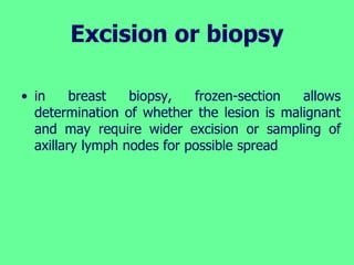
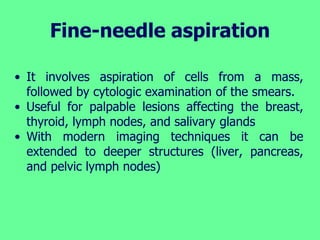
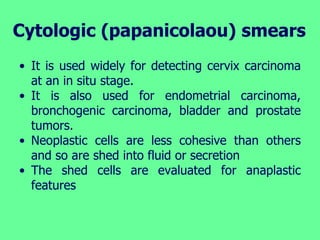
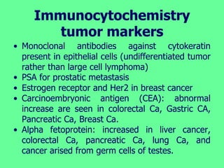
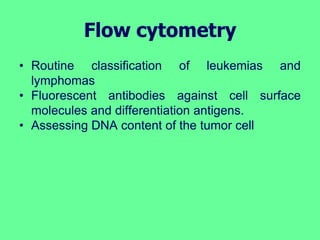
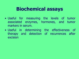
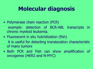
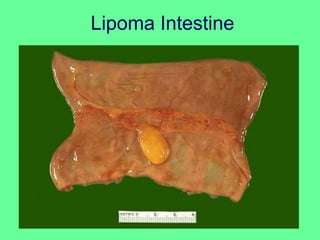
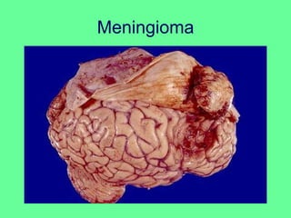
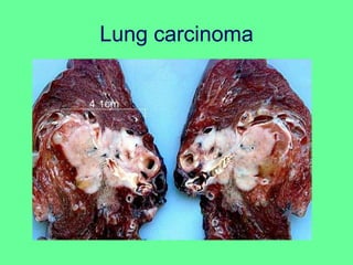
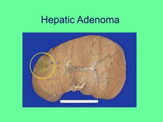
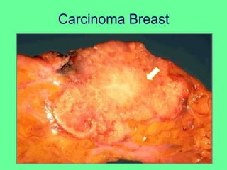
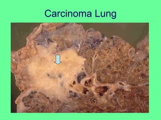
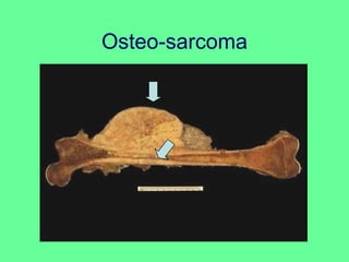
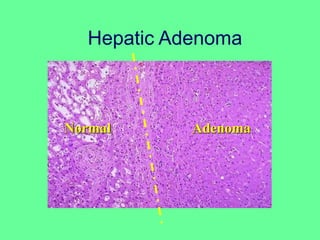
Faculty of Medicine
NEOPLSIA
2021 Cancer Lab
diagnosis
King Abdulaziz University
Rabigh Branch
Tumor Diagnosis:
• History and Clinical examination
• Imaging: X-Ray, US, CT, MRI
• Biochemical assays
– Tumor markers Laboratory analysis
• Morphologic methods (Histological diagnosis)
– Cytology: Pap smear, FNAB
– Biopsy: Histopathology, markers.
• Molecular Tech: Gene detection.
Histological diagnosis
Microscopic tissue examination is the gold
standard of cancer diagnosis.
• The specimen must be adequate,
representative and properly preserved.
Several sampling approaches are available:
• Excision or biopsy
• fine-needle aspiration
• Cytologic (papanicolaou) smears
• Immunocytochemistry
• Flow cytometry
Excision or biopsy
• Awareness in selecting appropriate site for biopsy
• In case of large mass: the margins may not be
representative and the center may be largely
necrotic.
• Frozen-section: quick technique permits histologic
evaluation within minutes
– determining the nature of lesion
– Evaluating the margins of an excised cancer to
ascertain that the entire neoplasm has been
removed.
Excision or biopsy
• in breast biopsy, frozen-section allows
determination of whether the lesion is malignant
and may require wider excision or sampling of
axillary lymph nodes for possible spread
Fine-needle aspiration
• It involves aspiration of cells from a mass,
followed by cytologic examination of the smears.
• Useful for palpable lesions affecting the breast,
thyroid, lymph nodes, and salivary glands
• With modern imaging techniques it can be
extended to deeper structures (liver, pancreas,
and pelvic lymph nodes)
Cytologic (papanicolaou) smears
• It is used widely for detecting cervix carcinoma
at an in situ stage.
• It is also used for endometrial carcinoma,
bronchogenic carcinoma, bladder and prostate
tumors.
• Neoplastic cells are less cohesive than others
and so are shed into fluid or secretion
• The shed cells are evaluated for anaplastic
features
Immunocytochemistry
tumor markers
• Monoclonal antibodies against cytokeratin
present in epithelial cells (undifferentiated tumor
rather than large cell lymphoma)
• PSA for prostatic metastasis
• Estrogen receptor and Her2 in breast cancer
• Carcinoembryonic antigen (CEA): abnormal
increase are seen in colorectal Ca, Gastric CA,
Pancreatic Ca, Breast Ca.
• Alpha fetoprotein: increased in liver cancer,
colorectal Ca, pancreatic Ca, lung Ca, and
cancer arised from germ cells of testes.
Flow cytometry
• Routine classification of leukemias and
lymphomas
• Fluorescent antibodies against cell surface
molecules and differentiation antigens.
• Assessing DNA content of the tumor cell
Biochemical assays
• Useful for measuring the levels of tumor
associated enzymes, hormones, and tumor
markers in serum.
• Useful in determining the effectiveness of
therapy and detection of recurrences after
excision
Molecular diagnosis
• Polymerase chain reaction (PCR)
example: detection of BCR-ABL transcripts in
chronic myeloid leukemia.
• Fluorescent in situ hybridization (fish)
it is useful for detecting translocation characteristic
of many tumors
• Both PCR and Fish can show amplification of
oncogenes (HER2 and N-MYC)
Bilateral Cystadenoma Ovary
Lipoma Intestine
Meningioma
Lung carcinoma
Hepatic Adenoma
Carcinoma Breast
Carcinoma Lung
Osteo-sarcoma
Colon Polyp
Hepatic Adenoma
Normal Adenoma
Metastatic Adenocarcinoma
Hepatic Adenocarcinoma

Cancer lab diagnosis

  • 1.
    Faculty of Medicine NEOPLSIA 2021Cancer Lab diagnosis King Abdulaziz University Rabigh Branch
  • 2.
    Tumor Diagnosis: • Historyand Clinical examination • Imaging: X-Ray, US, CT, MRI • Biochemical assays – Tumor markers Laboratory analysis • Morphologic methods (Histological diagnosis) – Cytology: Pap smear, FNAB – Biopsy: Histopathology, markers. • Molecular Tech: Gene detection.
  • 3.
    Histological diagnosis Microscopic tissueexamination is the gold standard of cancer diagnosis. • The specimen must be adequate, representative and properly preserved. Several sampling approaches are available: • Excision or biopsy • fine-needle aspiration • Cytologic (papanicolaou) smears • Immunocytochemistry • Flow cytometry
  • 4.
    Excision or biopsy •Awareness in selecting appropriate site for biopsy • In case of large mass: the margins may not be representative and the center may be largely necrotic. • Frozen-section: quick technique permits histologic evaluation within minutes – determining the nature of lesion – Evaluating the margins of an excised cancer to ascertain that the entire neoplasm has been removed.
  • 5.
    Excision or biopsy •in breast biopsy, frozen-section allows determination of whether the lesion is malignant and may require wider excision or sampling of axillary lymph nodes for possible spread
  • 6.
    Fine-needle aspiration • Itinvolves aspiration of cells from a mass, followed by cytologic examination of the smears. • Useful for palpable lesions affecting the breast, thyroid, lymph nodes, and salivary glands • With modern imaging techniques it can be extended to deeper structures (liver, pancreas, and pelvic lymph nodes)
  • 7.
    Cytologic (papanicolaou) smears •It is used widely for detecting cervix carcinoma at an in situ stage. • It is also used for endometrial carcinoma, bronchogenic carcinoma, bladder and prostate tumors. • Neoplastic cells are less cohesive than others and so are shed into fluid or secretion • The shed cells are evaluated for anaplastic features
  • 8.
    Immunocytochemistry tumor markers • Monoclonalantibodies against cytokeratin present in epithelial cells (undifferentiated tumor rather than large cell lymphoma) • PSA for prostatic metastasis • Estrogen receptor and Her2 in breast cancer • Carcinoembryonic antigen (CEA): abnormal increase are seen in colorectal Ca, Gastric CA, Pancreatic Ca, Breast Ca. • Alpha fetoprotein: increased in liver cancer, colorectal Ca, pancreatic Ca, lung Ca, and cancer arised from germ cells of testes.
  • 9.
    Flow cytometry • Routineclassification of leukemias and lymphomas • Fluorescent antibodies against cell surface molecules and differentiation antigens. • Assessing DNA content of the tumor cell
  • 10.
    Biochemical assays • Usefulfor measuring the levels of tumor associated enzymes, hormones, and tumor markers in serum. • Useful in determining the effectiveness of therapy and detection of recurrences after excision
  • 11.
    Molecular diagnosis • Polymerasechain reaction (PCR) example: detection of BCR-ABL transcripts in chronic myeloid leukemia. • Fluorescent in situ hybridization (fish) it is useful for detecting translocation characteristic of many tumors • Both PCR and Fish can show amplification of oncogenes (HER2 and N-MYC)
  • 12.
  • 13.
  • 14.
  • 15.
  • 16.
  • 17.
  • 18.
  • 19.
  • 20.
  • 21.
  • 22.
  • 23.