
Recommended
More Related Content
What's hot
What's hot (20)
Viewers also liked
Viewers also liked (20)
Similar to Agarose gel electrophoresis
Similar to Agarose gel electrophoresis (20)
More from Marudhar Kesari Jain College for Women Vaniyambadi - 635 751, Tamil Nadu, INDIA.
More from Marudhar Kesari Jain College for Women Vaniyambadi - 635 751, Tamil Nadu, INDIA. (20)
Recently uploaded
Recently uploaded (20)
Agarose gel electrophoresis
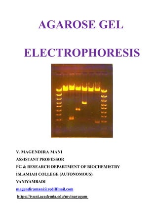
- 1. AGAROSE GEL ELECTROPHORESIS V. MAGENDIRA MANI ASSISTANT PROFESSOR PG & RESEARCH DEPARTMENT OF BIOCHEMISTRY ISLAMIAH COLLEGE (AUTONOMOUS) VANIYAMBADI magendiramani@rediffmail.com https://tvuni.academia.edu/mvinayagam
- 3. Agarose gel electrophoresis is the easiest and most popular way of separating and analyzing DNA. Here DNA molecules are separated on the basis of charge by applying an electric field to the electrophoretic apparatus. Shorter molecules migrate more easily and move faster than longermolecules through the pores of the gel and this process is called sieving. The gel might be used to look at the DNA in order to quantify it or to isolate a particular band. The DNA can be visualized in the gel by the addition of ethidium bromide.
- 4. Agarose is a polysaccharide obtained from the red algae Porphyra umbilicalis. Its systematic name is (1 4)-3, 6- anhydro-a-L-galactopyranosyl-(1,3)-β-D-galactopyranan. Agarose makes an inert matrix. Most agarose gels are made between 0.7% and 2% of agarose. A 0.7% gel will show good separation for large DNA fragments (5-10kb) and a 2% gel will show good resolution for small fragments with size range of 0.2-1kb.
- 5. Materials Required Gel casting trays, which are available in a variety of sizes and composed of Uv transparent plastic. Sample combs, around which molten agarose is poured to form sample wells in the gel. Electrophoresis buffer, usually Tris-acetate-EDTA (TAE) or Tris-borate-EDTA (TBE).
- 6. The migration rate of DNA fragments in both of these buffers is somewhat different due to the differences in ionic strength. These buffers provide the ions for supporting conductivity. Loading buffer, which contains something dense (e.g. glycerol) to allow the sample to "fall" into the sample wells, and one or two tracking dyes, which migrate in the gel and allow visual monitoring or how far the electrophoresis has proceeded.
- 7. Ethidium Bromide (EtBr) Ethidium bromide, a fluorescent dye used for staining nucleic acids. It is an intercalating agent which intercalates between nucleic acid bases and allows the convenient detection of DNA fragments in gel. When exposed to UV light, it will fluoresce with an orange colour. After the running of DNA through an EtBr-treated gel, any band containing more than ~20 ng DNA becomes distinctly visible under UV light. EtBr is a known "mutagen", however, safer alternatives are available. It can be incorporated with agarose gels or DNA samples before loading, for visualization of the fragments. Binding of Ethidium bromide to DNA alters its mass and rigidity, and thereby its mobility. Transilluminator (an ultraviolet light box), which is used to visualize ethidium bromide stained DNA in gels.
- 9. General procedure Casting of gel The gel is prepared by dissolving the agarose powder in an appropriate buffer, such as TAE or TBE, to be used in electrophoresis. The agarose is dispersed in the buffer before heating it to near-boiling point, but avoid boiling. The melted agarose is allowed to cool sufficiently before pouring the solution into a cast as the cast may warp or crack if the agarose solution is too hot. A comb is placed in the cast to create wells for loading sample, and the gel should be completely set before use. The concentration of gel affects the resolution of DNA separation. For a standard agarose gel electrophoresis, a 0.8% gives good separation or resolution of large 5-10kb DNA fragments, while 2% gel gives good resolution for small 0.2-1kb fragments. 1% gels are common for many applications
- 11. Loading of samples Once the gel has set, the comb is removed, leaving wells where DNA samples can be loaded. Loading buffer is mixed with the DNA sample before the mixture is loaded into the wells. The loading buffer contains a dense compound, which may be glycerol, sucrose, or Ficoll, that raises the density of the sample so that the DNA sample may sink to the bottom of the well. If the DNA sample contains residual ethanol after its preparation, it may float out of the well. The loading buffer also include colored dyes such as xylene cyanol and bromophenol blue used to monitor the progress of the electrophoresis. The DNA samples are loaded using a micropipette.
- 12. Electrophoresis Agarose gel electrophoresis is most commonly done horizontally in a submarine mode whereby the slab gel is completely submerged in buffer during electrophoresis. For optimal resolution of DNA greater than 2 kb in size in standard gel electrophoresis, 5 to 8 V/cm is recommended (the distance in cm refers to the distance between electrodes, therefore this recommended voltage would be 5 to 8 multiplied by the distance between the electrodes in cm). Voltage may also be limited by the fact that it heats the gel and may cause the gel to melt if it is run at high voltage for a prolonged period, especially if the gel used is LMP agarose gel.
- 13. Too high a voltage may also reduce resolution, as well as causing band streaking for large DNA molecules. Too low a voltage may lead to broadening of band for small DNA fragments due to dispersion and diffusion. A DNA marker is also run together for the estimation of the molecular weight of the DNA fragments.
- 14. Visualization / Staining DNA as well as RNA is normally visualized by staining with ethidium bromide, which intercalates into the major grooves of the DNA and fluoresces under UV light. The ethidium bromide may be added to the agarose solution before it gels, or the DNA gel may be stained later after electrophoresis. Destaining of the gel is not necessary but may produce better images.
- 15. Other methods of staining are available; examples are SYBR Green, GelRed, methylene blue, brilliant cresyl blue, Nile bluesulphate, and crystal violet. SYBR Green, GelRed and other similar commercial products are sold as safer alternatives to ethidium bromide as it has been shown to be mutagenic in Ames test, although the carcinogenicity of ethidium bromide has not actually been established. SYBR Green requires the use of a blue-light transilluminator. DNA stained with crystal violet can be viewed under natural light without the use of a UV transilluminator which is an advantage; however it may not produce a strong band.
- 16. When stained with ethidium bromide, the gel is viewed with an ultraviolet (UV) transilluminator. Standard transilluminators use wavelengths of 302/312-nm UV-B. The transilluminator apparatus may also contain image capture devices, such as a digital or Polaroid camera that allow an image of the gel to be taken or printed. Applications Estimation of the size of DNA molecules following restriction enzyme digestion, e.g. in restriction mapping of cloned DNA. Analysis of PCR products, e.g. in molecular genetic diagnosis or genetic fingerprinting Separation of restricted genomic DNA prior to Southern transfer or of RNA prior to Northern transfer.
- 17. Agarose gel electrophoresis is a method of gel electrophoresis used in biochemistry, molecular biology, and clinical chemistry to separate a mixed population of DNA or proteins in a matrix of agarose. The proteins may be separated by charge and/or size (IEF agarose, essentially size independent), and the DNA and RNA fragments by length. Biomolecules are separated by applying an electric field to move the charged molecules through an agarose matrix, and the biomolecules are separated by size in the agarose gel matrix.
- 18. V. MAGENDIRA MANI ASSISTANT PROFESSOR PG & RESEARCH DEPARTMENT OF BIOCHEMISTRY ISLAMIAH COLLEGE (AUTONOMOUS) VANIYAMBADI magendiramani@rediffmail.com https://tvuni.academia.edu/mvinayagam