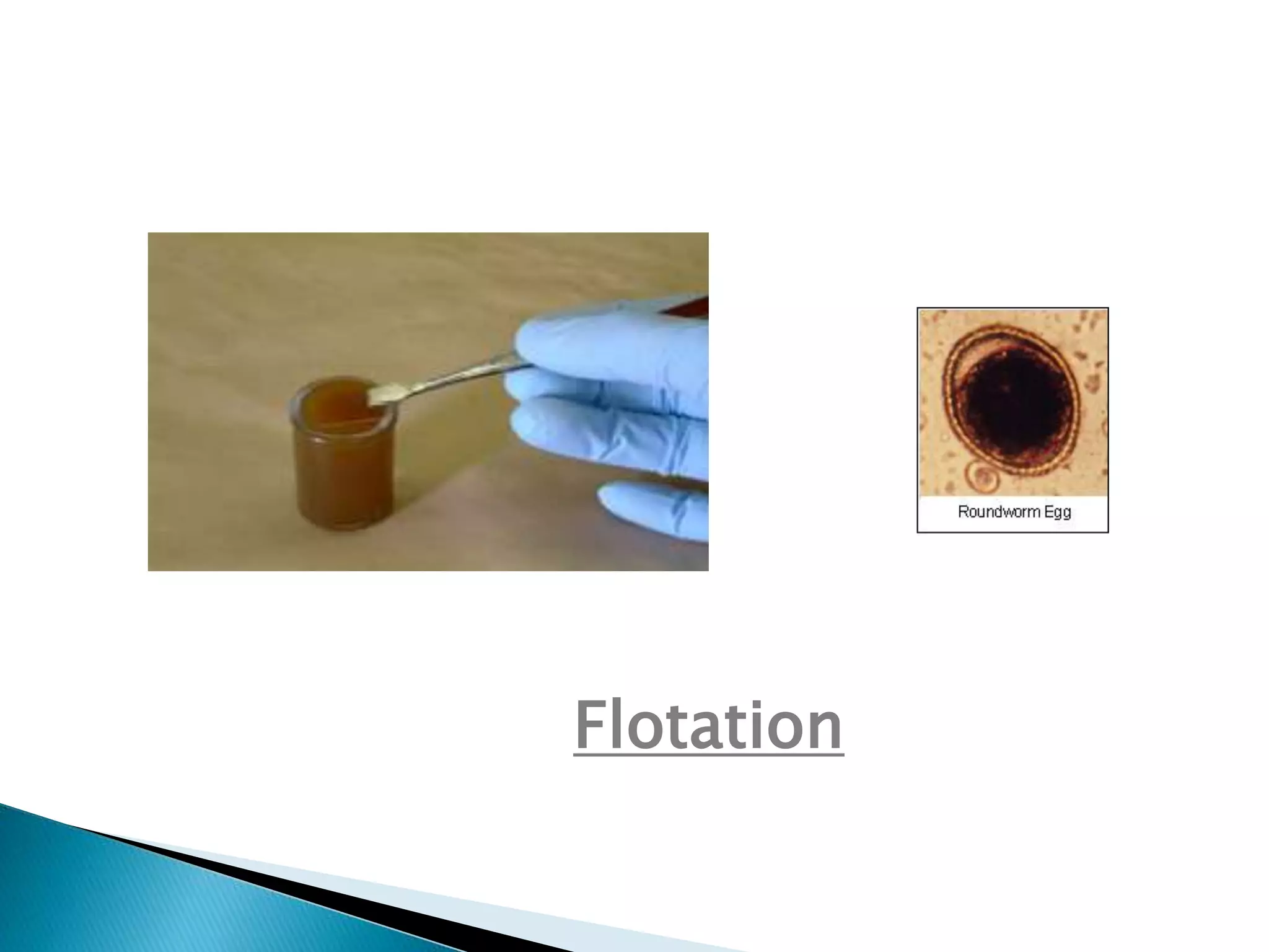
Dr. Urfeya Mirza
Department of Vety.
Surgery and Radiology
 Examining fecal samples for the presence of worm eggs
and larvae and protozoa trophozoites and cysts is a
common practice in most veterinary clinics.
Veterinarians try to prevent continued parasitic diseases
in animals, which decrease production; reduce growth
rates; cause infertility, abortions, and deaths; and
require costly treatment medications.
 The veterinary assistant should know how to collect and
examine fecal samples. However, the veterinarian will
give the assistant specific instructions for when and
what veterinary procedures need to be performed.
Methods for testing fecal samples include direct smear,
flotation, and gross examinations.
 When collecting fecal samples, first make certain that
the feces is from the animal in question. Secondly,
secure a fresh sample that is free from rocks, soil,
bedding, and other foreign materials. Place the fecal
sample in a plastic vial, glass jar, waxed cup, or plastic
bag. If the examination does not follow closely after
collection, preserve the sample in a refrigerator.
 Fecal examination procedures likely to be accepted and
implemented in most veterinary practices include
flotation (centrifugal or passive), sedimentation, and
direct examination (direct smear). Only flotation and
sedimentation are concentration procedures.
 Direct smears have poor sensitivity because of the
small amount of feces examined, but may be useful for
demonstrating motile organisms. CAPC recommends
that feces be routinely screened by a centrifugal
flotation method, which is consistently more sensitive
than simple flotation. Accuracy of centrifugal flotation
techniques depends on procedural details and
specimen attributes.
 Gross examination. Specimens should be examined
grossly for the presence of blood, mucus, intact worms, or
tapeworm segments.
 Sample size and preparation. Specimen size should be at
least 1 gram of formed feces (1 cubic centimeter or a cube
about one-half inch on a side). If feces are soft, sample size
should be 2 grams. If it is slurry-like, the sample should be 4
grams. For liquid feces, a sample of 6 grams or greater might
be appropriate. Inadequate sample size (e.g., fecal loop
sample) may result in false-negative results. To remove large
fecal debris, sieving is recommended prior to centrifugation.
The sample is sieved through cheesecloth or a tea strainer
after mixing with water or flotation solution. Passive flotation
kits typically include a device that prevents larger particles
from floating to the surface.
 Flotation solution.
Both the type and concentration of sugar or salt solutions
used can affect recovery of diagnostic stages of parasites
from feces. Common flotation solutes include sodium nitrate,
zinc sulfate, sucrose (usually granulated sugar), magnesium
sulfate, and sodium chloride. These solutes can be mixed at
varying concentrations with water to achieve flotation
solutions with different densities. Flotation solutions with
higher densities are capable of floating heavier (denser)
parasite stages. However, higher density flotation solutions
also float many other fecal particles that can render
preparations more difficult to examine and can collapse thin-
shelled parasite stages, making them difficult to identify or
causing them to float poorly. More viscous solutions, such as
Sheather's sugar (sucrose) solution, are more efficient for
centrifugation. Most salt solutions dry very quickly after
crystallizing on slides and obscuring observation.

Flotation
 Centrifugation.
Centrifugation of sieved feces may be performed in flotation
solution either with a coverslip placed on top of a filled tube
or with the coverslip added after the centrifuge has stopped.
In the later case, the tube is spun near-full, and then the
tube is filled to form a reverse meniscus, the coverslip is
added, and the tube is allowed to sit a few minutes longer.
Centrifugation with the coverslip on the tube works best
when a sugar flotation medium is used. Alternate methods
for sampling the reverse meniscus include loops or glass
rods that can be flamed between samples; however, this
approach is less efficient than centrifuging with the
coverslip in place.
 Slide examination.
The entire area under the coverslip should be examined. It is
helpful to focus on a small air bubble to obtain the correct
focal plane. The edge of the coverslip can be sealed with nail
polish to prevent drying and to allow examination of the
specimen under oil immersion. Sucrose preparations can be
stored in high humidity in a refrigerator for hours to days
without significantly altering the morphology of most
common helminth eggs.
 Although routine fecal examination should always include
centrifugation, at times, other examination methods are
needed to reach a diagnosis. For example, motile
trophozoites and nematode larvae can be observed using the
direct smear method. Certain nematode, trematode, and
tapeworm eggs will not float in less dense flotation solutions
and are better demonstrated using sedimentation.
 The Baermann funnel method may aid in diagnosis of a feline
lungworm (Aelurostrongylus abstrusus) infection. Stained
direct smears are useful for diagnosis of a protozoal infection
such as giardiasis or trichomoniasis. Specimens to be
examined for protozoa can first be fixed using a commercial
fixative such as Proto-fix™ or a fixating stain such as MIF
(merthiolate-iodine-formalin). Fecal antigen detection tests
are useful for diagnosis of giardiasis and cryptosporidiosis.
“Fecal Examination for
Diagnosis of Parasitic
Infections”
The sequence for the diagnosis of fecal parasitic infections
follows:
 Fecal examination should be done on fresh samples. If fecal
samples are used after being in the environment for hours or
days accurate reading of parasite indicators cannot be
guaranteed.
 Also, free- living nematodes rapidly invade a fecal sample
on the ground and can confuse diagnosis. Several grams of
feces should be collected immediately after observing
defecation.
 The sample is then prepared for flotation or centrifuging. This
procedure entails grinding if needed, mixing with clean water,
straining and mixing with a prepared flotation medium.
 There are several choices for the flotation medium used in fecal
material flotation. There are several commercial flotation mediums
available.
 A supersaturated medium of sugar or salt can be made by heating
water just below boiling then adding salt or sugar until no more will
dissolve. This will raise the specific gravity of the mixture. The
sample then undergoes fecal flotation.
 This procedure is based on the fact that parasitic material is less
dense than the flotation medium allowing it to float to the top of the
container where it can be collected for microscopic evaluation.
 The flotation method takes several hours.
 The procedure can be sped up by the use of a centrifuge. This
method spins the mixture in a horizontal attitude which forces the
heavier medium to the bottom of the tube and allows the lighter
parasite eggs to rise to the top.
 A hand crank or powered centrifuge may be used with like results.
 To prepare the sample for flotation it is mixed with clean water and
strained through a tea strainer or cheese cloth.
 The strained material is mixed with the flotation medium and
allowed to sit overnight or centrifuged.
 A cover slip is placed on top of the test tube in contact with the
mixture and then lifted straight up. The cover slip is placed on the
slide and then onto the microscope table.
 Depending on the parasite and the skill of the technician a parasite
load can be determined.
 Parasite loads vary with weather, season, rainfall, height and
abundance of feed on offer, age and/or size of the host animal,
extenuating health problems and nutrition of both the soil where the
host grazes and the host.
 In some situations the host animals in a herd can be parasitized and
symptoms do not stand out on individuals because all animals are
infected.
Faecal examination

Faecal examination

  • 1.
    Dr. Urfeya Mirza Departmentof Vety. Surgery and Radiology
  • 2.
     Examining fecalsamples for the presence of worm eggs and larvae and protozoa trophozoites and cysts is a common practice in most veterinary clinics. Veterinarians try to prevent continued parasitic diseases in animals, which decrease production; reduce growth rates; cause infertility, abortions, and deaths; and require costly treatment medications.  The veterinary assistant should know how to collect and examine fecal samples. However, the veterinarian will give the assistant specific instructions for when and what veterinary procedures need to be performed. Methods for testing fecal samples include direct smear, flotation, and gross examinations.
  • 3.
     When collectingfecal samples, first make certain that the feces is from the animal in question. Secondly, secure a fresh sample that is free from rocks, soil, bedding, and other foreign materials. Place the fecal sample in a plastic vial, glass jar, waxed cup, or plastic bag. If the examination does not follow closely after collection, preserve the sample in a refrigerator.
  • 5.
     Fecal examinationprocedures likely to be accepted and implemented in most veterinary practices include flotation (centrifugal or passive), sedimentation, and direct examination (direct smear). Only flotation and sedimentation are concentration procedures.  Direct smears have poor sensitivity because of the small amount of feces examined, but may be useful for demonstrating motile organisms. CAPC recommends that feces be routinely screened by a centrifugal flotation method, which is consistently more sensitive than simple flotation. Accuracy of centrifugal flotation techniques depends on procedural details and specimen attributes.
  • 6.
     Gross examination.Specimens should be examined grossly for the presence of blood, mucus, intact worms, or tapeworm segments.  Sample size and preparation. Specimen size should be at least 1 gram of formed feces (1 cubic centimeter or a cube about one-half inch on a side). If feces are soft, sample size should be 2 grams. If it is slurry-like, the sample should be 4 grams. For liquid feces, a sample of 6 grams or greater might be appropriate. Inadequate sample size (e.g., fecal loop sample) may result in false-negative results. To remove large fecal debris, sieving is recommended prior to centrifugation. The sample is sieved through cheesecloth or a tea strainer after mixing with water or flotation solution. Passive flotation kits typically include a device that prevents larger particles from floating to the surface.
  • 7.
     Flotation solution. Boththe type and concentration of sugar or salt solutions used can affect recovery of diagnostic stages of parasites from feces. Common flotation solutes include sodium nitrate, zinc sulfate, sucrose (usually granulated sugar), magnesium sulfate, and sodium chloride. These solutes can be mixed at varying concentrations with water to achieve flotation solutions with different densities. Flotation solutions with higher densities are capable of floating heavier (denser) parasite stages. However, higher density flotation solutions also float many other fecal particles that can render preparations more difficult to examine and can collapse thin- shelled parasite stages, making them difficult to identify or causing them to float poorly. More viscous solutions, such as Sheather's sugar (sucrose) solution, are more efficient for centrifugation. Most salt solutions dry very quickly after crystallizing on slides and obscuring observation. 
  • 8.
  • 9.
     Centrifugation. Centrifugation ofsieved feces may be performed in flotation solution either with a coverslip placed on top of a filled tube or with the coverslip added after the centrifuge has stopped. In the later case, the tube is spun near-full, and then the tube is filled to form a reverse meniscus, the coverslip is added, and the tube is allowed to sit a few minutes longer. Centrifugation with the coverslip on the tube works best when a sugar flotation medium is used. Alternate methods for sampling the reverse meniscus include loops or glass rods that can be flamed between samples; however, this approach is less efficient than centrifuging with the coverslip in place.
  • 10.
     Slide examination. Theentire area under the coverslip should be examined. It is helpful to focus on a small air bubble to obtain the correct focal plane. The edge of the coverslip can be sealed with nail polish to prevent drying and to allow examination of the specimen under oil immersion. Sucrose preparations can be stored in high humidity in a refrigerator for hours to days without significantly altering the morphology of most common helminth eggs.
  • 11.
     Although routinefecal examination should always include centrifugation, at times, other examination methods are needed to reach a diagnosis. For example, motile trophozoites and nematode larvae can be observed using the direct smear method. Certain nematode, trematode, and tapeworm eggs will not float in less dense flotation solutions and are better demonstrated using sedimentation.  The Baermann funnel method may aid in diagnosis of a feline lungworm (Aelurostrongylus abstrusus) infection. Stained direct smears are useful for diagnosis of a protozoal infection such as giardiasis or trichomoniasis. Specimens to be examined for protozoa can first be fixed using a commercial fixative such as Proto-fix™ or a fixating stain such as MIF (merthiolate-iodine-formalin). Fecal antigen detection tests are useful for diagnosis of giardiasis and cryptosporidiosis.
  • 12.
    “Fecal Examination for Diagnosisof Parasitic Infections”
  • 13.
    The sequence forthe diagnosis of fecal parasitic infections follows:  Fecal examination should be done on fresh samples. If fecal samples are used after being in the environment for hours or days accurate reading of parasite indicators cannot be guaranteed.  Also, free- living nematodes rapidly invade a fecal sample on the ground and can confuse diagnosis. Several grams of feces should be collected immediately after observing defecation.
  • 14.
     The sampleis then prepared for flotation or centrifuging. This procedure entails grinding if needed, mixing with clean water, straining and mixing with a prepared flotation medium.  There are several choices for the flotation medium used in fecal material flotation. There are several commercial flotation mediums available.  A supersaturated medium of sugar or salt can be made by heating water just below boiling then adding salt or sugar until no more will dissolve. This will raise the specific gravity of the mixture. The sample then undergoes fecal flotation.  This procedure is based on the fact that parasitic material is less dense than the flotation medium allowing it to float to the top of the container where it can be collected for microscopic evaluation.  The flotation method takes several hours.  The procedure can be sped up by the use of a centrifuge. This method spins the mixture in a horizontal attitude which forces the heavier medium to the bottom of the tube and allows the lighter parasite eggs to rise to the top.  A hand crank or powered centrifuge may be used with like results.
  • 15.
     To preparethe sample for flotation it is mixed with clean water and strained through a tea strainer or cheese cloth.  The strained material is mixed with the flotation medium and allowed to sit overnight or centrifuged.  A cover slip is placed on top of the test tube in contact with the mixture and then lifted straight up. The cover slip is placed on the slide and then onto the microscope table.  Depending on the parasite and the skill of the technician a parasite load can be determined.  Parasite loads vary with weather, season, rainfall, height and abundance of feed on offer, age and/or size of the host animal, extenuating health problems and nutrition of both the soil where the host grazes and the host.  In some situations the host animals in a herd can be parasitized and symptoms do not stand out on individuals because all animals are infected.