SlideShare a Scribd company logo
Dept. of Medicine , Surgery
and Obstetrics
A Presentation On
Anal Atresia
Rectovaginal fistula
Dermoid Cyst
Anal Atresia
Anal atresia is a
congenital anorectal
malformation (ARM)
where a normal anal
opening is absent at birth.
ARMs comprise of a broad spectrum
of defects ranging from minor (e.g.,
membranous covering) to complex
cloacal malformations involving the
urinary and genital tracts as well
Four types of atresia ani have been reported,
including:
 Congenital anal stenosis (Type I);
 Imperforate anus alone (Type II)
 Combined with more cranial termination
of the rectum as a blind pouch (Type III)
 Discontinuity of the proximal rectum with
normal anal and terminal rectal
development (Type IV).
Types of atresia ani
The cause is not known but may have a genetic component or may
be from an insult to the fetus in critical developmental stages in
utero. Environmental teratogens, plant toxins and a few viruses are
recognized complicating factors in calves. Atresia ani is the most
common intestinal defect in sheep and usually arises during the
embryonic period which results from autosomal recessive gene.
Etiology
Absence of an anal opening
Depression
Anorexia Colic
Marked gradual
abdominal distension
Tenesmus
Lack of feces.
Vomiting.
Clinical signs
Atresia ani can be diagnosed by
 simple visual examination
 By digital palpation and
 The clinical signs
Diagnosis
Surgical Procedure:
1. The Calf was placed in a lateral recumbency with its hind quarter raised high.
2. The perineal region was prepared for aseptic surgery.
Treatment
3. For Local infiltration : 2% Lignocaine hydrochloride(4-5ml)/calf was done at
the perineal region below the tail. Low epidural anaesthesia (3ml).
4. A circular incision was made on the bulge of the anus to remove a circular
piece of skin.
5. The rectum was exposed by separation of the perineal muscles.
6. Hold the rectum with stay suture and remove the feces using dushcane ( a pipe
containing water source , use to remove debris ).
Treatment
7.Suture ( simple interrupted ) the rectum with skin
like urethrostomy.
4 interrupted suture – ventral, dorsal, right, left
8. The new opening line should be cleaned with
finger using coconut oil 2-3 times per day to
prevent auto healing
Treatment
POST OPERATIVE CARE
The animal was administered Enrofloxacin @
5mg/kg for five days.
Meloxicam @0.5 ml for 3 days
Routine dressing with povidone iodine
ointment and application of topicure spray as a
fly repellant.
The calf showed normal in defecation with
minimum tenesmus and active in nature on the
3rd postoperative day and therefore the sutures
were removed on 10th post-operative day and
the animal made a noticeable recovery.
 Fecal incontinence
 Persistent megacolon
 Anal stricture
 recurrent cystitis.
Complications after
surgery
in
Rectovaginal fistula
A rectovaginal fistula in a cow
is a rare condition where there is
an abnormal connection or
passage way between the rectum
and the vagina.This can lead to
the passage of fecal material into
the reproductive tract and can
result in health issues for the
cow.
1.Congenital
2.Local infection
3.Traumatic :
A)Post-partum
B) Secondary to surgery :
• following low anterior rectal resection
• following procedures for pelvic floor
dysfunction
• following haemorrhoid surgery
• following drainage of local infection.
Etiology
C)Resulting from violent acts.
4.Chronic inflammatory bowel disease :
-crohn's disease
-ulcerative colitis
-indeterminate colitis
5.following radiation therapy of tumors in the
lesser pelvis
6.resulting from carcinoma
Etiology
CLINICAL SIGNS
• 1.passage of gas or stool from vagina
• 2. Foul smelling vaginal discharge
• 3.chronic vaginal infection
• 4.pain during intercourse
• 5.incontinence
• 6.tenesmus
• 7.abdominal distention
• 8.irritation of the vulva
Diagnosis
Diagnosis of a rectovaginal fistula is typically made by
the history of the passage of stool and/or gas from the
vagina, in addition to a physical exam.
A vaginal exam with a speculum and a rectal exam
should be performed.
Ultrasound
CT scan
MRI
Treatment
Surgical techniques:
A)In case of internal attachment of rectum
to vagina:
1.After adequate restraint and preparation, a
transverse incision is made between the
rectum and vagina.
2. By using a combination of sharp and
blunt dissection in a horizontal plane, the
fistula is exposed.
Treatment
3 .Ideally, two thirds of the thickness of the shelf
should be with the rectum and one third with the
vaginal shelf.
4. Most fistulas measure 3 to 5 cm. Dissection is
continued 3 to 4 cm rostral to the fistula.
5.The rectal defect is closed transversely by using
No. 0 or 1 absorbable sutures in a simple interrupted
pattern placed in the submucosa.
Treatment
6.The first suture should divide the defect in half ,the next
two sutures should be placed to bisect the halves, and so
on.
Alternatively, one can pre place the sutures.
7.The vaginal defect is closed next. A continuous
horizontal mattress pattern in a longitudinal direction is
used so that the two suture rows are at right angles to each
other and the vaginal mucosa is everted.
8.The incised perineal body is closed with multiple
interrupted sutures of 2-0 suture; the skin is closed
routinely.
Treatment
B) In case of secondary complication of Atrisia anii
surgery:
1.Animal is restrained laterally by the owner and low
epidural anesthesia is performed using 2% lignocaine
HCl.
2.Then clean the opening with water.
3 .Make a stretch over the visceral part of the skin
area to make a fresh wound that will induce the
healing quickly.
4.Then the rectal wall is closed with purse string
suture using 3/0 catgut.
Post operative care:
Antibiotics -The animal was administered Ciprofloxacin
@ 5mg/kg for five days.
Meloxicam @0.5 ml for 3 days
Stop movement of heifers for 2-3 day
Clear the opening of rectum 4-5 times daily with
coconut oil to make sure that the rectum doesn’t get
closed.
Dermoid cyst
Dermoid cysts are a
relatively rare congenital
anomaly in cattle.
Dermoid cysts are congenital
cysts and occur due to failure of
the embryonic cavity to close or
defective epidermal closure
Location
Dermoid cysts are reported in many species of animals at
different anatomical location: Tail, Neck, Skull, Nasal cavity
• But ocular dermoid are common in ruminants and they may
be present unilaterally or bilaterally.
• Usually dermoids are located in limbus, cornea.
conjunctiva, corneo-conjunctiva they have also been
noticed in membrana nictitans
• eyelid
Age of predisposition
 Congenital, thus usually noticed in young
animals
 But may occur also in large adult animals.
Breed/species predisposition
 May affect all breeds.
 Has been reported in Hereford and Jersey cattle
as well as Holstein crossbred
Etiology
 Failure of the embryonic cavity to close or defective epidermal closure.
 Degenerative changes in hair follicles
 Cystic changes in ducts or cells of sebaceous glands
 Traumatic displacement of epidermal fragments.
 Cysts originated from incarceration and subsequent growth of embryonic
epithelial cells during the closure of neural tube and therefore most of these
lesions occur along the median line.
Classification of dermoid cyst
Ventrolateral corneo-conjunctival dermoid,
Supero-temporal corneal limbal dermoid,
Medial canthal dermoid
Membrana nictitans dermoid
ocular dermoid in dairy cattle include: Corneal dermoid, corneo-scleral dermoid,
corneo-conjunctival dermoid and conjunctival dermoid
PATHOLOGY & PATHOGENESIS
• Dermoid cysts are composed of keratinized stratified squamous epithelium with
dermal appendages and adnexal structures, including hair follicles, sebaceous
glands, sweat glands, smooth muscle, and fibro adipose tissue. The lumen
contains keratin and hair. Cysts that are only lined with epithelium without
adnexal elements are termed epidermoid cysts
• Dermoid cyst arises from the entrapped embryonal ectodermal cells during the
fetal development and are generally formed in the embryonic lines of fusion.
CLINICAL SIGNS
Mass on/near eye
Blepharospasm
Lacrimation
Visual impairment
Tearing
Inflammation
Irritation in the eye
Corneal opacity in Holstein crossbred
DIAGNOSIS
1. Physical Examination: Painless swelling that
may be freely mobile or fixed to the skin &
deeper structure.
2. Radiological Examination:
MRI,ULTRASOUND,CT SCAN
SURGICAL TREATMENT
1. Restraining: The animal should be controlled in lateral recumbency by casting.
The animal should be sedated with 0.1mg/kg Xylazine intramuscularly.
2. Anaesthesia: Local analgesia was performed by 2% Lignocaine HCl. The
anaesthetic was injected through the stalk after fixing eyelid with eye speculum.
Then Ketamine HCl @ 5 to 6 mg/kg body weight was injected through IM.
3. Operative Procedure:
o The dermoid cyst should be lifted as much as possible and curved artery forceps
was used to hold the stalk.
SURGICAL TREATMENT
o A chromic catgut is used to ligate the blood vessels along with the stalk by anchoring it. Sometimes two
horizontal mattress stiches may be required.
o Excision of the dermoid cyst or stalk is made after crushing with artery forceps.
o Cauterization: For excision electric thermocauterizer is used worldwide,it will help to perform the
surgery with less bleeding.
o Keratectomy: In this procedure,eyelashes was trimmed with scissors and whole eye washed with 0.9%
povidone iodine to remove contaminates. After attainment of adequate sedation superficial keratectomy
was performed. Then the mass of dermoid was grasped with forceps gently and the tissue mass was
completely excised by surgical blade. Bleeding from surgical wound was stopped by applying povidone
ointment.
Fig: Dermoid cyst mass after surgery
Fig: Performing the surgery
POST OPERATIVE CARE
Ciprofloxacin eye drop T.I.D in a day for 5 days.
Gentamicin 4mg /kg intra muscular for 6 days.
Meloxicam 0.4mg /kg intramuscular for 3 days.
Boric acid 0.3% solution should be used for eye wash
Ointment: Chloramphenicol or Neobacterin 3 times in
a day up to recovery.
Atresi ani and Recto-vaginal fistula.pptx

More Related Content

What's hot

Abscess hematoma & maggot wound
Abscess hematoma & maggot woundAbscess hematoma & maggot wound
Abscess hematoma & maggot wound
Dr.Jigdrel Dorji
 
Fetotomy in bovines by Dr Dushyant Yadav BASU, Patna INDIA
Fetotomy  in bovines by  Dr Dushyant Yadav BASU, Patna INDIAFetotomy  in bovines by  Dr Dushyant Yadav BASU, Patna INDIA
Fetotomy in bovines by Dr Dushyant Yadav BASU, Patna INDIA
DrDushyant Yadav
 
Vet obst lecture 6 Peurperium in domestic animals
Vet obst lecture 6 Peurperium in domestic animalsVet obst lecture 6 Peurperium in domestic animals
Vet obst lecture 6 Peurperium in domestic animals
DrGovindNarayanPuroh
 
Estrus cycle of bitch
Estrus cycle of bitchEstrus cycle of bitch
Estrus cycle of bitch
Dr. Muhammad Awais
 
Caesarean section in cow
Caesarean section in cowCaesarean section in cow
Caesarean section in cow
Rekha Pathak
 
Cesarean section in bovines
Cesarean section in bovinesCesarean section in bovines
Cesarean section in bovines
Arvinder singh
 
Presetation on rumen impaction lactic acidosis final osr
Presetation on rumen impaction lactic acidosis final osrPresetation on rumen impaction lactic acidosis final osr
Presetation on rumen impaction lactic acidosis final osr
Harshit Saxena
 
Eclampsia in dogs
Eclampsia in dogsEclampsia in dogs
Eclampsia in dogs
Dr Alok Bharti
 
Teat surgery
Teat surgeryTeat surgery
Teat surgery
Vikash Babu Rajput
 
Surgical Approaches to thorax in small animals
 Surgical Approaches to   thorax in small animals Surgical Approaches to   thorax in small animals
Surgical Approaches to thorax in small animals
baljotjot
 
Management of dystocia in bovines
Management of dystocia in bovinesManagement of dystocia in bovines
Management of dystocia in bovines
Bharat Regmi
 
Veterinary Gastrointestinal surgery Part-II
Veterinary Gastrointestinal surgery Part-II Veterinary Gastrointestinal surgery Part-II
Veterinary Gastrointestinal surgery Part-II Rekha Pathak
 
Paraphimosis in Cattle
Paraphimosis in CattleParaphimosis in Cattle
Paraphimosis in Cattle
Abdullah Al Mubin
 
Repeat breeding Dr Ashiq
Repeat breeding Dr AshiqRepeat breeding Dr Ashiq
Repeat breeding Dr Ashiq
Megha Kose
 
Sub-clinical endometritis and its effect on the fertility of dairy cattle
Sub-clinical endometritis and its effect on the fertility of dairy cattleSub-clinical endometritis and its effect on the fertility of dairy cattle
Sub-clinical endometritis and its effect on the fertility of dairy cattle
habtamu kenide
 
Obstectrical instruments and their uses
Obstectrical instruments and their usesObstectrical instruments and their uses
Obstectrical instruments and their uses
Tanmay Tewari
 
Rajeev mishra ,castration of small animal(dog and cat).
Rajeev mishra ,castration of small animal(dog and cat).Rajeev mishra ,castration of small animal(dog and cat).
Rajeev mishra ,castration of small animal(dog and cat).
Raaz Eve Mishra
 
Backyard Farming: Dystocia in Sheep & Goats
Backyard Farming: Dystocia in Sheep & GoatsBackyard Farming: Dystocia in Sheep & Goats
Backyard Farming: Dystocia in Sheep & Goats
University of Maryland Extension Small Ruminant Program
 
Surgical affection of oesophagus
Surgical affection of oesophagusSurgical affection of oesophagus
Surgical affection of oesophagus
Bikas Puri
 

What's hot (20)

Abscess hematoma & maggot wound
Abscess hematoma & maggot woundAbscess hematoma & maggot wound
Abscess hematoma & maggot wound
 
Fetotomy in bovines by Dr Dushyant Yadav BASU, Patna INDIA
Fetotomy  in bovines by  Dr Dushyant Yadav BASU, Patna INDIAFetotomy  in bovines by  Dr Dushyant Yadav BASU, Patna INDIA
Fetotomy in bovines by Dr Dushyant Yadav BASU, Patna INDIA
 
Vet obst lecture 6 Peurperium in domestic animals
Vet obst lecture 6 Peurperium in domestic animalsVet obst lecture 6 Peurperium in domestic animals
Vet obst lecture 6 Peurperium in domestic animals
 
Estrus cycle of bitch
Estrus cycle of bitchEstrus cycle of bitch
Estrus cycle of bitch
 
Caesarean section in cow
Caesarean section in cowCaesarean section in cow
Caesarean section in cow
 
Cesarean section in bovines
Cesarean section in bovinesCesarean section in bovines
Cesarean section in bovines
 
Presetation on rumen impaction lactic acidosis final osr
Presetation on rumen impaction lactic acidosis final osrPresetation on rumen impaction lactic acidosis final osr
Presetation on rumen impaction lactic acidosis final osr
 
Eclampsia in dogs
Eclampsia in dogsEclampsia in dogs
Eclampsia in dogs
 
Teat surgery
Teat surgeryTeat surgery
Teat surgery
 
Surgical Approaches to thorax in small animals
 Surgical Approaches to   thorax in small animals Surgical Approaches to   thorax in small animals
Surgical Approaches to thorax in small animals
 
Management of dystocia in bovines
Management of dystocia in bovinesManagement of dystocia in bovines
Management of dystocia in bovines
 
Veterinary Gastrointestinal surgery Part-II
Veterinary Gastrointestinal surgery Part-II Veterinary Gastrointestinal surgery Part-II
Veterinary Gastrointestinal surgery Part-II
 
Paraphimosis in Cattle
Paraphimosis in CattleParaphimosis in Cattle
Paraphimosis in Cattle
 
Repeat breeding Dr Ashiq
Repeat breeding Dr AshiqRepeat breeding Dr Ashiq
Repeat breeding Dr Ashiq
 
Sub-clinical endometritis and its effect on the fertility of dairy cattle
Sub-clinical endometritis and its effect on the fertility of dairy cattleSub-clinical endometritis and its effect on the fertility of dairy cattle
Sub-clinical endometritis and its effect on the fertility of dairy cattle
 
Obstectrical instruments and their uses
Obstectrical instruments and their usesObstectrical instruments and their uses
Obstectrical instruments and their uses
 
Rajeev mishra ,castration of small animal(dog and cat).
Rajeev mishra ,castration of small animal(dog and cat).Rajeev mishra ,castration of small animal(dog and cat).
Rajeev mishra ,castration of small animal(dog and cat).
 
Hernia
HerniaHernia
Hernia
 
Backyard Farming: Dystocia in Sheep & Goats
Backyard Farming: Dystocia in Sheep & GoatsBackyard Farming: Dystocia in Sheep & Goats
Backyard Farming: Dystocia in Sheep & Goats
 
Surgical affection of oesophagus
Surgical affection of oesophagusSurgical affection of oesophagus
Surgical affection of oesophagus
 

Similar to Atresi ani and Recto-vaginal fistula.pptx

Congenital Benign Neck masses
Congenital Benign Neck masses Congenital Benign Neck masses
Congenital Benign Neck masses
Haya Taha
 
3-Vulvar dystrophies. Erosion of cervix. Ectropion. Cervical intraepithelial ...
3-Vulvar dystrophies. Erosion of cervix. Ectropion. Cervical intraepithelial ...3-Vulvar dystrophies. Erosion of cervix. Ectropion. Cervical intraepithelial ...
3-Vulvar dystrophies. Erosion of cervix. Ectropion. Cervical intraepithelial ...
HalaAlzamel
 
Vet obst lecture 11 Genital surgeries in domestic animals
Vet obst lecture 11 Genital surgeries in domestic animalsVet obst lecture 11 Genital surgeries in domestic animals
Vet obst lecture 11 Genital surgeries in domestic animals
DrGovindNarayanPuroh
 
Branchial arch fistulae, thyroglossal duct anomalies& lymphangioma
Branchial arch fistulae, thyroglossal duct anomalies& lymphangiomaBranchial arch fistulae, thyroglossal duct anomalies& lymphangioma
Branchial arch fistulae, thyroglossal duct anomalies& lymphangiomaShekhar Krishna Debnath
 
Tracheotomy, By Dr. Rekha Pathak, Senior scientist IVRI
Tracheotomy, By Dr. Rekha Pathak, Senior scientist IVRITracheotomy, By Dr. Rekha Pathak, Senior scientist IVRI
Tracheotomy, By Dr. Rekha Pathak, Senior scientist IVRI
Rekha Pathak
 
ventral hernias
ventral herniasventral hernias
ventral hernias
arshad abbas
 
Hydatidcystofliverby hegazy
Hydatidcystofliverby hegazyHydatidcystofliverby hegazy
Hydatidcystofliverby hegazy
mostafa hegazy
 
Hydatidcystofliverby hegazy
Hydatidcystofliverby hegazyHydatidcystofliverby hegazy
Hydatidcystofliverby hegazy
mostafa hegazy
 
Umbilical hernia by Dr. kiran maindale
Umbilical hernia by Dr. kiran maindaleUmbilical hernia by Dr. kiran maindale
Umbilical hernia by Dr. kiran maindale
kiran Maindale
 
Endometriosis
EndometriosisEndometriosis
Endometriosis
Gurkirat Kaur
 
Cervical Ectopy or cervical erosion.pptx
Cervical Ectopy  or cervical erosion.pptxCervical Ectopy  or cervical erosion.pptx
Cervical Ectopy or cervical erosion.pptx
taruntiwari12355
 
Rectovaginal fistula and Perineal tear
Rectovaginal fistula and Perineal tearRectovaginal fistula and Perineal tear
Rectovaginal fistula and Perineal tear
Abdullah Almazyad
 
Umbilical cord.pptx
Umbilical cord.pptxUmbilical cord.pptx
Umbilical cord.pptx
Dhruv Saini
 
Umbilical cord.pptx
Umbilical cord.pptxUmbilical cord.pptx
Umbilical cord.pptx
Dhruv Saini
 
Case presentation on scar endometriosis
Case presentation on scar endometriosisCase presentation on scar endometriosis
Case presentation on scar endometriosis
mukulika3
 
C SECTION IN SMALL AND LARGE ANIMALSpptx
C SECTION IN SMALL AND LARGE ANIMALSpptxC SECTION IN SMALL AND LARGE ANIMALSpptx
C SECTION IN SMALL AND LARGE ANIMALSpptx
meenupm2
 
Male reproductive system disorders
Male reproductive system disordersMale reproductive system disorders
Male reproductive system disorders
Khushi Devgan
 
03,04 Embryology & congenital anomalies.pdf
03,04 Embryology & congenital anomalies.pdf03,04 Embryology & congenital anomalies.pdf
03,04 Embryology & congenital anomalies.pdf
UthMh
 
Pediatric Circumcision lecture
Pediatric Circumcision lecturePediatric Circumcision lecture
Pediatric Circumcision lecture
Amro Ahmed Fouad Abdellatif
 

Similar to Atresi ani and Recto-vaginal fistula.pptx (20)

Congenital Benign Neck masses
Congenital Benign Neck masses Congenital Benign Neck masses
Congenital Benign Neck masses
 
Benign neck disease
Benign neck diseaseBenign neck disease
Benign neck disease
 
3-Vulvar dystrophies. Erosion of cervix. Ectropion. Cervical intraepithelial ...
3-Vulvar dystrophies. Erosion of cervix. Ectropion. Cervical intraepithelial ...3-Vulvar dystrophies. Erosion of cervix. Ectropion. Cervical intraepithelial ...
3-Vulvar dystrophies. Erosion of cervix. Ectropion. Cervical intraepithelial ...
 
Vet obst lecture 11 Genital surgeries in domestic animals
Vet obst lecture 11 Genital surgeries in domestic animalsVet obst lecture 11 Genital surgeries in domestic animals
Vet obst lecture 11 Genital surgeries in domestic animals
 
Branchial arch fistulae, thyroglossal duct anomalies& lymphangioma
Branchial arch fistulae, thyroglossal duct anomalies& lymphangiomaBranchial arch fistulae, thyroglossal duct anomalies& lymphangioma
Branchial arch fistulae, thyroglossal duct anomalies& lymphangioma
 
Tracheotomy, By Dr. Rekha Pathak, Senior scientist IVRI
Tracheotomy, By Dr. Rekha Pathak, Senior scientist IVRITracheotomy, By Dr. Rekha Pathak, Senior scientist IVRI
Tracheotomy, By Dr. Rekha Pathak, Senior scientist IVRI
 
ventral hernias
ventral herniasventral hernias
ventral hernias
 
Hydatidcystofliverby hegazy
Hydatidcystofliverby hegazyHydatidcystofliverby hegazy
Hydatidcystofliverby hegazy
 
Hydatidcystofliverby hegazy
Hydatidcystofliverby hegazyHydatidcystofliverby hegazy
Hydatidcystofliverby hegazy
 
Umbilical hernia by Dr. kiran maindale
Umbilical hernia by Dr. kiran maindaleUmbilical hernia by Dr. kiran maindale
Umbilical hernia by Dr. kiran maindale
 
Endometriosis
EndometriosisEndometriosis
Endometriosis
 
Cervical Ectopy or cervical erosion.pptx
Cervical Ectopy  or cervical erosion.pptxCervical Ectopy  or cervical erosion.pptx
Cervical Ectopy or cervical erosion.pptx
 
Rectovaginal fistula and Perineal tear
Rectovaginal fistula and Perineal tearRectovaginal fistula and Perineal tear
Rectovaginal fistula and Perineal tear
 
Umbilical cord.pptx
Umbilical cord.pptxUmbilical cord.pptx
Umbilical cord.pptx
 
Umbilical cord.pptx
Umbilical cord.pptxUmbilical cord.pptx
Umbilical cord.pptx
 
Case presentation on scar endometriosis
Case presentation on scar endometriosisCase presentation on scar endometriosis
Case presentation on scar endometriosis
 
C SECTION IN SMALL AND LARGE ANIMALSpptx
C SECTION IN SMALL AND LARGE ANIMALSpptxC SECTION IN SMALL AND LARGE ANIMALSpptx
C SECTION IN SMALL AND LARGE ANIMALSpptx
 
Male reproductive system disorders
Male reproductive system disordersMale reproductive system disorders
Male reproductive system disorders
 
03,04 Embryology & congenital anomalies.pdf
03,04 Embryology & congenital anomalies.pdf03,04 Embryology & congenital anomalies.pdf
03,04 Embryology & congenital anomalies.pdf
 
Pediatric Circumcision lecture
Pediatric Circumcision lecturePediatric Circumcision lecture
Pediatric Circumcision lecture
 

More from Hajee Mohammad Danesh Science and Technology University, Dinajpur-5200,Bangladesh

Avian Pathology.pptx
Avian Pathology.pptxAvian Pathology.pptx
Immunology and Serology.ppt
Immunology and Serology.pptImmunology and Serology.ppt
Zoo and Wild Animal.pptx
Zoo and Wild Animal.pptxZoo and Wild Animal.pptx
Castration of animal .pptx
Castration of animal .pptxCastration of animal .pptx
SOUNDNESS .pptx
SOUNDNESS .pptxSOUNDNESS .pptx
HERD HEALTH MANAGEMENT.ppt
HERD HEALTH MANAGEMENT.pptHERD HEALTH MANAGEMENT.ppt
Avian Pathology.pptx
Avian Pathology.pptxAvian Pathology.pptx

More from Hajee Mohammad Danesh Science and Technology University, Dinajpur-5200,Bangladesh (17)

Tail Amputation.pptx
Tail Amputation.pptxTail Amputation.pptx
Tail Amputation.pptx
 
Subcutaneous Cyst and its correction.pptx
Subcutaneous Cyst and its correction.pptxSubcutaneous Cyst and its correction.pptx
Subcutaneous Cyst and its correction.pptx
 
Rectovaginal fistula and correction.pptx
Rectovaginal fistula and correction.pptxRectovaginal fistula and correction.pptx
Rectovaginal fistula and correction.pptx
 
Ocular dermoid cyst.pdf
Ocular dermoid cyst.pdfOcular dermoid cyst.pdf
Ocular dermoid cyst.pdf
 
Navel ill and joint ill.pptx
Navel ill and joint ill.pptxNavel ill and joint ill.pptx
Navel ill and joint ill.pptx
 
Limb amputation and disarticulation.pptx
Limb amputation and disarticulation.pptxLimb amputation and disarticulation.pptx
Limb amputation and disarticulation.pptx
 
Gid disease in goat and surgical correction.pptx
Gid disease in goat and surgical correction.pptxGid disease in goat and surgical correction.pptx
Gid disease in goat and surgical correction.pptx
 
Castration of animal.pptx
Castration of animal.pptxCastration of animal.pptx
Castration of animal.pptx
 
Caecal dilatation and torsion and correction.pdf
Caecal dilatation and torsion and correction.pdfCaecal dilatation and torsion and correction.pdf
Caecal dilatation and torsion and correction.pdf
 
A PRESENTATION ON MYIASIS.pptx
A PRESENTATION ON MYIASIS.pptxA PRESENTATION ON MYIASIS.pptx
A PRESENTATION ON MYIASIS.pptx
 
Avian Pathology.pptx
Avian Pathology.pptxAvian Pathology.pptx
Avian Pathology.pptx
 
Immunology and Serology.ppt
Immunology and Serology.pptImmunology and Serology.ppt
Immunology and Serology.ppt
 
Zoo and Wild Animal.pptx
Zoo and Wild Animal.pptxZoo and Wild Animal.pptx
Zoo and Wild Animal.pptx
 
Castration of animal .pptx
Castration of animal .pptxCastration of animal .pptx
Castration of animal .pptx
 
SOUNDNESS .pptx
SOUNDNESS .pptxSOUNDNESS .pptx
SOUNDNESS .pptx
 
HERD HEALTH MANAGEMENT.ppt
HERD HEALTH MANAGEMENT.pptHERD HEALTH MANAGEMENT.ppt
HERD HEALTH MANAGEMENT.ppt
 
Avian Pathology.pptx
Avian Pathology.pptxAvian Pathology.pptx
Avian Pathology.pptx
 

Recently uploaded

UNIVERSAL HUMAN VALUES- Harmony in the Nature
UNIVERSAL HUMAN VALUES- Harmony in the NatureUNIVERSAL HUMAN VALUES- Harmony in the Nature
UNIVERSAL HUMAN VALUES- Harmony in the Nature
Chandrakant Divate
 
SOCIOLOGY PPT. SOCIAL SECURITY POWER POINT
SOCIOLOGY PPT. SOCIAL SECURITY POWER POINTSOCIOLOGY PPT. SOCIAL SECURITY POWER POINT
SOCIOLOGY PPT. SOCIAL SECURITY POWER POINT
ssuser8d5e2d1
 
Program Your Destiny eBook - Destiny University.pdf
Program Your Destiny eBook - Destiny University.pdfProgram Your Destiny eBook - Destiny University.pdf
Program Your Destiny eBook - Destiny University.pdf
Michael Herlache, MBA
 
Collocation thường gặp trong đề thi THPT Quốc gia.pdf
Collocation thường gặp trong đề thi THPT Quốc gia.pdfCollocation thường gặp trong đề thi THPT Quốc gia.pdf
Collocation thường gặp trong đề thi THPT Quốc gia.pdf
ngochaavk33a
 
Ethical_dilemmas_MDI_Gurgaon-Business Ethics Case 1.pptx
Ethical_dilemmas_MDI_Gurgaon-Business Ethics Case 1.pptxEthical_dilemmas_MDI_Gurgaon-Business Ethics Case 1.pptx
Ethical_dilemmas_MDI_Gurgaon-Business Ethics Case 1.pptx
TANMAYJAIN511570
 
What Is The Psychology Behind Reborn Dolls_.pptx
What Is The Psychology Behind Reborn Dolls_.pptxWhat Is The Psychology Behind Reborn Dolls_.pptx
What Is The Psychology Behind Reborn Dolls_.pptx
Lloyd Dobson Artist
 

Recently uploaded (6)

UNIVERSAL HUMAN VALUES- Harmony in the Nature
UNIVERSAL HUMAN VALUES- Harmony in the NatureUNIVERSAL HUMAN VALUES- Harmony in the Nature
UNIVERSAL HUMAN VALUES- Harmony in the Nature
 
SOCIOLOGY PPT. SOCIAL SECURITY POWER POINT
SOCIOLOGY PPT. SOCIAL SECURITY POWER POINTSOCIOLOGY PPT. SOCIAL SECURITY POWER POINT
SOCIOLOGY PPT. SOCIAL SECURITY POWER POINT
 
Program Your Destiny eBook - Destiny University.pdf
Program Your Destiny eBook - Destiny University.pdfProgram Your Destiny eBook - Destiny University.pdf
Program Your Destiny eBook - Destiny University.pdf
 
Collocation thường gặp trong đề thi THPT Quốc gia.pdf
Collocation thường gặp trong đề thi THPT Quốc gia.pdfCollocation thường gặp trong đề thi THPT Quốc gia.pdf
Collocation thường gặp trong đề thi THPT Quốc gia.pdf
 
Ethical_dilemmas_MDI_Gurgaon-Business Ethics Case 1.pptx
Ethical_dilemmas_MDI_Gurgaon-Business Ethics Case 1.pptxEthical_dilemmas_MDI_Gurgaon-Business Ethics Case 1.pptx
Ethical_dilemmas_MDI_Gurgaon-Business Ethics Case 1.pptx
 
What Is The Psychology Behind Reborn Dolls_.pptx
What Is The Psychology Behind Reborn Dolls_.pptxWhat Is The Psychology Behind Reborn Dolls_.pptx
What Is The Psychology Behind Reborn Dolls_.pptx
 

Atresi ani and Recto-vaginal fistula.pptx

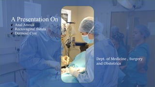
  • 1. Dept. of Medicine , Surgery and Obstetrics A Presentation On Anal Atresia Rectovaginal fistula Dermoid Cyst
  • 2. Anal Atresia Anal atresia is a congenital anorectal malformation (ARM) where a normal anal opening is absent at birth. ARMs comprise of a broad spectrum of defects ranging from minor (e.g., membranous covering) to complex cloacal malformations involving the urinary and genital tracts as well
  • 3. Four types of atresia ani have been reported, including:  Congenital anal stenosis (Type I);  Imperforate anus alone (Type II)  Combined with more cranial termination of the rectum as a blind pouch (Type III)  Discontinuity of the proximal rectum with normal anal and terminal rectal development (Type IV). Types of atresia ani
  • 4. The cause is not known but may have a genetic component or may be from an insult to the fetus in critical developmental stages in utero. Environmental teratogens, plant toxins and a few viruses are recognized complicating factors in calves. Atresia ani is the most common intestinal defect in sheep and usually arises during the embryonic period which results from autosomal recessive gene. Etiology
  • 5. Absence of an anal opening Depression Anorexia Colic Marked gradual abdominal distension Tenesmus Lack of feces. Vomiting. Clinical signs
  • 6. Atresia ani can be diagnosed by  simple visual examination  By digital palpation and  The clinical signs Diagnosis
  • 7. Surgical Procedure: 1. The Calf was placed in a lateral recumbency with its hind quarter raised high. 2. The perineal region was prepared for aseptic surgery. Treatment
  • 8. 3. For Local infiltration : 2% Lignocaine hydrochloride(4-5ml)/calf was done at the perineal region below the tail. Low epidural anaesthesia (3ml). 4. A circular incision was made on the bulge of the anus to remove a circular piece of skin. 5. The rectum was exposed by separation of the perineal muscles. 6. Hold the rectum with stay suture and remove the feces using dushcane ( a pipe containing water source , use to remove debris ). Treatment
  • 9. 7.Suture ( simple interrupted ) the rectum with skin like urethrostomy. 4 interrupted suture – ventral, dorsal, right, left 8. The new opening line should be cleaned with finger using coconut oil 2-3 times per day to prevent auto healing Treatment
  • 10. POST OPERATIVE CARE The animal was administered Enrofloxacin @ 5mg/kg for five days. Meloxicam @0.5 ml for 3 days Routine dressing with povidone iodine ointment and application of topicure spray as a fly repellant. The calf showed normal in defecation with minimum tenesmus and active in nature on the 3rd postoperative day and therefore the sutures were removed on 10th post-operative day and the animal made a noticeable recovery.
  • 11.  Fecal incontinence  Persistent megacolon  Anal stricture  recurrent cystitis. Complications after surgery
  • 12. in Rectovaginal fistula A rectovaginal fistula in a cow is a rare condition where there is an abnormal connection or passage way between the rectum and the vagina.This can lead to the passage of fecal material into the reproductive tract and can result in health issues for the cow.
  • 13. 1.Congenital 2.Local infection 3.Traumatic : A)Post-partum B) Secondary to surgery : • following low anterior rectal resection • following procedures for pelvic floor dysfunction • following haemorrhoid surgery • following drainage of local infection. Etiology
  • 14. C)Resulting from violent acts. 4.Chronic inflammatory bowel disease : -crohn's disease -ulcerative colitis -indeterminate colitis 5.following radiation therapy of tumors in the lesser pelvis 6.resulting from carcinoma Etiology
  • 15. CLINICAL SIGNS • 1.passage of gas or stool from vagina • 2. Foul smelling vaginal discharge • 3.chronic vaginal infection • 4.pain during intercourse • 5.incontinence • 6.tenesmus • 7.abdominal distention • 8.irritation of the vulva
  • 16. Diagnosis Diagnosis of a rectovaginal fistula is typically made by the history of the passage of stool and/or gas from the vagina, in addition to a physical exam. A vaginal exam with a speculum and a rectal exam should be performed. Ultrasound CT scan MRI
  • 17. Treatment Surgical techniques: A)In case of internal attachment of rectum to vagina: 1.After adequate restraint and preparation, a transverse incision is made between the rectum and vagina. 2. By using a combination of sharp and blunt dissection in a horizontal plane, the fistula is exposed.
  • 18. Treatment 3 .Ideally, two thirds of the thickness of the shelf should be with the rectum and one third with the vaginal shelf. 4. Most fistulas measure 3 to 5 cm. Dissection is continued 3 to 4 cm rostral to the fistula. 5.The rectal defect is closed transversely by using No. 0 or 1 absorbable sutures in a simple interrupted pattern placed in the submucosa.
  • 19. Treatment 6.The first suture should divide the defect in half ,the next two sutures should be placed to bisect the halves, and so on. Alternatively, one can pre place the sutures. 7.The vaginal defect is closed next. A continuous horizontal mattress pattern in a longitudinal direction is used so that the two suture rows are at right angles to each other and the vaginal mucosa is everted. 8.The incised perineal body is closed with multiple interrupted sutures of 2-0 suture; the skin is closed routinely.
  • 20. Treatment B) In case of secondary complication of Atrisia anii surgery: 1.Animal is restrained laterally by the owner and low epidural anesthesia is performed using 2% lignocaine HCl. 2.Then clean the opening with water. 3 .Make a stretch over the visceral part of the skin area to make a fresh wound that will induce the healing quickly. 4.Then the rectal wall is closed with purse string suture using 3/0 catgut.
  • 21. Post operative care: Antibiotics -The animal was administered Ciprofloxacin @ 5mg/kg for five days. Meloxicam @0.5 ml for 3 days Stop movement of heifers for 2-3 day Clear the opening of rectum 4-5 times daily with coconut oil to make sure that the rectum doesn’t get closed.
  • 22. Dermoid cyst Dermoid cysts are a relatively rare congenital anomaly in cattle. Dermoid cysts are congenital cysts and occur due to failure of the embryonic cavity to close or defective epidermal closure
  • 23. Location Dermoid cysts are reported in many species of animals at different anatomical location: Tail, Neck, Skull, Nasal cavity • But ocular dermoid are common in ruminants and they may be present unilaterally or bilaterally. • Usually dermoids are located in limbus, cornea. conjunctiva, corneo-conjunctiva they have also been noticed in membrana nictitans • eyelid
  • 24. Age of predisposition  Congenital, thus usually noticed in young animals  But may occur also in large adult animals.
  • 25. Breed/species predisposition  May affect all breeds.  Has been reported in Hereford and Jersey cattle as well as Holstein crossbred
  • 26. Etiology  Failure of the embryonic cavity to close or defective epidermal closure.  Degenerative changes in hair follicles  Cystic changes in ducts or cells of sebaceous glands  Traumatic displacement of epidermal fragments.  Cysts originated from incarceration and subsequent growth of embryonic epithelial cells during the closure of neural tube and therefore most of these lesions occur along the median line.
  • 27. Classification of dermoid cyst Ventrolateral corneo-conjunctival dermoid, Supero-temporal corneal limbal dermoid, Medial canthal dermoid Membrana nictitans dermoid ocular dermoid in dairy cattle include: Corneal dermoid, corneo-scleral dermoid, corneo-conjunctival dermoid and conjunctival dermoid
  • 28. PATHOLOGY & PATHOGENESIS • Dermoid cysts are composed of keratinized stratified squamous epithelium with dermal appendages and adnexal structures, including hair follicles, sebaceous glands, sweat glands, smooth muscle, and fibro adipose tissue. The lumen contains keratin and hair. Cysts that are only lined with epithelium without adnexal elements are termed epidermoid cysts • Dermoid cyst arises from the entrapped embryonal ectodermal cells during the fetal development and are generally formed in the embryonic lines of fusion.
  • 29. CLINICAL SIGNS Mass on/near eye Blepharospasm Lacrimation Visual impairment Tearing Inflammation Irritation in the eye Corneal opacity in Holstein crossbred
  • 30. DIAGNOSIS 1. Physical Examination: Painless swelling that may be freely mobile or fixed to the skin & deeper structure. 2. Radiological Examination: MRI,ULTRASOUND,CT SCAN
  • 31. SURGICAL TREATMENT 1. Restraining: The animal should be controlled in lateral recumbency by casting. The animal should be sedated with 0.1mg/kg Xylazine intramuscularly. 2. Anaesthesia: Local analgesia was performed by 2% Lignocaine HCl. The anaesthetic was injected through the stalk after fixing eyelid with eye speculum. Then Ketamine HCl @ 5 to 6 mg/kg body weight was injected through IM. 3. Operative Procedure: o The dermoid cyst should be lifted as much as possible and curved artery forceps was used to hold the stalk.
  • 32. SURGICAL TREATMENT o A chromic catgut is used to ligate the blood vessels along with the stalk by anchoring it. Sometimes two horizontal mattress stiches may be required. o Excision of the dermoid cyst or stalk is made after crushing with artery forceps. o Cauterization: For excision electric thermocauterizer is used worldwide,it will help to perform the surgery with less bleeding. o Keratectomy: In this procedure,eyelashes was trimmed with scissors and whole eye washed with 0.9% povidone iodine to remove contaminates. After attainment of adequate sedation superficial keratectomy was performed. Then the mass of dermoid was grasped with forceps gently and the tissue mass was completely excised by surgical blade. Bleeding from surgical wound was stopped by applying povidone ointment.
  • 33. Fig: Dermoid cyst mass after surgery Fig: Performing the surgery
  • 34. POST OPERATIVE CARE Ciprofloxacin eye drop T.I.D in a day for 5 days. Gentamicin 4mg /kg intra muscular for 6 days. Meloxicam 0.4mg /kg intramuscular for 3 days. Boric acid 0.3% solution should be used for eye wash Ointment: Chloramphenicol or Neobacterin 3 times in a day up to recovery.