This document provides an overview of stool analysis procedures, including:
- Specimen collection guidelines such as using leak-proof containers and avoiding refrigeration.
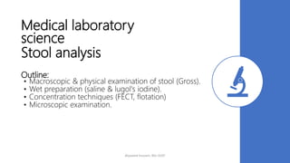
- Macroscopic examination of stool color, consistency, and presence of blood or mucus.
- Wet preparation techniques like saline and lugol's iodine slides.
- Concentration methods like formol-ether and flotation that recover parasites.
- Microscopic examination of samples for parasites, eggs, cysts, and trophozoites using 10x and 40x objectives.