
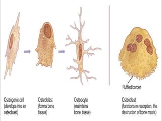
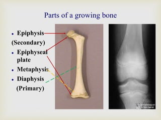
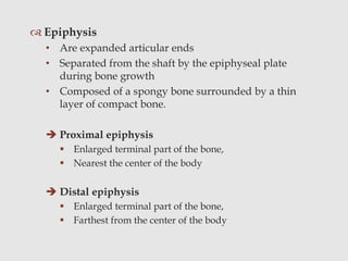
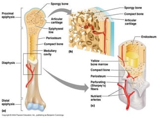
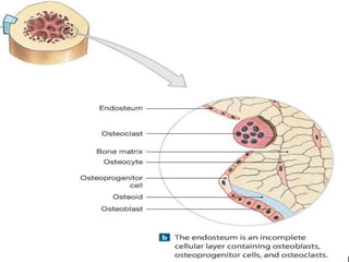
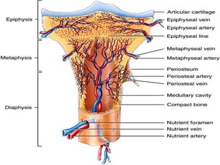


This document provides information on bone structure and composition. It discusses the different types of bones and bone cells. The key parts of long bones are described, including the epiphysis, metaphysis, and diaphysis. Both compact and spongy bone tissue are examined. Other structures like the periosteum, endosteum, and bone marrow are also summarized. The document concludes with sections on blood supply and nerve innervation of bones.


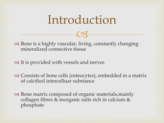
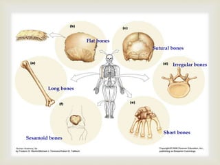
![CTEV [ clubfoot] DR ARUN LAL ,DR MOHAMED ASHRAF travancore medical college k...](https://cdn.slidesharecdn.com/ss_thumbnails/ctevclubfootdrarunlaldrmohamedashraftravancoremedicalcollegekollamkeralaindia-260208063247-18fc466c-thumbnail.jpg?width=640&height=640&fit=bounds)


