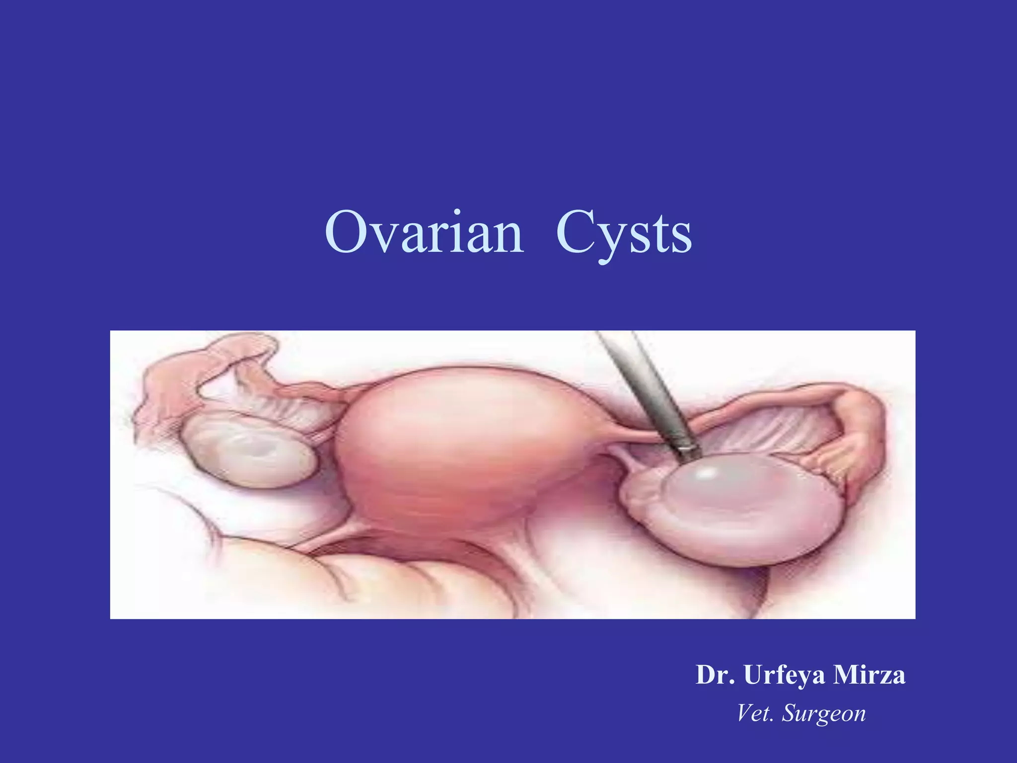
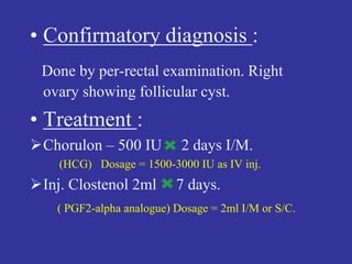
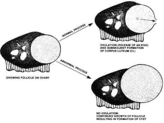
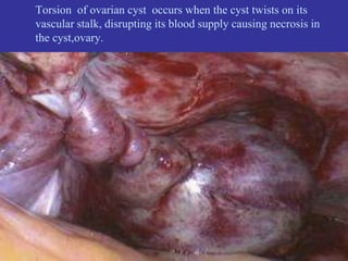
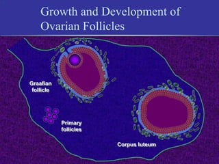
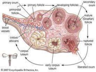
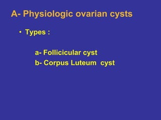
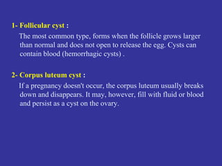
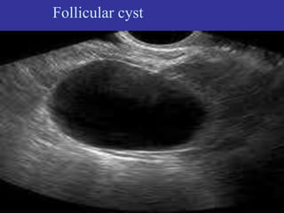
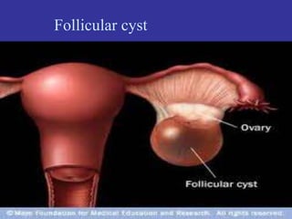
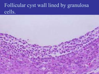
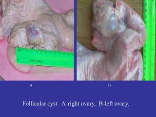
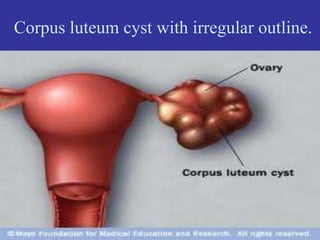
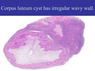
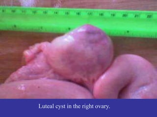
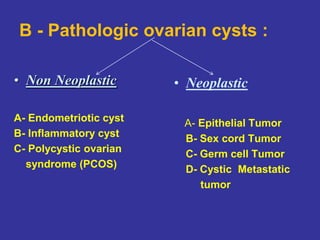
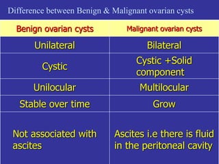
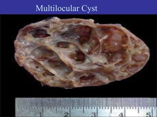
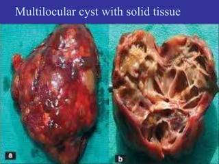
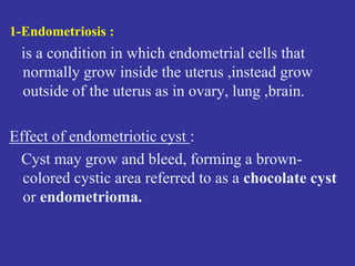
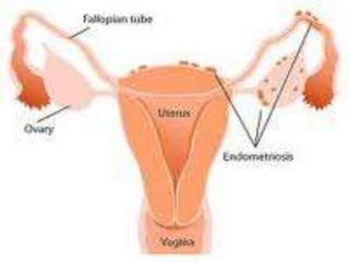
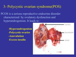
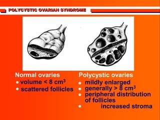
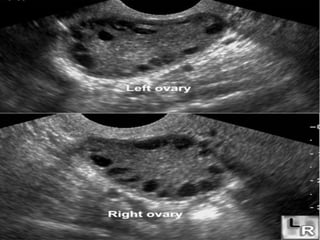
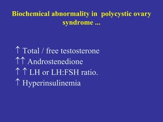
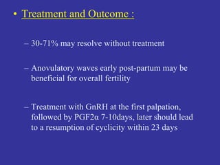
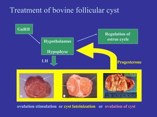
Ovarian Cysts
Dr. Urfeya Mirza
Vet. Surgeon
INTRODUCTION
Disorders of the rumen and fore stomach in cattle can result
from variety of causes:
 Dietary
 Inflammatory
 Mechanical
The bovine species does not have highly sensitive prehensile
organs such as lips and tongues that discriminating sense of
taste. As a consequence, cattle kept in farm yards stables or
at other sites close to human mechanical activities are prone
to swallow metallic objects such as nails and pieces of wires
that have been carelessly left in their feeding areas.
Epidemiology
Occurrences: Cattle are more likely to ingest foreign bodies than small
ruminant since they don’t use their lips for prehension and are more likely to
eat a chopped feed. The majority of affected cattle (87%) are dairy cattle and
93% are older than two years of age. It is hypothesized that dairy cattle are
more commonly affected than beef cattle since they are more likely to feed a
chopped feed, such as silage or hay.
Influencing Factors: According to Schipper, there are a number of
influencing factors. These includes; remodeling of livestock housing,
careless handling of baling wires, pins, feed sack bags and wires, using old
buildings sites for hay fields, often occur in female shortly after calving,
often occur in male shortly after or during extensive uses for breeding.
Pathogenesis
• Ingestion of Foreign Body: Lack of oral discrimination by cattle
leads to the ingestion of foreign bodies. Swallowed foreign body may
lodge in the upper esophagus or in the esophageal groove; but most
instances they pass to the reticulum.
• Penetration of Reticulum: If the reticular wall is injured without
penetration to the serous surfaces, no detectable illness occur. Most
perforation occurs in the lower part of the cranial wall of reticulum but
some occur laterally in the direction of the spleen and medially towards the
liver. In the sites for long period and gradually be corroded away. Many
foreign bodies may not remain embedded but are commonly found free in
the reticulum if surgery is carried out about 72 hours after illness
commences.
• Acute Local Peritonitis: The initial reaction to perforation is acute
local peritonitis and it depends on the severity. The ventral aspects of the
reticulum becomes adherent to varying degrees to the abdominal floor and
diaphragm. This result in decreased reticular motility and reticular abscess
formation which is a common complication and may be located between
the reticulum and the ventral body wall, between the reticulum and the
right thoracic wall and between the reticulum and the spleen.
• Generalized Peritonitis and Extension Diseases: Spread of the
inflammation causing generalized peritonitis may occur in the cows that
calves at time of perforation and in cattle that are forced to exercise.
Peritonitis results in toxemia, alimentary tract stasis, dehydration and
shock. During the initial penetration of the reticulum, the foreign body may
penetrate beyond the peritoneal cavity and into the pericardial sacks or
pleural.
Hematological Findings:
 Increases in the concentration of fibrinogen and plasma proteins.
 In acute local peritonitis, neutrophils (mature neutrophils above 400/ul)
and (immature neutrophils above 200/ul) are common. This is regenerative
left shift.
 Significant increase in total leukocyte (WBC) & in the percentage of
neutrophils while decrease in the percentage of lymphocytes.
Biochemical findings:
 Calcium concentration is significantly low in the cattle with TRP whereas
phosphorous concentration is slightly high.
 Plasma fibrinogen levels are often elevated (>1000 mg/ dl, normal 300-
600mg/dl).
 The most common abnormality associated with TRP is hyperproteinemia
with hyperglobulinemia.
 Total serum protein concentration greater than 10mg/dl.
Case Record :
• Species : Bovine
• Sex : Female
• Age : 5 years
• Temperature : 101.5 0 F
• Pulse : 55 beats/min
• Mucous Memb : Pink
• History :
Cow, 5 years of age, cross-bred, parturated 6
months back. Animal is showing signs of estrus
and on enquiry owner reported that animal is
showing irregular estrus since one month.
• Tentative diagnosis :
Follicular cyst.
• Confirmatory diagnosis :
Done by per-rectal examination. Right
ovary showing follicular cyst.
• Treatment :
Chorulon – 500 IU 2 days I/M.
(HCG) Dosage = 1500-3000 IU as IV inj.
Inj. Clostenol 2ml 7 days.
( PGF2-alpha analogue) Dosage = 2ml I/M or S/C.
ovarian cyst
• Definition : It is a fluid-filled sac (>25mm
diameter) arising from the ovary.
• Most types of ovarian cysts are harmless
(specially the physiologic types )and go
away without any treatment.
What are the Symptoms of Ovarian Cysts?
It depend on its type -
1- Physiologic cysts : usually Asymptomatic due to their small size
and usually resolve spontaneously.
2- Pathologic cysts : ovarian cysts produce :
A- Pain is caused by :
- large size
- bleeding
- bursting of a cyst
- twisting of cyst (called torsion), which blocks blood flow to cyst.
B- Infertility as with endometriotic cyst, polycystic ovarian syndrome.
Torsion of ovarian cyst occurs when the cyst twists on its
vascular stalk, disrupting its blood supply causing necrosis in
the cyst,ovary.
Classification of ovarian cysts
• A - Physiologic ovarian cysts
• B - Pathologic ovarian cysts
Physiologic ovarian cysts
What Causes physiologic Ovarian Cysts?
• Normally during ovulation, the mature follicle ruptures
so an egg is released and a corpus luteum forms from
the empty follicle, and if pregnancy does not occur, the
corpus luteum dissolves.
• Sometimes, however, this process does not conclude
appropriately, causing the most common type of ovarian
cyst -- functional ovarian cysts.
1
Corpus luteum
Graafian
follicle
Primary
follicles
Growth and Development of
Ovarian Follicles
A- Physiologic ovarian cysts
• Types :
a- Follicicular cyst
b- Corpus Luteum cyst
1- Follicular cyst :
The most common type, forms when the follicle grows larger
than normal and does not open to release the egg. Cysts can
contain blood (hemorrhagic cysts) .
2- Corpus luteum cyst :
If a pregnancy doesn't occur, the corpus luteum usually breaks
down and disappears. It may, however, fill with fluid or blood
and persist as a cyst on the ovary.
Follicular cyst
Follicular cyst
Follicular cyst wall lined by granulosa
cells.
A B
Follicular cyst A-right ovary, B-left ovary.
Corpus luteum cyst with irregular outline.
Corpus luteum cyst has irregular wavy wall.
Luteal cyst in the right ovary.
B - Pathologic ovarian cysts :
• Non Neoplastic
A- Endometriotic cyst
B- Inflammatory cyst
C- Polycystic ovarian
syndrome (PCOS)
• Neoplastic
A- Epithelial Tumor
B- Sex cord Tumor
C- Germ cell Tumor
D- Cystic Metastatic
tumor
Neoplastic ovarian cysts
Difference between Benign & Malignant ovarian cysts
Benign ovarian cysts Malignant ovarian cysts
Unilateral Bilateral
Cystic
Cystic +Solid
component
Unilocular Multilocular
Stable over time Grow
Not associated with
ascites
Ascites i.e there is fluid
in the peritoneal cavity
Multilocular Cyst
Multilocular cyst with solid tissue
Dermoid ovarian cyst showing hair.
Dermoid cyst lined by stratified squamous epithelium
with sebacous glands in the dermis.
“Cystadenoma of ovary” is an ovarian cysts that develop
from cells on the outer surface of the ovaries.
Non-neoplastic ovarian Cyst
1-Endometriosis :
is a condition in which endometrial cells that
normally grow inside the uterus ,instead grow
outside of the uterus as in ovary, lung ,brain.
Effect of endometriotic cyst :
Cyst may grow and bleed, forming a brown-
colored cystic area referred to as a chocolate cyst
or endometrioma.
2-Tubo-ovarian abscesses :
Infections of the pelvic organs can involve the
ovaries and Fallopian tubes .
In severe cases, pus-filled cystic spaces may be
present on or around the ovary or tubes .
3- Polycystic ovarian syndrome(POS)
PCOS is a serious reproductive endocrine disorder
characterized by ovulatory dysfunction and
hyperandrogenism. It leads to :
-Hyperandrogenism
-Polycystic ovaries
-Anovulation
-Excess insulin
POLYCYSTIC OVARIAN SYNDROME


Normal ovaries
volume < 8 cm3
scattered follicles
mildly enlarged
generally > 8 cm3
peripheral distribution
of follicles
increased stroma
Polycystic ovaries






Biochemical abnormality in polycystic ovary
syndrome ...
 Total / free testosterone
 Androstenedione
  LH or LH:FSH ratio.
 Hyperinsulinemia
• Treatment and Outcome :
– 30-71% may resolve without treatment
– Anovulatory waves early post-partum may be
beneficial for overall fertility
– Treatment with GnRH at the first palpation,
followed by PGF2α 7-10days, later should lead
to a resumption of cyclicity within 23 days
GnRH
Hypothalamus
Hypophyse
LH Progesterone
Regulation of
estrus cycle
Treatment of bovine follicular cyst
ovulation stimulation or cyst luteinization or ovulation of cyst
• Prostaglandin Products
– Lutalyse® Pharmacia Animal Health
– Estrumate® Schering-Plough
– ProstaMate® Phoenix Scientific
– In-Synch
• Gonadotropin Releasing Hormone –
GnRH
– Cystorelin® Merial
– Fertagyl® Intervet
– Factrel® Fort Dodge
– Ovacyst
A Happy End !!

Ovarian Cysts

  • 1.
    Ovarian Cysts Dr. UrfeyaMirza Vet. Surgeon
  • 2.
    INTRODUCTION Disorders of therumen and fore stomach in cattle can result from variety of causes:  Dietary  Inflammatory  Mechanical The bovine species does not have highly sensitive prehensile organs such as lips and tongues that discriminating sense of taste. As a consequence, cattle kept in farm yards stables or at other sites close to human mechanical activities are prone to swallow metallic objects such as nails and pieces of wires that have been carelessly left in their feeding areas.
  • 3.
    Epidemiology Occurrences: Cattle aremore likely to ingest foreign bodies than small ruminant since they don’t use their lips for prehension and are more likely to eat a chopped feed. The majority of affected cattle (87%) are dairy cattle and 93% are older than two years of age. It is hypothesized that dairy cattle are more commonly affected than beef cattle since they are more likely to feed a chopped feed, such as silage or hay. Influencing Factors: According to Schipper, there are a number of influencing factors. These includes; remodeling of livestock housing, careless handling of baling wires, pins, feed sack bags and wires, using old buildings sites for hay fields, often occur in female shortly after calving, often occur in male shortly after or during extensive uses for breeding.
  • 4.
    Pathogenesis • Ingestion ofForeign Body: Lack of oral discrimination by cattle leads to the ingestion of foreign bodies. Swallowed foreign body may lodge in the upper esophagus or in the esophageal groove; but most instances they pass to the reticulum. • Penetration of Reticulum: If the reticular wall is injured without penetration to the serous surfaces, no detectable illness occur. Most perforation occurs in the lower part of the cranial wall of reticulum but some occur laterally in the direction of the spleen and medially towards the liver. In the sites for long period and gradually be corroded away. Many foreign bodies may not remain embedded but are commonly found free in the reticulum if surgery is carried out about 72 hours after illness commences.
  • 5.
    • Acute LocalPeritonitis: The initial reaction to perforation is acute local peritonitis and it depends on the severity. The ventral aspects of the reticulum becomes adherent to varying degrees to the abdominal floor and diaphragm. This result in decreased reticular motility and reticular abscess formation which is a common complication and may be located between the reticulum and the ventral body wall, between the reticulum and the right thoracic wall and between the reticulum and the spleen. • Generalized Peritonitis and Extension Diseases: Spread of the inflammation causing generalized peritonitis may occur in the cows that calves at time of perforation and in cattle that are forced to exercise. Peritonitis results in toxemia, alimentary tract stasis, dehydration and shock. During the initial penetration of the reticulum, the foreign body may penetrate beyond the peritoneal cavity and into the pericardial sacks or pleural.
  • 6.
    Hematological Findings:  Increasesin the concentration of fibrinogen and plasma proteins.  In acute local peritonitis, neutrophils (mature neutrophils above 400/ul) and (immature neutrophils above 200/ul) are common. This is regenerative left shift.  Significant increase in total leukocyte (WBC) & in the percentage of neutrophils while decrease in the percentage of lymphocytes. Biochemical findings:  Calcium concentration is significantly low in the cattle with TRP whereas phosphorous concentration is slightly high.  Plasma fibrinogen levels are often elevated (>1000 mg/ dl, normal 300- 600mg/dl).  The most common abnormality associated with TRP is hyperproteinemia with hyperglobulinemia.  Total serum protein concentration greater than 10mg/dl.
  • 7.
    Case Record : •Species : Bovine • Sex : Female • Age : 5 years • Temperature : 101.5 0 F • Pulse : 55 beats/min • Mucous Memb : Pink
  • 8.
    • History : Cow,5 years of age, cross-bred, parturated 6 months back. Animal is showing signs of estrus and on enquiry owner reported that animal is showing irregular estrus since one month. • Tentative diagnosis : Follicular cyst.
  • 9.
    • Confirmatory diagnosis: Done by per-rectal examination. Right ovary showing follicular cyst. • Treatment : Chorulon – 500 IU 2 days I/M. (HCG) Dosage = 1500-3000 IU as IV inj. Inj. Clostenol 2ml 7 days. ( PGF2-alpha analogue) Dosage = 2ml I/M or S/C.
  • 10.
    ovarian cyst • Definition: It is a fluid-filled sac (>25mm diameter) arising from the ovary. • Most types of ovarian cysts are harmless (specially the physiologic types )and go away without any treatment.
  • 12.
    What are theSymptoms of Ovarian Cysts? It depend on its type - 1- Physiologic cysts : usually Asymptomatic due to their small size and usually resolve spontaneously. 2- Pathologic cysts : ovarian cysts produce : A- Pain is caused by : - large size - bleeding - bursting of a cyst - twisting of cyst (called torsion), which blocks blood flow to cyst. B- Infertility as with endometriotic cyst, polycystic ovarian syndrome.
  • 13.
    Torsion of ovariancyst occurs when the cyst twists on its vascular stalk, disrupting its blood supply causing necrosis in the cyst,ovary.
  • 14.
    Classification of ovariancysts • A - Physiologic ovarian cysts • B - Pathologic ovarian cysts
  • 15.
  • 16.
    What Causes physiologicOvarian Cysts? • Normally during ovulation, the mature follicle ruptures so an egg is released and a corpus luteum forms from the empty follicle, and if pregnancy does not occur, the corpus luteum dissolves. • Sometimes, however, this process does not conclude appropriately, causing the most common type of ovarian cyst -- functional ovarian cysts.
  • 17.
  • 19.
    A- Physiologic ovariancysts • Types : a- Follicicular cyst b- Corpus Luteum cyst
  • 20.
    1- Follicular cyst: The most common type, forms when the follicle grows larger than normal and does not open to release the egg. Cysts can contain blood (hemorrhagic cysts) . 2- Corpus luteum cyst : If a pregnancy doesn't occur, the corpus luteum usually breaks down and disappears. It may, however, fill with fluid or blood and persist as a cyst on the ovary.
  • 21.
  • 22.
  • 23.
    Follicular cyst walllined by granulosa cells.
  • 24.
    A B Follicular cystA-right ovary, B-left ovary.
  • 25.
    Corpus luteum cystwith irregular outline.
  • 26.
    Corpus luteum cysthas irregular wavy wall.
  • 27.
    Luteal cyst inthe right ovary.
  • 28.
    B - Pathologicovarian cysts : • Non Neoplastic A- Endometriotic cyst B- Inflammatory cyst C- Polycystic ovarian syndrome (PCOS) • Neoplastic A- Epithelial Tumor B- Sex cord Tumor C- Germ cell Tumor D- Cystic Metastatic tumor
  • 29.
  • 30.
    Difference between Benign& Malignant ovarian cysts Benign ovarian cysts Malignant ovarian cysts Unilateral Bilateral Cystic Cystic +Solid component Unilocular Multilocular Stable over time Grow Not associated with ascites Ascites i.e there is fluid in the peritoneal cavity
  • 31.
  • 32.
  • 33.
    Dermoid ovarian cystshowing hair.
  • 34.
    Dermoid cyst linedby stratified squamous epithelium with sebacous glands in the dermis.
  • 35.
    “Cystadenoma of ovary”is an ovarian cysts that develop from cells on the outer surface of the ovaries.
  • 36.
  • 37.
    1-Endometriosis : is acondition in which endometrial cells that normally grow inside the uterus ,instead grow outside of the uterus as in ovary, lung ,brain. Effect of endometriotic cyst : Cyst may grow and bleed, forming a brown- colored cystic area referred to as a chocolate cyst or endometrioma.
  • 40.
    2-Tubo-ovarian abscesses : Infectionsof the pelvic organs can involve the ovaries and Fallopian tubes . In severe cases, pus-filled cystic spaces may be present on or around the ovary or tubes .
  • 41.
    3- Polycystic ovariansyndrome(POS) PCOS is a serious reproductive endocrine disorder characterized by ovulatory dysfunction and hyperandrogenism. It leads to : -Hyperandrogenism -Polycystic ovaries -Anovulation -Excess insulin
  • 42.
    POLYCYSTIC OVARIAN SYNDROME POLYCYSTICOVARIAN SYNDROME POLYCYSTIC OVARIAN SYNDROME   Normal ovaries volume < 8 cm3 scattered follicles mildly enlarged generally > 8 cm3 peripheral distribution of follicles increased stroma Polycystic ovaries      
  • 44.
    Biochemical abnormality inpolycystic ovary syndrome ...  Total / free testosterone  Androstenedione   LH or LH:FSH ratio.  Hyperinsulinemia
  • 45.
    • Treatment andOutcome : – 30-71% may resolve without treatment – Anovulatory waves early post-partum may be beneficial for overall fertility – Treatment with GnRH at the first palpation, followed by PGF2α 7-10days, later should lead to a resumption of cyclicity within 23 days
  • 46.
    GnRH Hypothalamus Hypophyse LH Progesterone Regulation of estruscycle Treatment of bovine follicular cyst ovulation stimulation or cyst luteinization or ovulation of cyst
  • 47.
    • Prostaglandin Products –Lutalyse® Pharmacia Animal Health – Estrumate® Schering-Plough – ProstaMate® Phoenix Scientific – In-Synch • Gonadotropin Releasing Hormone – GnRH – Cystorelin® Merial – Fertagyl® Intervet – Factrel® Fort Dodge – Ovacyst
  • 48.