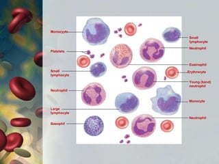
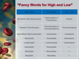
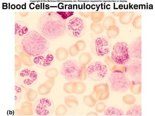
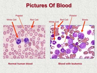
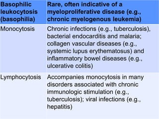
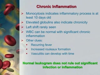
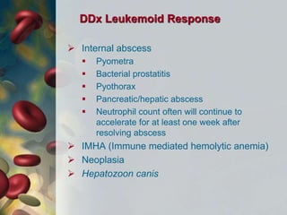
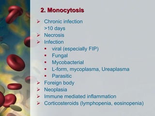
“Leukocytosis”
Dr. Urfeya Mirza
Department of Vety. Surgery and
Radiology
“Blood”
 Connective Tissue
 Mesodermal Origin
 Composition:
 Cells
 Aqueous Plasma Matrix
 Proteins
 Organic and inorganic salts
“Blood Cells”
 Erythrocytes: Red Blood Cells
(RBCs)
 Platelets: Thrombocytes (clotting
cells)
Cell fragments from special cell in bone
marrow
 Leukocytes: white blood cells
(WBCs) -
Neutrophil
Erythrocyte
Eosinophil
Monocyte
Neutrophil
Basophil
Neutrophil
Platelets
Monocyyte
Small
lymphocyte
Young (band)
neutrophil
Small
lymphocyte
Large
lymphocyte
How Are Blood Cells Formed?
“Fancy Words for High and Low”
Cell Type Too High Too Low
Red Blood Cells (Erythrocytes)
Erythrocytosis or
Polycythemia
Anemia
Platelets (Thrombocytes)
Thrombocytosis or
Thrombocythemia
Thrombocytopenia
White Blood Cells (Leukocytes) Leukocytosis Leukopenia
Neutrophils Neutrophilia Neutropenia
Lymphocytes Lymphocytosis Lymphopenia
Eosinophils Eosinophilia ---
Monocytes Monocytosis ---
Basophils Basophilia ---
“Plasma”
 Plasma is the liquid
component of blood
 Comprised mostly of
water, but also
includes:
 Protein (albumin,
globulin, fibrinogen)
 Lipids (cholesterol,
triglycerides)
 Dissolved salts and
minerals (sodium,
calcium, potassium)
 Glucose
“Leukocytes”
 White blood cells
 Defend body through:
• the inflammatory process
• phagocytosis
• removal of cell debris
• immune reactions
White Blood Cell Types :
Granulocytes and Agranulocytes
 Granulocytes – visible granules in
the cytoplasm
 Granules contain:
• Enzymes
• Other biochemicals that serve as
signals and mediators of the
inflammatory response
Granulocyte cell types:
 Neutrophils (60% to 70%) –
Phagocytes polymorphonuclear
leukocytes, Barely visible granules
in cytoplasm; three- to five-lobed
nucleus
 Eosinophils (2% to 4%) – Large
red-orange granules, associated
with allergic response and parasitic
worms
 Basophils (less than 1%) – Large,
abundant, violet granules granules -
Release heparin, histamine and
serotonin
Agranulocytes
 Granules too small to be visible
 Monocytes (3% to 8%) – Largest WBC;
generally ovoid, kidney, or horseshoe-shaped
nucleus, become macrophages
 Lymphocytes (25% to 33%) – B cells and
T cells = immune functions, Variable amounts
of bluish cytoplasm (scanty to abundant);
ovoid/round, uniform dark violet nucleus
15
• WBC’s originate in red bone marrow
from stem cells
• Granulocytes mature in the marrow and
have a lifespan of hours to days
• Agranulocytes finish maturing in blood,
or in other locations. Monocytes live
about 2 - 3 months, lymphocytes for
years
18-18
Leukopoiesis
leaves
Lymphoblast
B prolymphocyte
B lymphocyte
T lymphocyte
NK cell
T prolymphocyte
NK prolymphocyte
Monoblast Promonocyte Monocyte
Basophil
Eosinophil
Neutrophil
Pluripotent
stem cell
Colony-forming
units (CFUs)
Precursor
cells
Mature
cells
Eosinophilic
myelocyte
Eosinophilic
promyelocyte
Eosinophilic
myeloblast
Eosinophilic
CFU
Basophilic
CFU
Neutrophilic
CFU
Monocytic
CFU
Lymphocytic
CFU
Basophilic
myeloblast
Neutrophilic
myeloblast
Neutrophilic
promyelocyte
Basophilic
promyelocyte
Basophilic
myelocyte
Neutrophilic
myelocyte
 Production of WBC’s increases
in response to :
• Infection
• Presence of steroids
• Decreased reserve of
leukocyte pool in bone
marrow
“WBC Abnormalities”
Disorders of white blood cells can be classified into two
broad categories:
 Proliferative disorders (Leukocytosis)
– increased numbers of WBC’s
 May be a normal protective response to
physiological stressors
 Or may signify a disease state – a
malignancy or hematologic disorder
 Leukopenias – decreased numbers of
WBC’s – this is never normal
 Increases the risk of infections
 Agranulocytosis = granulocytopenia
“Leukopenia”
 Leukemia—cancer of hemopoietic
tissue that usually produces an
extraordinary high number of
circulating leukocytes and their
precursors
 Myeloid leukemia: uncontrolled
granulocyte production
 Lymphoid leukemia: uncontrolled
lymphocyte or monocyte production
Leukopenia may be due to:
• High level radiation/toxin exposure
• Anaphylactic shock
• Autoimmune disease
• Chemotherapeutic agents
• Idiosyncratic drug reactions
• Splenomegaly
• Infections
• Mostly unknown
Causes
Types of Leukemia
 All originate from the bone marrow
 Aleukemic leukemia – no cancer cells
in circulation
 Subleukemic leukemia – small
amounts of cancer cells in circulation
 Leukemic leukemia – many cancer
cells in circulation
 Maturity of leukemia
 Acute leukemia – proliferation of
blasts, tends to be more severe
 Chronic leukemia – proliferation of
more mature blood cells, tends to be
less severe
Main Types
 Acute Lymphocytic Leukemia (ALL)
 Acute Myelogenous Leukemia (AML)
 Chronic Lymphocytic Leukemia (CLL)
 Chronic Myelogenous Leukemia (CML)
Signs and Symptoms
 Insidious nonspecific onset
 Pallor due to anemia
 Febrile (fever) due to ineffective WBC
 Petechiae (skin bruising) due to
thrombocytopenia
 Bone pain
 Hepatosplenomegaly
 Lymphadenopathy
 Fever, weight loss
 Symptoms of cytopenias
Pictures Of Blood
Normal human blood
White Cell Red Cell
Platelet
Blood with leukemia
BlastsRed Cell
Platelet
White Cell
Treatment
• Chemotherapy
• Blood transfusions and antimicrobial,
antifungal and antiviral medications
• Bone marrow transplants
“Reactive Proliferations of
White Cells (Leukocytosis)”
 Definition:
Leukocytosis refers to an increase in
the number of blood leukocytes
 It is a common reaction to a variety of
inflammatory states and is sometimes
the first indication of neoplastic growth
of leukocytes
 Usually affects a specific type of
leukocyte- neutrophil, basophil,
eosinophil, lymphocyte
 Normal level is 4,400 to 10,000 WBC per mm3
 This can result from many causes,
principally infections, inflammatory
disorders, and medications
 Cancer and myeloproliferative disorders can
also cause high, sometimes extremely high,
WBC counts
 Treatment is aimed at the underlying cause
 Death may result from the underlying cause
such as severe infection or cancer
(leukemia)
Pathogenesis
The peripheral blood leukocyte count is
influenced by several factors, including:
1. The size of the myeloid (for granulocytes and
monocytes) and lymphoid (for lymphocytes)
precursor and storage cell pools in the bone
marrow, circulation, and peripheral tissues
2. Leukocyte homeostasis is maintained by
cytokines, growth factors, and adhesion
molecules through their effects on the
commitment, proliferation, differentiation, and
extravasation of leukocytes and their progenitors
3. Other growth factors preferentially
stimulate other types of leukocytosis.
4. For example, IL-5 causes eosinophilia by
enhancing the growth, survival, and
differentiation of eosinophils, while IL-7
plays a central role in lymphopoiesis.
5. Such factors are differentially produced in
response to various pathogenic stimuli.
Pathogenesis
 In most instances, it is not difficult to
distinguish reactive leukocytosis from
leukocytosis caused by flooding of the
peripheral blood by neoplastic white blood
cells (leukemia).
 Uncertainties may arise in two settings:
1. Particularly in children, acute viral infections
can produce the appearance of activated
lymphocytes in the peripheral blood and
marrow that resemble neoplastic lymphoid
cells.
2. At other times, particularly in inflammatory
states and severe chronic infections, many
immature granulocytes appear in the blood,
simulating a picture of myelogenous
leukemia (leukemoid reaction)
“Types of Leukocytosis”
 There are five principal types of
leukocytosis :
 Neutrophilia (the most common form)
 Lymphocytosis
 Monocytosis
 Eosinophilia
 Basophilia
“Causes of Leukocytosis”
Neutrophilic
leukocytosis
Acute bacterial infections, especially
those caused by pyogenic organisms;
sterile inflammation(myocardial
infarction, burns)
Eosinophilic
leukocytosis
(eosinophilia)
Allergic disorders such as asthma,
allergic skin diseases; parasitic
infestations; drug reactions; certain
malignancies (e.g., Hodgkin disease and
some non-Hodgkin lymphomas); collagen
vascular disorders and some vasculitides;
atheroembolic disease
Basophilic
leukocytosis
(basophilia)
Rare, often indicative of a
myeloproliferative disease (e.g.,
chronic myelogenous leukemia)
Monocytosis Chronic infections (e.g., tuberculosis),
bacterial endocarditis and malaria;
collagen vascular diseases (e.g.,
systemic lupus erythematosus) and
inflammatory bowel diseases (e.g.,
ulcerative colitis)
Lymphocytosis Accompanies monocytosis in many
disorders associated with chronic
immunologic stimulation (e.g.,
tuberculosis); viral infections (e.g.,
hepatitis)
1. DDx Neutrophilia
• Infection
• Sterile inflammation
• Necrosis
• Stress/corticosteroids
• Exercise/epinephrine
• Neutrophilic leukemia
• Neoplasia
Acute Inflammation (Left Shift)
 Left shift indicates acute, intense inflammation
 >1000/ul bands = left shift
 300-1000/ul = mild left shift
 Immature unsegmented neutrophils indicates a more
intense inflammation
 Metamyelocytes
 Myelocytes
 promyelocytes
 Peripheral myeloblasts often indicates leukemia
 “Degenerative Left shift” = overwhelming inflammation
 Normal pyramid of maturation is interrupted
 Usually, the more mature forms are more plentiful
Segs > bands > meta > myelo > pro > blast
Chronic Inflammation
 Monocytosis indicates inflammatory process is at
least 10 days old
 Elevated globulins also indicate chronicity
 Left shift rarely seen
 WBC can be normal with significant chronic
inflammation
 Other clues:
 Recurring fever
 Increased rouleaux formation
 Vasculitis can develop with time
Normal leukogram does not rule out significant
infection or inflammation
Prognosis for Neutrophilia
 Poor prognostic indicators :
 Progressive degenerative left shift
 WBC > 60,000/ul correlated with increased
risk of sudden death in dogs
 Extremely high mature neutrophilia
“Leukemoid response”
 Marked toxic changes in the neutrophils
Graded 1+ to 4+
 Obvious infection without fever
 Severe persistent lymphopenia
 Sustained stress on the body
 Magnitude of feline neutrophilic response is
less than canine
DDx Leukemoid Response
 Internal abscess
 Pyometra
 Bacterial prostatitis
 Pyothorax
 Pancreatic/hepatic abscess
 Neutrophil count often will continue to
accelerate for at least one week after
resolving abscess
 IMHA (Immune mediated hemolytic anemia)
 Neoplasia
 Hepatozoon canis
Stress/Corticosteroid Response
 <40,000/ul in the dog
 <30,000/ul in the cat
 Lymphopenia
 Eosinopenia
 Monocytosis
 Mature neutrophilia
 Increased hypersegmented segs
 “right shift”
 Onset within 4-13 hours
 Resolves within 24 hours
Epinephrine/Exercise Response
 <40,000/ul in the dog
 <30,000/ul in the cat
 More of a problem in cats
 Lymphocytosis
 Increased HCT (hematocrit)
2. Monocytosis
 Chronic infection
>10 days
 Necrosis
 Infection
 viral (especially FIP)
 Fungal
 Mycobacterial
 L-form, mycoplasma, Ureaplasma
 Parasitic
 Foreign body
 Neoplasia
 Immune mediated inflammation
 Corticosteroids (lymphopenia, eosinopenia)
3. Lymphocytosis
 Stress/corticosteroid response
 Chronic infection
viremia
 Immune mediated disease
 Recent vaccination
 Lymphoid neoplasia
 Ehrlichia spp.
 Addison’s Disease
 Activated lymphocytes
 Large, immunostimulated lymphocytes
 Dark blue cytoplasm with perinuclear clear
zone
 Irregular, scalloped or cleaved nuclei
 Not terribly clinically significant
 Immunoblasts
 Lighter, more lacy chromatin
 Prominent nucleoli or nucleolar rings
 Atypical lymphocytes
 Characteristics of malignancy
 Darkly basophilic cytoplasm
 Large and atypical nucleolus
 Immature granular chromatin
Lymphocyte
Lymphocytosis
Activated lymphocyte
Lymphocytosis
Activated lymphocyte
Lymphocytosis
Immunoblast
Atypical lymphocytes
Lymphocytosis
4. Eosinophilia
 Infection
 Parasitic
 Fungal
 Viral – FeLV
 Streptococcus, Staphylococcus spp.
 Allergy/asthma
 Immune mediated disease
 Hypereosinophilic syndrome
 Eosinophilic granuloma
 Mast Cell Tumor
 Other neoplasia
 Lymphoma
 Mucinous carcinoma
 Canine estrus
Hypereosinophilic Syndrome (HES)
 Primarily a disease of cats
 Persistent eosinophilia
 Organ infiltration with eosinophils
 Bone marrow
 Spleen
 Liver
 Lymph nodes (often mesenteric)
 Gut
 Skin
 Clinical Signs
 Diarrhea, vomiting
 Anorexia, weight loss
 Fever
 Pruritus, lymphadenopathy
 Abdominal masses are possible
 Eventually causes organ failure and death
 Difficult to distinguish from eosinophilic
leukemia (EL)
 May be two forms of the same disease
 More immature eos in circulation with EL
 Treatment
 No known effective treatment
 Cortisteroids – immunosuppressive
 Hydroxyurea
 Alpha interferon
 Gleevec (imatinib) has been used in
people
5. Basophilia
 Basophils can be difficult to identify
 Mistaken for monocytes or eos
 Parasites
 Allergy
 Mast Cell Tumor
 Lipemia
 Basophilic leukemia (very rare).
“References”
 Fundamentals of Veterinary Clinical Pathology,
2nd Edition (Steven L. Stockham, Michael A.
Scott)
 Practical Diagnostic Imaging for the Veterinary
Technician (Connie M. Han)
 BSAVA Manual of Canine and Feline Clinical
Pathology
 Diagnostic Cytology and Hematology of the
Dog and Cat (Rick L. Cowell)
“A Happy End” !!

Leucocytosis

  • 1.
    “Leukocytosis” Dr. Urfeya Mirza Departmentof Vety. Surgery and Radiology
  • 2.
    “Blood”  Connective Tissue Mesodermal Origin  Composition:  Cells  Aqueous Plasma Matrix  Proteins  Organic and inorganic salts
  • 3.
    “Blood Cells”  Erythrocytes:Red Blood Cells (RBCs)  Platelets: Thrombocytes (clotting cells) Cell fragments from special cell in bone marrow  Leukocytes: white blood cells (WBCs) -
  • 4.
  • 5.
    How Are BloodCells Formed?
  • 6.
    “Fancy Words forHigh and Low” Cell Type Too High Too Low Red Blood Cells (Erythrocytes) Erythrocytosis or Polycythemia Anemia Platelets (Thrombocytes) Thrombocytosis or Thrombocythemia Thrombocytopenia White Blood Cells (Leukocytes) Leukocytosis Leukopenia Neutrophils Neutrophilia Neutropenia Lymphocytes Lymphocytosis Lymphopenia Eosinophils Eosinophilia --- Monocytes Monocytosis --- Basophils Basophilia ---
  • 7.
    “Plasma”  Plasma isthe liquid component of blood  Comprised mostly of water, but also includes:  Protein (albumin, globulin, fibrinogen)  Lipids (cholesterol, triglycerides)  Dissolved salts and minerals (sodium, calcium, potassium)  Glucose
  • 8.
    “Leukocytes”  White bloodcells  Defend body through: • the inflammatory process • phagocytosis • removal of cell debris • immune reactions
  • 9.
    White Blood CellTypes : Granulocytes and Agranulocytes  Granulocytes – visible granules in the cytoplasm  Granules contain: • Enzymes • Other biochemicals that serve as signals and mediators of the inflammatory response
  • 10.
    Granulocyte cell types: Neutrophils (60% to 70%) – Phagocytes polymorphonuclear leukocytes, Barely visible granules in cytoplasm; three- to five-lobed nucleus  Eosinophils (2% to 4%) – Large red-orange granules, associated with allergic response and parasitic worms  Basophils (less than 1%) – Large, abundant, violet granules granules - Release heparin, histamine and serotonin
  • 14.
    Agranulocytes  Granules toosmall to be visible  Monocytes (3% to 8%) – Largest WBC; generally ovoid, kidney, or horseshoe-shaped nucleus, become macrophages  Lymphocytes (25% to 33%) – B cells and T cells = immune functions, Variable amounts of bluish cytoplasm (scanty to abundant); ovoid/round, uniform dark violet nucleus
  • 15.
  • 17.
    • WBC’s originatein red bone marrow from stem cells • Granulocytes mature in the marrow and have a lifespan of hours to days • Agranulocytes finish maturing in blood, or in other locations. Monocytes live about 2 - 3 months, lymphocytes for years
  • 18.
    18-18 Leukopoiesis leaves Lymphoblast B prolymphocyte B lymphocyte Tlymphocyte NK cell T prolymphocyte NK prolymphocyte Monoblast Promonocyte Monocyte Basophil Eosinophil Neutrophil Pluripotent stem cell Colony-forming units (CFUs) Precursor cells Mature cells Eosinophilic myelocyte Eosinophilic promyelocyte Eosinophilic myeloblast Eosinophilic CFU Basophilic CFU Neutrophilic CFU Monocytic CFU Lymphocytic CFU Basophilic myeloblast Neutrophilic myeloblast Neutrophilic promyelocyte Basophilic promyelocyte Basophilic myelocyte Neutrophilic myelocyte
  • 19.
     Production ofWBC’s increases in response to : • Infection • Presence of steroids • Decreased reserve of leukocyte pool in bone marrow
  • 20.
    “WBC Abnormalities” Disorders ofwhite blood cells can be classified into two broad categories:  Proliferative disorders (Leukocytosis) – increased numbers of WBC’s  May be a normal protective response to physiological stressors  Or may signify a disease state – a malignancy or hematologic disorder  Leukopenias – decreased numbers of WBC’s – this is never normal  Increases the risk of infections  Agranulocytosis = granulocytopenia
  • 21.
    “Leukopenia”  Leukemia—cancer ofhemopoietic tissue that usually produces an extraordinary high number of circulating leukocytes and their precursors  Myeloid leukemia: uncontrolled granulocyte production  Lymphoid leukemia: uncontrolled lymphocyte or monocyte production
  • 22.
    Leukopenia may bedue to: • High level radiation/toxin exposure • Anaphylactic shock • Autoimmune disease • Chemotherapeutic agents • Idiosyncratic drug reactions • Splenomegaly • Infections • Mostly unknown Causes
  • 23.
    Types of Leukemia All originate from the bone marrow  Aleukemic leukemia – no cancer cells in circulation  Subleukemic leukemia – small amounts of cancer cells in circulation  Leukemic leukemia – many cancer cells in circulation  Maturity of leukemia  Acute leukemia – proliferation of blasts, tends to be more severe  Chronic leukemia – proliferation of more mature blood cells, tends to be less severe
  • 24.
    Main Types  AcuteLymphocytic Leukemia (ALL)  Acute Myelogenous Leukemia (AML)  Chronic Lymphocytic Leukemia (CLL)  Chronic Myelogenous Leukemia (CML)
  • 25.
    Signs and Symptoms Insidious nonspecific onset  Pallor due to anemia  Febrile (fever) due to ineffective WBC  Petechiae (skin bruising) due to thrombocytopenia  Bone pain  Hepatosplenomegaly  Lymphadenopathy  Fever, weight loss  Symptoms of cytopenias
  • 29.
    Pictures Of Blood Normalhuman blood White Cell Red Cell Platelet Blood with leukemia BlastsRed Cell Platelet White Cell
  • 30.
    Treatment • Chemotherapy • Bloodtransfusions and antimicrobial, antifungal and antiviral medications • Bone marrow transplants
  • 31.
    “Reactive Proliferations of WhiteCells (Leukocytosis)”
  • 32.
     Definition: Leukocytosis refersto an increase in the number of blood leukocytes  It is a common reaction to a variety of inflammatory states and is sometimes the first indication of neoplastic growth of leukocytes  Usually affects a specific type of leukocyte- neutrophil, basophil, eosinophil, lymphocyte
  • 33.
     Normal levelis 4,400 to 10,000 WBC per mm3  This can result from many causes, principally infections, inflammatory disorders, and medications  Cancer and myeloproliferative disorders can also cause high, sometimes extremely high, WBC counts  Treatment is aimed at the underlying cause  Death may result from the underlying cause such as severe infection or cancer (leukemia)
  • 34.
    Pathogenesis The peripheral bloodleukocyte count is influenced by several factors, including: 1. The size of the myeloid (for granulocytes and monocytes) and lymphoid (for lymphocytes) precursor and storage cell pools in the bone marrow, circulation, and peripheral tissues 2. Leukocyte homeostasis is maintained by cytokines, growth factors, and adhesion molecules through their effects on the commitment, proliferation, differentiation, and extravasation of leukocytes and their progenitors
  • 35.
    3. Other growthfactors preferentially stimulate other types of leukocytosis. 4. For example, IL-5 causes eosinophilia by enhancing the growth, survival, and differentiation of eosinophils, while IL-7 plays a central role in lymphopoiesis. 5. Such factors are differentially produced in response to various pathogenic stimuli.
  • 36.
    Pathogenesis  In mostinstances, it is not difficult to distinguish reactive leukocytosis from leukocytosis caused by flooding of the peripheral blood by neoplastic white blood cells (leukemia).  Uncertainties may arise in two settings: 1. Particularly in children, acute viral infections can produce the appearance of activated lymphocytes in the peripheral blood and marrow that resemble neoplastic lymphoid cells.
  • 37.
    2. At othertimes, particularly in inflammatory states and severe chronic infections, many immature granulocytes appear in the blood, simulating a picture of myelogenous leukemia (leukemoid reaction)
  • 38.
    “Types of Leukocytosis” There are five principal types of leukocytosis :  Neutrophilia (the most common form)  Lymphocytosis  Monocytosis  Eosinophilia  Basophilia
  • 39.
  • 40.
    Neutrophilic leukocytosis Acute bacterial infections,especially those caused by pyogenic organisms; sterile inflammation(myocardial infarction, burns) Eosinophilic leukocytosis (eosinophilia) Allergic disorders such as asthma, allergic skin diseases; parasitic infestations; drug reactions; certain malignancies (e.g., Hodgkin disease and some non-Hodgkin lymphomas); collagen vascular disorders and some vasculitides; atheroembolic disease
  • 41.
    Basophilic leukocytosis (basophilia) Rare, often indicativeof a myeloproliferative disease (e.g., chronic myelogenous leukemia) Monocytosis Chronic infections (e.g., tuberculosis), bacterial endocarditis and malaria; collagen vascular diseases (e.g., systemic lupus erythematosus) and inflammatory bowel diseases (e.g., ulcerative colitis) Lymphocytosis Accompanies monocytosis in many disorders associated with chronic immunologic stimulation (e.g., tuberculosis); viral infections (e.g., hepatitis)
  • 42.
    1. DDx Neutrophilia •Infection • Sterile inflammation • Necrosis • Stress/corticosteroids • Exercise/epinephrine • Neutrophilic leukemia • Neoplasia
  • 43.
    Acute Inflammation (LeftShift)  Left shift indicates acute, intense inflammation  >1000/ul bands = left shift  300-1000/ul = mild left shift  Immature unsegmented neutrophils indicates a more intense inflammation  Metamyelocytes  Myelocytes  promyelocytes  Peripheral myeloblasts often indicates leukemia  “Degenerative Left shift” = overwhelming inflammation  Normal pyramid of maturation is interrupted  Usually, the more mature forms are more plentiful Segs > bands > meta > myelo > pro > blast
  • 44.
    Chronic Inflammation  Monocytosisindicates inflammatory process is at least 10 days old  Elevated globulins also indicate chronicity  Left shift rarely seen  WBC can be normal with significant chronic inflammation  Other clues:  Recurring fever  Increased rouleaux formation  Vasculitis can develop with time Normal leukogram does not rule out significant infection or inflammation
  • 45.
    Prognosis for Neutrophilia Poor prognostic indicators :  Progressive degenerative left shift  WBC > 60,000/ul correlated with increased risk of sudden death in dogs  Extremely high mature neutrophilia “Leukemoid response”  Marked toxic changes in the neutrophils Graded 1+ to 4+  Obvious infection without fever  Severe persistent lymphopenia  Sustained stress on the body  Magnitude of feline neutrophilic response is less than canine
  • 46.
    DDx Leukemoid Response Internal abscess  Pyometra  Bacterial prostatitis  Pyothorax  Pancreatic/hepatic abscess  Neutrophil count often will continue to accelerate for at least one week after resolving abscess  IMHA (Immune mediated hemolytic anemia)  Neoplasia  Hepatozoon canis
  • 47.
    Stress/Corticosteroid Response  <40,000/ulin the dog  <30,000/ul in the cat  Lymphopenia  Eosinopenia  Monocytosis  Mature neutrophilia  Increased hypersegmented segs  “right shift”  Onset within 4-13 hours  Resolves within 24 hours
  • 48.
    Epinephrine/Exercise Response  <40,000/ulin the dog  <30,000/ul in the cat  More of a problem in cats  Lymphocytosis  Increased HCT (hematocrit)
  • 49.
    2. Monocytosis  Chronicinfection >10 days  Necrosis  Infection  viral (especially FIP)  Fungal  Mycobacterial  L-form, mycoplasma, Ureaplasma  Parasitic  Foreign body  Neoplasia  Immune mediated inflammation  Corticosteroids (lymphopenia, eosinopenia)
  • 50.
    3. Lymphocytosis  Stress/corticosteroidresponse  Chronic infection viremia  Immune mediated disease  Recent vaccination  Lymphoid neoplasia  Ehrlichia spp.  Addison’s Disease
  • 51.
     Activated lymphocytes Large, immunostimulated lymphocytes  Dark blue cytoplasm with perinuclear clear zone  Irregular, scalloped or cleaved nuclei  Not terribly clinically significant  Immunoblasts  Lighter, more lacy chromatin  Prominent nucleoli or nucleolar rings  Atypical lymphocytes  Characteristics of malignancy  Darkly basophilic cytoplasm  Large and atypical nucleolus  Immature granular chromatin
  • 52.
  • 53.
  • 54.
  • 55.
  • 56.
    4. Eosinophilia  Infection Parasitic  Fungal  Viral – FeLV  Streptococcus, Staphylococcus spp.  Allergy/asthma  Immune mediated disease  Hypereosinophilic syndrome  Eosinophilic granuloma  Mast Cell Tumor  Other neoplasia  Lymphoma  Mucinous carcinoma  Canine estrus
  • 57.
    Hypereosinophilic Syndrome (HES) Primarily a disease of cats  Persistent eosinophilia  Organ infiltration with eosinophils  Bone marrow  Spleen  Liver  Lymph nodes (often mesenteric)  Gut  Skin  Clinical Signs  Diarrhea, vomiting  Anorexia, weight loss  Fever  Pruritus, lymphadenopathy
  • 58.
     Abdominal massesare possible  Eventually causes organ failure and death  Difficult to distinguish from eosinophilic leukemia (EL)  May be two forms of the same disease  More immature eos in circulation with EL  Treatment  No known effective treatment  Cortisteroids – immunosuppressive  Hydroxyurea  Alpha interferon  Gleevec (imatinib) has been used in people
  • 59.
    5. Basophilia  Basophilscan be difficult to identify  Mistaken for monocytes or eos  Parasites  Allergy  Mast Cell Tumor  Lipemia  Basophilic leukemia (very rare).
  • 60.
    “References”  Fundamentals ofVeterinary Clinical Pathology, 2nd Edition (Steven L. Stockham, Michael A. Scott)  Practical Diagnostic Imaging for the Veterinary Technician (Connie M. Han)  BSAVA Manual of Canine and Feline Clinical Pathology  Diagnostic Cytology and Hematology of the Dog and Cat (Rick L. Cowell)
  • 61.