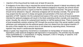
2. ANATOMY:
•In the ox, the spinal cord ends in the region of the last lumbar vertebra, but the
meningeal
•sac goes to the 3rd/4th sacral segments.
•INJECTION SITE:
•• For caudal and epidural anesthesia the injection site used is between
coccygeal C1 and
•C2 (located by raising tail in “pump handle” fashion, the first obvious
articulation behind
•the sacrum being C1 /C2).
3. ANESTHESIA:
• For a 500 kg bovine; 5-10 ml 2% lidocaine will give caudal
anesthesia without causinghind limb ataxia or paralysis.
• Onset of paralysis of the tail should occur in 1-2 mins.
The block will last 1-2 hours.
Larger doses will produce increasingly anterior effects.
4. • METHOD:
By the time 100-150 mls 2% lidocaine is injected, the block will be
sufficiently anterior to allow surgery of the hindlimbs, mammary
tissue, flanks and abdominal wall.
• However, the bovine will be recumbent. Injection of local
anesthetics can be carried out at the lumbosacral junction in order
to produce an anterior block with less anesthetic.
• However, there is a danger of accidental subarachnoid injection.
Segmental epidural anesthesia, where the anesthetic is injected into
the epidural space at the region required can be used for analgesia
of any ‘segment’ with less overall side effects. It is more difficult to
perform; penetration of the meninges is likely, but in skilled hands it
is a very useful technique.
8. • INDICATIONS:
Hind limb ataxia is a serious problem, so only caudal epidural
techniques are used. These are useful for various obstetrical
manipulations and surgery on the rectum, vagina and tail.
• ANATOMY:
The technique is less reliable than in cattle. Site of injection is
usually sacrococcygeal junction but can be between C1 and
C2.
• ANESTHESIA:
For 500 kg horse, a mixture of 50 mg of xylazine and 6 ml of
2% mepivacaine (may be repeated for another dose) may
prove very effective
9. • Three classes of drugs can be used for caudal epidurals:
• 1. Local anesthetics block not only sensory fibers but also motor fibers and
sympathetic fibers. Loss of motor control can cause ataxia and even collapse of the
hindlimbs; this is a rare occurrence but can be disastrous if it does occur.
• 2. Alpha-2 agonists selectively block sensory fibers, with minimal effects on hindlimb
function.
• 3. Opioids selectively block sensory fibers, with minimal effects on hind limb function.
• Caudal epidural anesthesia is easily performed on large animals. The site of
injection is between the first and second coccygeal vertebrae, on the dorsal midline
(Fig. 8-4). An estimate of this location is made by moving the tail up and down while
palpating for the first movable intercoccygeal space caudal to the sacrum. The
patient should be restrained, and personnel should stand to the side of the patient to
avoid being kicked.
10. • The area at the base of the tail and caudal sacrum are clipped and surgically prepped. The
anesthetist should wear sterile gloves. The clinician inserts an 18-gauge (ga) × image-inch
needle (large patients may require an 18-ga × image-inch spinal needle) at a 90-degree
angle to the skin and advances it into the epidural space. Anesthetic solution is injected with
a sterile syringe. Because sterile injection technique is required, the anesthetic solution
should be taken from a new, previously unopened container. Sometimes the needle is left in
place in case the patient needs more anesthetic later in the procedure. The needle should
not be withdrawn until the clinician approves because bleeding and swelling (after removing
the needle) can make repeating the epidural procedure difficult. Complications are unusual,
and aftercare involves imply cleaning the area and applying an antibiotic or antimicrobial
ointment to the puncture site.
• Epidurals are not performed more cranially than the first intercoccygeal space in awake
patients. This is because the risk of creating ataxia and collapse of the hindlimbs increases
as the site of administering the epidural moves cranially.
11.
12. • Advantages Can negate the need for general anesthesia Anesthesia: general -
overview for small scale surgical procedures.
• Labour and time saving technique.
• Minimal equipment and drugs required.
• Can be performed in the field.
• Cheap.
• Long-term catheterization may be feasible for repeated administration of morphine-
detomidine in cases of chronic pain.
• Disadvantages
• Requires degree of competence/experience for correct placement of needle in epidural
space.
• Restraint of patient may be difficult.
• The use of alpha-2 adrenoceptor agonists as sedative agents can induce regular and
profuse urination which can interfere with the surgical site.
• Occasionally severe ataxia and loss of motor control of hindlimbs can result.
• Instances of excitement have been reported necessitating induction of general anesthesia
14. ANATOMY:
• In both sheep and goats, anterior epidural anesthesia,
induced by injection at the lumbosacral junction is easily
performed and provides excellent analgesia and muscle
relaxation for abdominal surgery.
• Recumbency may occur but is not a problem in these
small animals. As in cattle, there is a risk of subarachnoid
injection.
15.
16. DOSE:
• Epidural block can be produced by injection of local anaesthetic solution into
the epidural space at the lumbosacral junction. Complete analgesia and
paralysis can be induced in the hindlimbs and abdomen to allow surgery,
depending on the volume of local anaesthetic injected . A lower dose of
lignocaine, such as 1 ml/7 kg, is sufficient for perineal or hindlimb surgical
procedures, and for caesarian section. The long duration of hindlimb
paralysis from bupivacaine block for caesarian section interferes with nursing
of the newborn, and for that reason lignocaine with adrenaline isusually
preferred.
17. • The lumbosacral junction is easy to palpate in thin animals but recognition of landmarks will
benecessary to identify the point of needle insertion in muscled or fat animals. Epidural block can be
performed with the goat or sheep standing or in lateral recumbency. An imaginary line between the
cranial borders of the ilium crosses between the spinous processes of the last lumbar vertebrae(Fig.
13.3). The caudal borders of the ilium, where the angle bends to parallel midline, are level withthe
cranial edge of the sacrum. The point of needle insertion is midline halfway between the spinousm
process of the seventh lumbar vertebra and the sacrum. If the spinous process of the last lumba
vertebra can be palpated, the next depression caudal to it is the lumbosacral space. This area must
be clipped and the skin prepared with a surgical scrub. A spinal needle should be used because it
has a stilette to prevent injection of a core of subcutaneous tissue into the epidural space. The notch
on the hub of the needle indicates the direction of the bevel. Thus the anaesthetist can ensure that
injection of local anaesthetic solution is towards the head of the animal. When epidural nerve block is
to be performed on the conscious animal, 1–3 ml 2% lignocaine should be injected subcutaneously
with a fine needle at the site intended for insertion of the spinal needle. For lambs, kids, and pygmy
goats, a 22 gauge 3.7 cm spinal needle can be used. For adult animals a sturdier needle, such as an
18 gauge 6.25cm spinal needle, is recommended.
18.
19.
20. • Considerable pressure may be needed to introduce the needle through the skin and supraspinous
ligament and it may be preferable to puncture the skin first with a larger, sharp hypodermic needle.
Once introduced, the spinal needle should be advanced gently for two reasons. First, to be able to
appreciate the resistance then penetration of the interarcuate ligament which lies over the epidural
space, described as a ‘pop’, and secondly, to control introduction of the tip of the needle into the
epidural space so that movement of the needle can be stopped immediately.
• If the tip of the needle strikes bone and the needle does not appear to be deep enough to be in the
epidural space, the needle should be withdrawn until the tip is just under the skin and redirected in a
cranial direction.
• After correct placement of the needle, the stilette should be removed and placed on a sterile surface.
A 3 ml syringe containing 0.5 ml air should be attached to the spinal needle and the plunger
withdrawn to test for aspiration of cerebrospinal fluid (CSF) or blood. After correct placement of the
needle, the 3 ml syringe should be detached and the syringe containing the local anaesthetic
solution attached
21. • .Injection of the drug should be made over at least 30 seconds.
• If analgesia of one side or leg is required the animal should be placed in lateral recumbency with
the side to be desensitized underneath. When bilateral analgesia is required, the animal should
be positioned either prone or supine so that the vertebral canal is horizontal. The goat or sheep
should not be allowed to ‘dog-sit’, otherwise analgesia will not develop cranially. The spinal cord
may project into the sacrum in sheep and goats and penetration of the dura will result in
aspiration of CSF. Injection into the subarachnoid space of the same volume of local anaesthetic
intended for epidural analgesia will result in the block extending further cranially and respiratory
arrest. Usually, the volume for subarachnoid injection is half the epidural dose. There is some risk
of local anaesthetic solution entering CSF through the puncture hole if the spinal needle is partly
withdrawn and redirected into the epidural space a few mm distant from the original insertion. If
the entry of the needle into a venous sinus is not detected by aspirating blood, intravenous
injection may result in cardiovascular depression.
Sheep or goats may respond by movement or vocalization to manipulation of viscera during
laparotomy under epidural analgesia with lignocaine or bupivacaine. The animals may be made
more comfortable by i.v. butorphanol, 0.1mg/kg, diazepam, 0.05–0.10mg/kg, or xylazine, 0.02
mg/kg. Disadvantages of adjunct drug