Downloaded 354 times

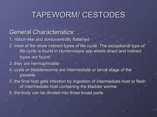
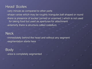

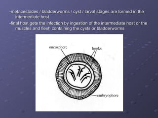
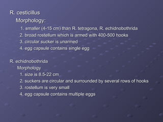
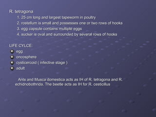
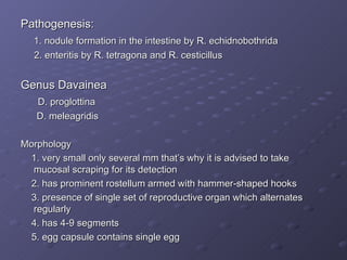
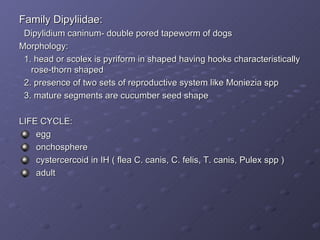
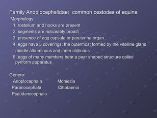
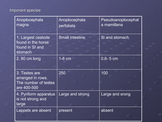
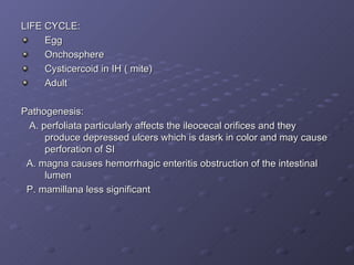
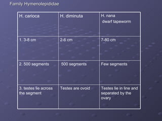
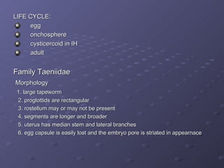

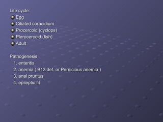

1. Tapeworms are ribbon-like parasites with three main body parts: head, neck, and segmented body. They have indirect lifecycles involving intermediate hosts. 2. Important tapeworm genera in poultry include Raillietina, Davainea, and Dipylidium. Raillietina species use ants as intermediate hosts. Davainea gravid segments actively exit the host. 3. Important equine tapeworms are in the Anoplocephalidae family. Anoplocephala perfoliata can cause intestinal ulcers and hemorrhage.


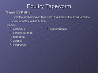
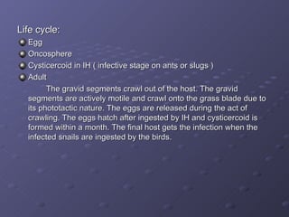
![[Micro] cestodes](https://cdn.slidesharecdn.com/ss_thumbnails/nvpl1fbyq2ofjfsbmped-signature-2127a2ca5368c7fdfd023e8d90dde3fc0b9fe7d91346a4189562c9f63dc0d19d-poli-150819190753-lva1-app6892-thumbnail.jpg?width=640&height=640&fit=bounds)


![5G Explained! A High Level Overview [Introduction]](https://cdn.slidesharecdn.com/ss_thumbnails/5gexplainedahighleveloverview-260119165306-cc137a3e-thumbnail.jpg?width=640&height=640&fit=bounds)


