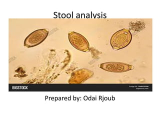
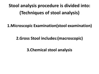
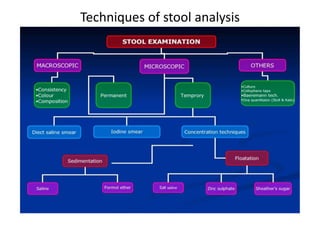
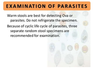
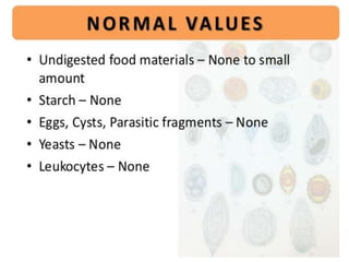
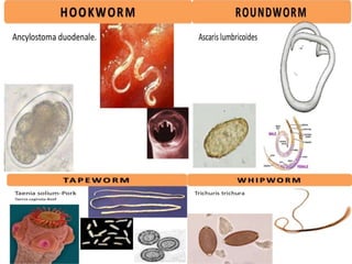
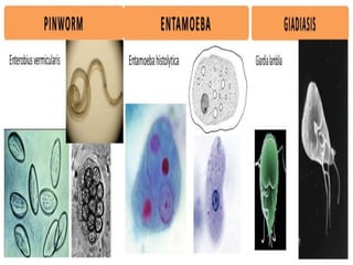
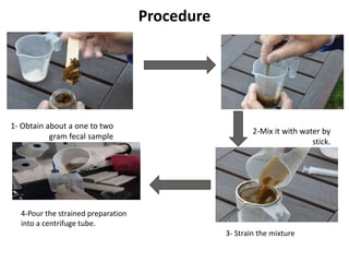
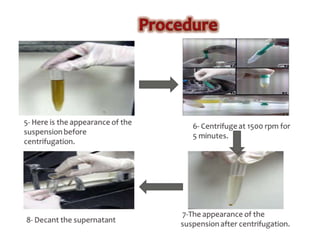
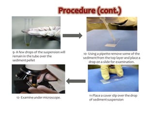
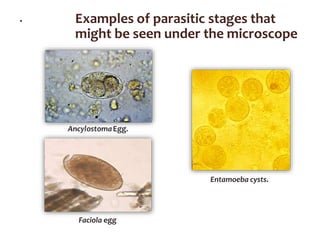
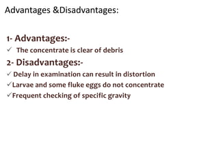
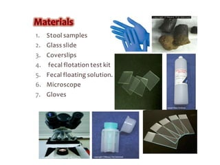
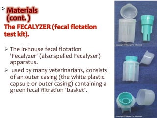
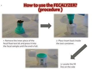
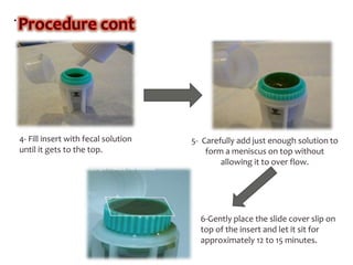
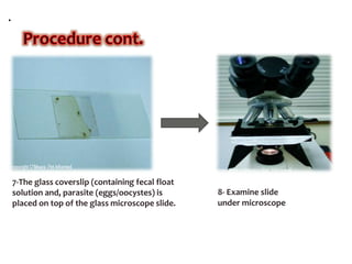
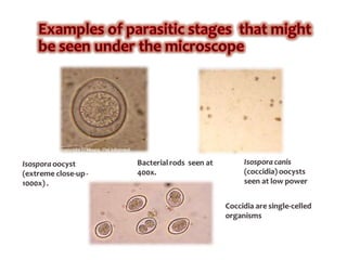
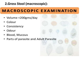
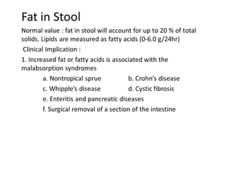
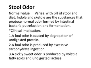
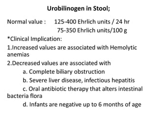
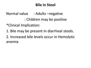
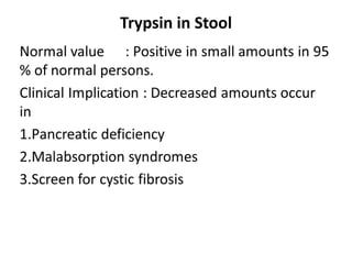
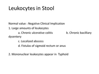
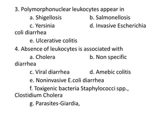
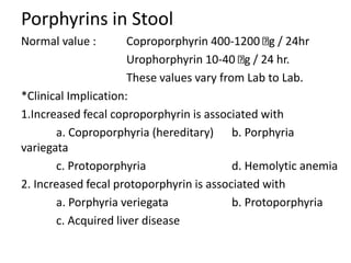
Faculty of Science and Technology
Hebron university
Department of Biology
Parasitology
Show: Stool analysis
Odai Rjoub
(STUDENT)
Stool analysis
Prepared by: Odai Rjoub
Stool analysis: is a series of tests done on
a stool (feces) sample for differential diagnosis
of certain diseases of digestive system and
include infection (such as from parasites,
viruses, or bacteria), poor nutrient absorption,
or cancer.
Clinical significance of stool analysis:
1-Diagnosis of digestive system infectious diseases:
Bacteria, parasites, virus, and fungi.
2-Diagnosis of pancreas disorders (inflammation); which
associated with malabsorption of nutrients.
3-Primary screening test for some types of digestive
system malignancy such as: Colon cancer.
4-Primary screening test for peptic ulcer disease, and
some types of anemia.
For whom Stool analysis is urgently required?
1-Patients with abdominal pain.
2-Patients with diarrhea.
3-Patients with anemia.
Other situations by which Stool analysis is non-
urgently required:
1-Patients who is too thin or do not grow well.
2-Patient with stool color that is changed to
abnormal color.
How to Take a Stool Sample:
*Preparing to Take the Sample:
1.Avoid medicine that will affect the sample
2. Consult with your doctor
3.Prepare your toilet with a hat
4.Cover your toilet bowl with plastic wrap
5.Lay a sheet of newspaper across your toilet
bowl.
6. Defecate into the collection device:
*Handling the Sample:
1. Deposit sample in the container
2.Dispose of your collection device
3.Refrigerate the sample
4.Return the samples to your doctor as soon as
possible.
Stool analysis procedure is divided into:
(Techniques of stool analysis)
1.Microscopic Examination(stool examination)
2.Gross Stool includes:(macroscopic)
3.Chemical stool analysis
Techniques of stool analysis
1.Microscopic Examination(stool examination)
Explain :
1.Presence of Red Blood Cells.
2.Ova and parasites.
3.Presence of meat fibers and muscle fibers.
4.Presence of fat.
5. Presence of leukocytes
Concentration methods of fecal parasites
There are two types of fecal Concentration
techniques:-
1.Sedimentation techniques.
2.Flotation techniques.
1- Sedimentation techniques:
Fecal sedimentation is used to detect large or
heavy ova such as many spirurid ova, many fluke
eggs, and many tapeworm eggs that will not
float in fecal flotation techniques.
The sediment contains a lot of debris so
examining these slides is quite tedious.
Note:- you can use formalin as a preservative
and ether or ethyl acetate as an extractor of fat
and debris from faeces after filtration to leave
the parasites in a sediment at the bottom of the
tube after centrifugation.
1- Advantages
1.Recovers most ova, cysts and larvae .
2. Morphology of most parasites is retained.
3. Less risk of infection from bacteria and
viruses.
2- Disadvantages
1.Preparation contains debris.
2. Some parasites do not concentrate well.
3.Ether is flammable and formalin is an irritant .
4.Liquid faeces do not concentrate well .
Materials:
1.Stool samples 2.Glassslides
3.Coverslips 4.Pipettes
5.Stick Gloves 6.Microscope
7.Plastic container (with small amount of
water).
8.Centrifuge. 9.Centrifuge tube
Procedure
4-Pour the strained preparation
into a centrifuge tube.
3- Strain the mixture
1- Obtain about a one to two
gram fecal sample
2-Mix it with water by
stick.
.
There are many variations on fecal
sedimentation technique, many of which
include a step using organic solvents (instead
of water) to remove some of the material
from the sediment.
at step (11) :-
 A drop of stain of your choice could be
mixed in before placing the cover slip.
 New Methylene blue or iodine solution
enhances visualization of the parasite
ova and oocytes.
 If there is a lot of debris, water may be
added first.
. Examples of parasitic stages that
might be seen under the microscope
Entamoeba cysts.
Faciola egg
2- Flotation techniques:
* A fecal flotation test uses the high specific
gravity of a solution to float the lighter ova and
cysts
*It is a diagnostic test commonly performed in-
house in most veterinary clinics as a way of
diagnosing parasitism in animals.
Advantages &Disadvantages:
1- Advantages:-
 The concentrate is clear of debris
2- Disadvantages:-
 Delay in examination can result in distortion
Larvae and some fluke eggs do not concentrate
Frequent checking of specific gravity
>
 The in-house fecal flotation
'Fecalyzer' (also spelled Fecalyser)
apparatus.
 used by many veterinarians, consists
of an outer casing (the white plastic
capsule or outer casing) containing a
green fecal filtration 'basket'.
.
1- Remove the inner piece of the
fecal float test kit and press it into
the fecal sample until the end is full.
2- Place insert back inside
the test container.
3- Locate the fill
line on the side
.
4- Fill insert with fecal solution
until it gets to the top.
5- Carefully add just enough solution to
form a meniscus on top without
allowing it to over flow.
6-Gently place the slide cover slip on
top of the insert and let it sit for
approximately 12 to 15 minutes.
.
7-The glass coverslip (containing fecal float
solution and, parasite (eggs/oocystes) is
placed on top of the glass microscope slide.
8- Examine slide
under microscope
The important points to be considered when
performing a concentration technique are:
2.Gross Stool (macroscopic):
This test help in discovered from many
things :
3.Chemical stool analysis;
Urobilinogen in Stool;
Bile in Stool
Trypsin in Stool
Porphyrins in Stool
Normal value : Coproporphyrin 400-1200 g / 24hr
Urophorphyrin 10-40 g / 24 hr.
These values vary from Lab to Lab.
*Clinical Implication:
1.Increased fecal coproporphyrin is associated with
a. Coproporphyria (hereditary) b. Porphyria
variegata
c. Protoporphyria d. Hemolytic anemia
2. Increased fecal protoporphyrin is associated with
a. Porphyria veriegata b. Protoporphyria
c. Acquired liver disease
Various factors can limit the accuracy
and significance of a faecal egg count.
(a) There is a fairly regular fluctuation in faecal egg
output.
(b) Eggs are not evenly distributed throughout the faeces.
(c) The quantity of faeces passed will affect the number
of eggs per unit weight.
(d) The egg output is influenced by the season of the year
(large infections may be acquired during rainy seasons).
(e) The resistance of the host can depress or entirely
inhibit the egg production of parasites.
(f) Immature worms do not indicate their presence
by producing eggs.
(g) Immunity may result in a marked extension of
the prepatent period and a lower egg output by
female parasites.
(h) An egg count often refers to the total number of
eggs of a mixture of species, which differ widely
both in their biotic potential and their
pathogenicity.
(i) Eggs may not be detected due to low numbers of
them or to a low test sensitivity.

stool analysis

  • 1.
    Faculty of Scienceand Technology Hebron university Department of Biology Parasitology Show: Stool analysis Odai Rjoub (STUDENT)
  • 2.
  • 3.
    Stool analysis: isa series of tests done on a stool (feces) sample for differential diagnosis of certain diseases of digestive system and include infection (such as from parasites, viruses, or bacteria), poor nutrient absorption, or cancer. Clinical significance of stool analysis: 1-Diagnosis of digestive system infectious diseases: Bacteria, parasites, virus, and fungi. 2-Diagnosis of pancreas disorders (inflammation); which associated with malabsorption of nutrients.
  • 4.
    3-Primary screening testfor some types of digestive system malignancy such as: Colon cancer. 4-Primary screening test for peptic ulcer disease, and some types of anemia. For whom Stool analysis is urgently required? 1-Patients with abdominal pain. 2-Patients with diarrhea. 3-Patients with anemia.
  • 5.
    Other situations bywhich Stool analysis is non- urgently required: 1-Patients who is too thin or do not grow well. 2-Patient with stool color that is changed to abnormal color. How to Take a Stool Sample: *Preparing to Take the Sample: 1.Avoid medicine that will affect the sample
  • 6.
    2. Consult withyour doctor 3.Prepare your toilet with a hat 4.Cover your toilet bowl with plastic wrap
  • 7.
    5.Lay a sheetof newspaper across your toilet bowl. 6. Defecate into the collection device:
  • 8.
    *Handling the Sample: 1.Deposit sample in the container 2.Dispose of your collection device
  • 9.
    3.Refrigerate the sample 4.Returnthe samples to your doctor as soon as possible.
  • 10.
    Stool analysis procedureis divided into: (Techniques of stool analysis) 1.Microscopic Examination(stool examination) 2.Gross Stool includes:(macroscopic) 3.Chemical stool analysis
  • 11.
  • 12.
    1.Microscopic Examination(stool examination) Explain: 1.Presence of Red Blood Cells. 2.Ova and parasites. 3.Presence of meat fibers and muscle fibers. 4.Presence of fat. 5. Presence of leukocytes
  • 20.
    Concentration methods offecal parasites There are two types of fecal Concentration techniques:- 1.Sedimentation techniques. 2.Flotation techniques.
  • 21.
    1- Sedimentation techniques: Fecalsedimentation is used to detect large or heavy ova such as many spirurid ova, many fluke eggs, and many tapeworm eggs that will not float in fecal flotation techniques. The sediment contains a lot of debris so examining these slides is quite tedious. Note:- you can use formalin as a preservative and ether or ethyl acetate as an extractor of fat and debris from faeces after filtration to leave the parasites in a sediment at the bottom of the tube after centrifugation.
  • 22.
    1- Advantages 1.Recovers mostova, cysts and larvae . 2. Morphology of most parasites is retained. 3. Less risk of infection from bacteria and viruses. 2- Disadvantages 1.Preparation contains debris. 2. Some parasites do not concentrate well. 3.Ether is flammable and formalin is an irritant . 4.Liquid faeces do not concentrate well .
  • 23.
    Materials: 1.Stool samples 2.Glassslides 3.Coverslips4.Pipettes 5.Stick Gloves 6.Microscope 7.Plastic container (with small amount of water). 8.Centrifuge. 9.Centrifuge tube
  • 24.
    Procedure 4-Pour the strainedpreparation into a centrifuge tube. 3- Strain the mixture 1- Obtain about a one to two gram fecal sample 2-Mix it with water by stick.
  • 27.
    . There are manyvariations on fecal sedimentation technique, many of which include a step using organic solvents (instead of water) to remove some of the material from the sediment. at step (11) :-  A drop of stain of your choice could be mixed in before placing the cover slip.  New Methylene blue or iodine solution enhances visualization of the parasite ova and oocytes.  If there is a lot of debris, water may be added first.
  • 28.
    . Examples ofparasitic stages that might be seen under the microscope Entamoeba cysts. Faciola egg
  • 29.
    2- Flotation techniques: *A fecal flotation test uses the high specific gravity of a solution to float the lighter ova and cysts *It is a diagnostic test commonly performed in- house in most veterinary clinics as a way of diagnosing parasitism in animals.
  • 30.
    Advantages &Disadvantages: 1- Advantages:- The concentrate is clear of debris 2- Disadvantages:-  Delay in examination can result in distortion Larvae and some fluke eggs do not concentrate Frequent checking of specific gravity
  • 32.
    >  The in-housefecal flotation 'Fecalyzer' (also spelled Fecalyser) apparatus.  used by many veterinarians, consists of an outer casing (the white plastic capsule or outer casing) containing a green fecal filtration 'basket'.
  • 33.
    . 1- Remove theinner piece of the fecal float test kit and press it into the fecal sample until the end is full. 2- Place insert back inside the test container. 3- Locate the fill line on the side
  • 34.
    . 4- Fill insertwith fecal solution until it gets to the top. 5- Carefully add just enough solution to form a meniscus on top without allowing it to over flow. 6-Gently place the slide cover slip on top of the insert and let it sit for approximately 12 to 15 minutes.
  • 35.
    . 7-The glass coverslip(containing fecal float solution and, parasite (eggs/oocystes) is placed on top of the glass microscope slide. 8- Examine slide under microscope
  • 37.
    The important pointsto be considered when performing a concentration technique are:
  • 38.
  • 47.
    This test helpin discovered from many things :
  • 48.
  • 51.
  • 52.
  • 53.
  • 56.
    Porphyrins in Stool Normalvalue : Coproporphyrin 400-1200 g / 24hr Urophorphyrin 10-40 g / 24 hr. These values vary from Lab to Lab. *Clinical Implication: 1.Increased fecal coproporphyrin is associated with a. Coproporphyria (hereditary) b. Porphyria variegata c. Protoporphyria d. Hemolytic anemia 2. Increased fecal protoporphyrin is associated with a. Porphyria veriegata b. Protoporphyria c. Acquired liver disease
  • 59.
    Various factors canlimit the accuracy and significance of a faecal egg count. (a) There is a fairly regular fluctuation in faecal egg output. (b) Eggs are not evenly distributed throughout the faeces. (c) The quantity of faeces passed will affect the number of eggs per unit weight. (d) The egg output is influenced by the season of the year (large infections may be acquired during rainy seasons). (e) The resistance of the host can depress or entirely inhibit the egg production of parasites.
  • 60.
    (f) Immature wormsdo not indicate their presence by producing eggs. (g) Immunity may result in a marked extension of the prepatent period and a lower egg output by female parasites. (h) An egg count often refers to the total number of eggs of a mixture of species, which differ widely both in their biotic potential and their pathogenicity. (i) Eggs may not be detected due to low numbers of them or to a low test sensitivity.