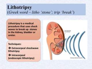
2. Introduction
Urinary calculi (also known as uroliths or stones) are common in small animal
patients. Although calculi vary in composition, most cause the same clinical signs
like dysuria, stranguria, pollakiuria, hematuria and incontinence; however,
calculi can exist without causing clinical signs.
The urinary tract is designed to remove wastes from the body in liquid form.
Calculi are formed when certain waste products precipitate and form crystals.
As microscopic crystals clump together, they can become macroscopic calculi.
Calculi can be described by their location, shape, size and mineral makeup.
Knowing the mineral composition of calculi is important because treatment and
prevention vary among types of calculi. More than 80% of calculi in dogs and
cats are either calcium oxalate or struvite.
3. Calcium Oxalate Urolithiasis
• Calcium oxalate calculi are often white and hard
with a jagged surface.
• No medical therapy or dietary change can
dissolve calcium oxalate calculi; therefore,
physical removal is the only option for
eliminating this type of calculus. Once calcium
oxalate calculi are removed, options are
available for prevention.
Struvite Urolithiasis
• Magnesium ammonium phosphate hexahydrate
calculi are commonly known as struvite calculi.
In dogs, most struvite calculi are caused by
urinary tract infection (i.e., infection induced).
In contrast, 95% of struvite calculi in cats are
present without infection (i.e., sterile struvite).
• These calculi can be eradicated by physical
removal or by medical or dietary dissolution.
4. Diagnosis
Imaging Techniques
Imaging techniques are used to determine the size, shape, number,
and location of calculi. Most calculi are radiodense - visible on
survey radiography.
Ultrasonography can provide information about kidney
architecture and the presence, location and number of calculi.
Urinalysis
Crystalluria can suggest crystalline oversaturation.
Urine specific gravity and urine pH can help assess the chemical
environment in the urinary bladder.
Urine culture and sensitivity testing is indicated because urinary
tract infections are common in patients with urolithiasis .
5. CONTD.,
Blood Chemistry
Blood chemistry results suggest the presence of underlying
diseases such as Cushing disease (hyperadrenocorticism) and
hypercalcemia, that can predispose patients to calculi.
Renal values and electrolytes are monitored because calculi
occasionally cause obstruction leading to renal or urinary tract
damage.
Hepatic function is evaluated to detect urate calculi, which are
caused by liver dysfunction.
Calculus Analysis
Determining the composition of calculus is essential for preventing
recurrence.
All removed or voided calculi should undergo chemical analysis to
determine the mineral composition and develop a successful
treatment and prevention plan.
6. Treatment
Dietary and Medical Dissolution:
• Reduce calculogenic substances in the urine by intake of specifically formulated
diet.
• Increase the solubility of calculogenic components in the urine by
administration of medications or diets that change the urine pH.
• Increase the volume of urine by adding water to the diet.
Surgical and Non-surgical Removal:
• Obstruction of urine outflow, an increase in the size or number of calculi,
persistent clinical signs and a lack of response to therapy are indications for
calculus removal.
• While traditional cystotomy is the most common method of calculus removal in
veterinary, calculi can be removed using less invasive options, including
catheter-assisted retrieval, urohydropropulsion and cystoscopic removal.
• Cystoscopic techniques are more efficient than surgical procedures, producing
magnified images, decreasing the risk of trauma and abdominal contamination.
7. Lithotripsy
(Greek word – litho ‘stone’; trip ‘break’)
Lithotripsy is a medical
procedure that uses shock
waves to break up stones
in the kidney, bladder or
ureter.
Techniques:
Extracorpeal shockwave
lithotripsy
Intracorpeal
(endoscopic lithotripsy)
8. Extracorpeal Shockwave
Lithotripsy (ESWL)
In 1985, Extracorpueral Shock Wave Lithotripsy (ESWL) technique
came into use.
It is the least invasive surgical stone treatment. A high frequency shock
wave is produced outside the patient from an external source, led
through the body and focused on the stone to break up stones into
smaller pieces and then allows them pass through the urinary tract.
It is not performed in pregnant animals, bleeding disorder or urinary
infection.
9.
10.
11. There are several disadvantages with Extracorpeal
shockwave lithotripsy (ESWL):
• ESWL involves some risks if the shock wave is not focused
exactly on the stone, damage will occur to the surrounding tissue.
• There is also a risk of developing high blood pressure, due to
disruptions in the blood circulation in the kidney.
12. Intracorpeal Lithotripsy (Endoscopic
Lithotripsy)
Endoscopic lithotripsy refers to the visualization of a calculus in the urinary
tract and the simultaneous application of energy to fragment the stones.
Many calculi in the upper urinary tract are treated with endoscopic
lithotripsy.
Ureteroscope is the most common means of visualizing an upper urinary
tract calculus.
14. Laser Lithotripsy
Laser lithotripsy is the fragmentation of stones using a laser. It was invented in
the 1980s to remove impacted urinary stones. Initially, flashlamp-pumped dye
lasers were used, then holmium lasers were studied in the 1990s.
Optical fibers carry light pulses that pulverize the stone. The energy needed to
destroy a stone is about 100 times less than the energy needed for stone
destruction with ESWL. With laser lithotripsy it only takes a few minutes to
destroy a stone.
The infrared Holmium:YAG laser is currently the clinical gold standard for
lithotripsy during ureteroscopy. Pulsed lasers are used because a sudden
temperature rise at the stone surface creates a shock wave which fragments
the stone into smaller pieces and also avoids heating of the surrounding tissue.
15.
16. Stone fragmentation by Laser Lithotripsy
The procedure is done under either local or general anesthesia. In transurethral
lithotripsy, a cystoscope is inserted into the urethra and passed into the urinary
bladder.
An optical fiber is inserted through the working channel of the scope and the
fiber emits light at an infrared wavelength to fragment the calculi. The stone is
fragmented and the remaining pieces are washed out of the urinary tract.
One way to be sure that the fibre is located at the correct position is to take a
spectrum at this location. The spectrum can either be a fluorescence spectrum
or an opto-acoustic spectrum (a spectrum of the shock waves emitted when laser
energy hits a tissue).
The resulting particles are removed transurethrally. If calculi are small enough,
they can be removed using baskets and graspers. For larger calculi, cystic
lithotripsy is indicated.
18. In this laser lithotripsy, a laser fiber has been passed through a
cystoscope into the bladder to fragment calculi (arrow).
19. CONTD.,
This procedure is preferred for use in female dogs and cats because it is less
invasive than other diagnostic and treatment methods.
A flexible urethroscope is necessary for transurethral cystoscopy in male dogs.
Calculi can be identified, but removal is limited because of the male urethral
anatomy.
In male dogs and cats, calculi can be removed by mini-cystotomy, a minimally
invasive procedure in which a cystoscope is passed into the bladder through a
small body wall incision and a smaller bladder incision.
Magnification allows complete removal of calculi. Bladder wall biopsies may
be obtained for histopathology, culture and sensitivity testing.
20. In this mini-cystotomy, the cystoscope has been inserted into the
bladder through a small abdominal incision.
21. A three-prong grasper has been passed through the cystoscope to
remove calculi from the bladder.
22.
23.
24. What Happens Upon Stone
Fragmentation?
This rapid expansion prohibits heat conduction from inside the plasma into the
surrounding tissue. Therefore, only that tissue which lies in the path of the
shock wave is affected thermally and is fully evaporated.
Upon plasma production, the temperature rises rapidly. The plasma shields the
surface from laser light by absorbing it. The interface between plasma and
adjacent medium represents a discontinuity surface in terms of density,
pressure and temperature. This discontinuity surface is called a shock wave and
is identical to the plasma boundary during the expansion phase.
When the laser energy hits the surface of the calculus, which is immersed in
liquid in the body, laser-induced breakdown (LIB) occurs and a plasma is
formed. The plasma expands and emits a shock wave which propagates into the
stone. This conversion of light energy into mechanical stress gradually destroys
the stone.
25. CONTD.,
The calculus is struck by the shock waves travelling through the liquid
medium. Mechanical stress occurs at the interface, and the calculus is
destroyed.
After laser emission, plasma motion slows down and the shock wave
propagates at supersonic speed. The cooling plasma left behind causes
cavitation to occur in liquids. The cavity is compressed so much by the
hydrostatic pressure of the surrounding liquid that the internal pressure
ultimately overcomes the external pressure. As a result of the two opposing
forces the cavity begins to oscillate, and after passing through the minimum
volume another shock wave of considerable amplitude is emitted. Due to
emission of shock waves and associated energy losses the oscillation of the
bubble rapidly attenuates.
26. Looking at it quantum mechanically, the laser beam constitutes a photon flux. A free
electron collides with an ion or an atom and absorbs photons in doing so. The electron gains
energy and is accelerated. When their energy is great enough, additional electrons are
stripped off when the electrons collide with atoms. The new electrons are also accelerated
and the number of free electrons increases in an avalanche fashion.
To trigger an electron avalanche, free electrons must be present, or else the first electrons
have to be produced by photoionization. However, the photon energy in a holmium laser is
1.2 eV, far below the necessary ionization energy. So several photons are needed
simultaneously, i.e. multiphoton ionization. This process is chiefly dependent on the
magnitude of the photon flux.
27. Laser Fibers
The fiber is the fragile laser power delivery tool and there are a great number
of demands, placed on the fiber in laser lithotripsy.
The fiber must be flexible and have a higher threshold for laser power
damage than calculi which is proportional to the fiber cross-sectional area.
Thus a fiber with a larger diameter can transmit more energy but will be less
flexible.
Other important factors that affect the damage threshold are focal length of
the coupling lens, fiber type and the nature of the fiber surface preparation.
30. Contact Laser Lithotripsy
The common step for both fragmentation and dusting strategies is contact
laser lithotripsy, where the stone is treated with the fiber touching the stone
surface.
The advent of multi-cavity high power holmium systems brought the ability
to achieve low Pulse Energy (<0.5 J) and high Fr’s (>20 Hz) settings (HiFr-
LoPE). This lead to the development of a “Dusting” technique to break stones
into fine (i.e., submillimeter) fragments.
Different combinations of Pulse Energy (PE), Frequency (Fr) and Pulse
Width (PW) during lithotripsy permit different effects on stone
fragmentation.
31. Holmium laser settings that are adjusted during laser lithotripsy
(from top to bottom): pulse energy (PE), pulse frequency (Fr), and pulse
width (PW).
Pulse Energy:
Holmium PE settings can range from 0.2 to 6.0 J
depending on the power of the system. Traditionally, PE
settings have been used at ranges between 0.6 and 1.2 J
to fragment stones.
Frequency:
Frequency is defined as the number of pulses emitted
from the laser fiber per second. The range of pulse
Frequencies available to the user depends on the power
of the holmium system. Initial 15- to 20-W systems were
limited to maximum frequencies of 15–20 Hz. Currently,
holmium systems are able to achieve frequencies as high
as 80 Hz.
Pulse Width:
Pulse width represents the time during which a single
pulse is emitted from the laser, measured in
microseconds. First-generation holmium systems
operated in a single PW mode of approximately 350 μs.
Recent systems have allowed the user to choose either
short or long PW modes (range 500–1,500 μs).
32. Non-contact Laser Lithotripsy
In a dusting technique, after the stone is debulked resulting in numerous
fragments, the next step is often non-contact laser lithotripsy. In this, stone
fragments are pulverized in a calyx with the laser fiber activated in bursts,
away from the stone fragments resulting in a whirlpool-like effect that
causes stones to collide and fragment further.
First described by Chawla et al., it is also commonly known as the
“popcorn” effect, and settings for this have traditionally employed
moderate-to-high Pulse Energy (1.0–1.5 J) and Fr (15–20 Hz).
With the 120-W system, using a high Fr (50–80 Hz) popcorn technique
utilizing a PE of 0.5 J is called “pop-dusting.” This results in fine fragments
without compromising fiber tip burnback.
33. Surgical schema for treating upper urinary tract stones with dusting
technique during ureteroscopic laser lithotripsy (HU: Hounsfield Unit;
UAS: Ureteral Access Sheath).
34.
35. Clinical Factors That Affect Laser
Lithotripsy Settings
Stone location:
Retropulsion is of greater concern when treating stones within the ureter. A
fragmentation technique for a mobile ureteral stone and dusting technique
for impacted stone might be a more efficient strategy.
Stone size:
Stones that are large are much easier to treat with a dusting technique using
painting and chipping methods during lithotripsy.
Stone composition:
With hard stones, higher pulse energy is needed to obtain smaller fragments
that can lead to fiber burnback and reduce lithotripsy efficiency.
Stone density:
Assessing the stone density (Hounsfield Unit) on computed tomography (CT)
may inform whether dusting is feasible.
36. Risks and Complications
Risks of general anesthesia include shortness of breath, nausea, vomiting,
urinary retention etc. More serious risks include heart attack, stroke and
pneumonia.
The most significant complication is the injury of the urothelium adjacent to
the stone. As the depth of tissue penetration of the Ho:YAG laser is 0.4 mm,
most injuries can be managed conservatively.
An absolute contraindication is the presence of untreated urinary tract
infection. In case of accidental laser fibre breakage, the detection of the
radiolucent fibre remainders may become troublesome.
Other complications include residual stones within the kidney or ureter, lost
stone, ureteral perforation, exravasation, avulsion, development of
subcapsular renal hematoma (SRH) and intra-renal arteriovenous bleeding
fistula .
37. Safety Implications
Eye protection is required for surgeons, although the operator’s cornea
would be damaged only if it was positioned at less than 10 cm from the laser
fiber. To avoid damaging the fibre, the laser light must be orthogonal to stone
surface.
One has to be careful regarding how much total power is applied in a ureter,
with careful attention paid to keep lithotripsy targeting the center of the
stone, not peripheral.
The clinical usage of the protective Flex-Guard sheath reduces the amount of
force required to insert the laser fibre through the working channel, thus
protective against mechanical damage caused to the laser fiber.
A novel endoscope protection system (EPS) uses an optical feedback from the
sensor of a digital flexible uteroscope to terminate laser energy on retraction
of the fiber.
38. Conclusions
Veterinary patients often have clinical signs associated with the lower
urinary tract. The possible presence of urinary calculi should be investigated
by non-invasive dissolution methods, minimally invasive procedures and
surgical procedures.
Laser lithotripsy is a procedure using scopes to reach the stone and a
powerful laser to break it up. It is a very safe and effective way to get rid of
painful stones.
An understanding of holmium laser settings will permit the surgeon to utilize
various techniques for uteroscopic lithotripsy.
Like any procedure, ureteroscopy with laser lithotripsy has risks. Though
these risks are rare, knowing about them may help find them and treat them
early.
39. References
• Chan KF, Pfefer TJ, Teichman JMH, Welch AJ. A perspective on laser lithotripsy:
the fragmentation process. J Endourol (2001); 15:257-273.
• Chawla SN, Chang MF, Chang A, Lenoir J, Bagley DH. Effectiveness of high-
frequency holmium:YAG laser stone fragmentation: the “popcorn effect”. J
Endourol (2008); 22:645-650.
• Jiang H, Wu Z, Ding Q, Zhang Y. Ureteroscopic treatment of ureteral calculi
with holmium: YAG laser lithotripsy. J Endourol (2007); 21:151-154.
• Kronenberg P, Traxer O. Update on lasers in urology 2014: current assessment on
holmium:yttrium-aluminum-garnet (Ho:YAG) laser lithotripter settings and laser
fibers. World J Urol (2015); 33(4):463-469.
• Lam JS, Greene TD, Gupta M. Treatment of proximal ureteral calculi:
holmium:YAG laser ureterolithotripsy versus extracorporeal shock wave
lithotripsy. J Urol (2002);16:1972–1976.
• Papatsoris AG, Skolarikos A, Buchholz N. Intracorporeal laser lithotripsy. Arab J
Urol (2012); in press.
• Teichman JMH, Schwesinger WH, Lackner J, Cossman RM. Holmium:YAG
lithotripsy for gallstones. Surg Endosc (2001); 15:1034-1037.
• https://en.wikipedia.org/wiki/Laser_lithotripsy
• http://www.endourology.org.za/endourology-library/radiological-
pictures/stones/laser-lithotripsy-dusting-the-stone
Editor's Notes
A high urine specific gravity can suggest an increase in the concentration of calculi precursors. Calcium oxalate calculi form in neutral to acidic urine, whereas struvite calculi are more common in alkaline urine.
Cystoscopy produces magnified images of the urinary tract, allowing identification of abnormalities such as strictures, masses and calculi.
1. Fragments stones with shock waves generated by an underwater electrical discharge
2. Pneumatic mechanical devices, such as the lithoclast ,are small endoscopic jackhammers. It is particularly useful for managing large and hard stones.
3. Probe tip causes the stone to resonate at high frequency and break.
A light guide is led through a stiff or flexible urethroscope and positioned by looking through the scope.
To be sure no tissue is damaged the laser must be used at a sufficiently low energy. A higher laser energy is then used to fragment the stone.
Most scopes are too big to be used transurethrally in male cats.
A plasma is defined as a medium consisting of neutral atoms, electrons, ions and quanta emitted from atoms or ions. The plasma is formed due to an avalanche effect.
The expanding plasma acts as a piston on the surrounding environment. The plasma-filled bubble propagates with a maximum velocity of about 8000 m/s.
If the LIB occurs only at the stone surface, the destruction is effectively promoted by cavitation effects.
Looking at it quantum mechanically, the laser beam constitutes a photon flux. A free electron collides with an ion or an atom and absorbs photons in doing so. The electron gains energy and is accelerated. At a high photon flux there is a considerable acceleration of the electrons. When their energy is great enough, additional electrons are stripped off when the electrons collide with atoms. The new electrons are also accelerated and the number of free electrons increases in an avalanche fashion.
To trigger an electron avalanche, free electrons must be present, or else the first electrons have to be produced by photoionization. However, the photon energy in a holmium laser is 1.2 eV, far below the necessary ionization energy. So several photons are needed simultaneously, i.e. multiphoton ionization. This process is chiefly dependent on the magnitude of the photon flux.
In addition, the size of the laser fiber influences irrigation flow during uteroscopy, which is important if an optimal vision, is needed to see through the “snow storm” of fragments during dusting.
Holmium lasers produce a thermal effect due to its strong absorption by water that causes stone vaporization.
The amount of energy provided during lithotripsy depends on the Pulse Energy and frequency (Fr) utilized; the total power (Watt) is a product of the PE (J) × Fr (Hz).
More recently, holmium systems have incorporated the option to alter the pulse width (PW).
Pulse width:
In a laboratory study assessing the effect of PW on stone fragmentation, the time needed to fragment an artificial stone using 1.0 J and 10 Hz was similar between short pulse (SP) and long pulse (LP) modes. However, when very high PE settings of 2.0 J were used, it took more time to fragment the stone using the SP mode. No significant differences in loss of stone mass between SP and LP modes were noted in a recent study by Wollin and coworkers (20). The main difference when utilizing PW is that LP results in less stone retropulsion (21–23). Kang and colleagues found that stones were displaced 30–50% more when SP was used compared to LP at comparable total power settings (22).
A further advantage of the LP mode is its protective effect on laser fiber tip degradation, known as “burnback,” which can result in a reduction in the energy emitted from the fiber and a loss in its length. Fiber burnback increases when high PE settings are used and when using SP compared to LP mode.
Ureteroscopy with laser lithotripsy is a very safe and effective operation. Risks and complications are rare, however, they may happen.
Some risks are related to anesthesia and others are related to procedures in general.
With the use of high flow irrigation excess heat generation in the confined spaces of the ureter is limited.