Downloaded 232 times


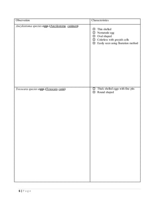
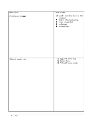
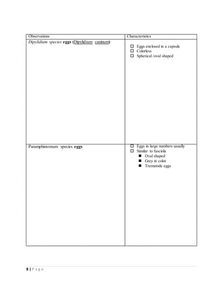
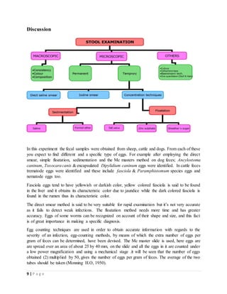

This document describes procedures for examining fecal samples to detect parasite eggs under a microscope. Several methods are discussed, including direct smear, McMaster technique, simple floatation, and sedimentation. Using these methods on samples from sheep, cattle, and dogs, several parasite eggs were observed, including Ancylostoma caninum, Toxocara canis, Fasciola eggs, Trichuris eggs, and Dipylidium caninum eggs enclosed in a capsule. The document concludes that multiple examination methods are needed to thoroughly detect parasites at different infection levels in fecal samples.


