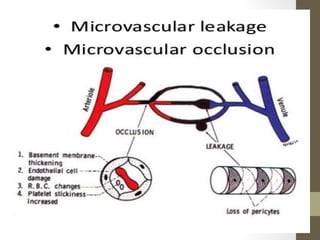
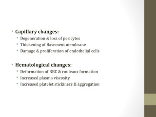
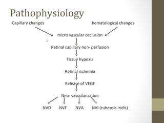
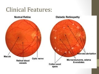
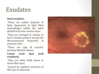
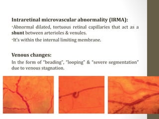
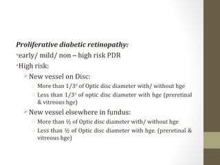
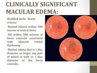
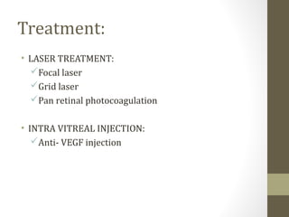
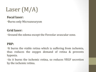
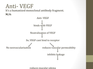
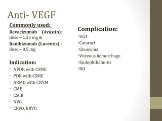
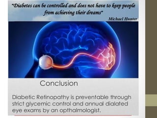
DIABETIC
RETINOPATHY
Dr Fahmida Hoque
DIABETIC RETINOPATHY
โ€ขIt is the โ€œdisease of the retinaโ€ caused by
microangiopathy due to long term effect of
diabetes leading to progressive damage of the
retina & blindness.
โ€ขMost common cause of severe bilateral visual
loss in working age group.
Risk factors
๏‚ง Age at diagnosis of diabetes
๏‚ง Duration of diabetes
๏‚ง Poor metabolic control
๏‚ง Pregnancy
๏‚ง Hypertension
๏‚ง Nephropathy
๏‚ง Others - smoking, obesity, hyperlipidemia, anemia
Pathophysiology
โ€ข DR is a microangiopathy primarily affecting the
precapillary arterioles, capillaries, venules and post
capillary venules.
โ€ข The basic component of damaging process are
microvascular occlusion & microvascular leakage.
โ€ข Capillary changes:
๏‚ง Degeneration & loss of pericytes
๏‚ง Thickening of Basement membrane
๏‚ง Damage & proliferation of endothelial cells
โ€ข Hematological changes:
๏‚ง Deformation of RBC & rouleaux formation
๏‚ง Increased plasma viscosity
๏‚ง Increased platelet stickiness & aggregation
Pathophysiology
Capillary changes hematological changes
micro vascular occlusion
Retinal capillary non- perfusion
Tissue hypoxia
Retinal ischemia
Release of VEGF
Neo- vascularization
NVD NVE NVA NVI (rubeosis iridis)
NVD, NVE
Pre โ€“ retinal hge fibro- vascular proliferation
Sub โ€“ hyaloid hge fibrous glial tissue proliferation
Vitreous hge contraction of F. P.
traction on Retina
TRD
rhegma forms
combined R. D.
NVA, NVI
NVG
Secondary open angle glaucoma
Secondary angle closure glaucoma
Optic atrophy
Absolute eye
Clinical Features:
Clinical features:
Microaneurysm:
๏‚ง Earliest sign of D. R.
๏‚ง Appear as tiny red dots.
๏‚ง It is focal saccular dilatation of capillary walls where pericytes
are absent.
๏‚ง Located in the outer plexiform layer & inner nuclear layer.
๏‚ง Usually at posterior pole, especially temporal to the fovea.
๏‚ง In FFA โ€“ present at the edge of area of capillary non- perfusion
(hyper fluorescence dots)
Retinal Hemorrhages:
Dot & blot hge:
โ€ขDue to rupture of wall of capillary or Microaneurysm, giving
rise to intra retinal hge.
โ€ขIf the hge is deep (i.e. in the inner layer of OPL) it is usually
is round or oval (dot/ blot hge)
Retinal Hemorrhages:
Flame shaped Hge:
โ€ขArise from superficial precapillary arterioles.
โ€ขthe hge is more superficial & in the nerve fiber layer, it takes
a flame/ splinter shape as they follow the architecture of the
nerve fiber layer.
Exudates
Hard exudates:
โ€ขThese are yellow deposits of
lipid, lipoprotein & lipid filled
macrophages within the outer
plexiform & inner nuclear layer.
โ€ขThey are arranged in clumps or
form circinate pattern around the
Microaneurysm frequently at
posterior pole.
โ€ขThese are sign of current/
previous Macular edema.
Cotton wool Spot (soft
exudates):
โ€ขThey are white fluffy lesion in
nerve fiber layer.
โ€ขCaused by capillary occlusion at
NFL due to infarction.
Intraretinal microvascular abnormality (IRMA):
โ€ขAbnormal dilated, tortuous retinal capillaries that act as a
shunt between arterioles & venules.
โ€ขItโ€™s within the internal limiting membrane.
Venous changes:
In the form of โ€œbeadingโ€, โ€œloopingโ€ & โ€œsevere segmentationโ€
due to venous stagnation.
Classification
ETDRS CLASSIFICATION:
NON PROLIFERATIVE DIABETIC RETINOPATHY
MILD NPDR MA, retinal hemorrhage, hard exudates
MODERATE NPDR Mild NPDR plus cotton wool spots, venous changes &
IRMA
SEVERE NPDR
4:2:1 rule
Moderate NPDR plus one of โ€“
โ€ขMA, retinal hge in all four quadrants
โ€ขVenous changes in 2/ more quadrants
โ€ขIRMA atleast in 1 quadrant
VERY SEVERE NPDR Two or more of the above features on severe NPDR
Proliferative diabetic retinopathy:
โ€ขearly/ mild/ non โ€“ high risk PDR
โ€ขHigh risk:
๏ƒ˜New vessel on Disc:
o More than 1/3rd
of Optic disc diameter with/ without hge
o Less than 1/3rd
of optic disc diameter with hge (preretinal
& vitreous hge)
๏ƒ˜New vessel elsewhere in fundus:
o More than ยฝ of Optic disc diameter with/ without hge
o Less than ยฝ of Optic disc diameter with hge. (preretinal &
vitreous hge)
CLINICALLY SIGNIFICANT
MACULAR EDEMA:
Modified Airlie- House
criteria:
โ€ขRetinal edema within 500
micron of central fovea
โ€ขHE within 500 micron of
fovea centralis associated
with adjacent retinal
thickening
โ€ขRetinal edema that is 1 disc
diameter or larger, any part
of which is with in 1 disc
diameter of the fovea
centralis.
Management
1) History from patient
2) Clinical features
3) Ocular examination
๏ถ BCVA
๏ถ Slit lamp examination
๏ƒ˜ Cornea
๏ƒ˜ Iris
๏ƒ˜ lens
๏ถ Funduscopy
Investigation:
๏‚ง Laboratory test :
๏ถ Blood sugar- FBS, RBS, 2HPPBS
๏ถ Serum lipid profile
๏ถ Medical evaluation of HbA1c
๏‚ง Ancillary test
๏ถ Color fundus photograph
๏ถ Fundus Fluorescein Angiography
๏ถ OCT
Treatment:
โ€ข LASER TREATMENT:
๏ƒผFocal laser
๏ƒผGrid laser
๏ƒผPan retinal photocoagulation
โ€ข INTRA VITREAL INJECTION:
๏ƒผAnti- VEGF injection
Laser
Indication:
โ€ขGrid + focal laser โ€“ CSME
โ€ขPRP:
โ€ข PDR --
๏ƒผHigh risk
๏ƒผEarly PDR โ€“
๏ƒ˜ In pts with poor compliance
๏ƒ˜ During pregnancy
๏ƒ˜ Pt with systemic disease
๏ƒ˜ Pending cataract surgery/ YAG laser capsulotomy
๏ƒ˜ Rubeosis iridis
๏ƒ˜ Severe/ very severe NPDR (irregular F/U)
Laser (M/A)
Focal laser:
โ€ขBurns only Microaneurysm
Grid laser:
โ€ขAround the edema except the Foveolar avascular zone.
PRP:
โ€ขIt burns the viable retina which is suffering from ischemia,
thus reduces the oxygen demand of retina & prevents
hypoxia.
โ€ขAs it burns the ischemic retina, so reduces VEGF secretion
by the ischemic retina.
Anti- VEGF
Itโ€™s a humanized monoclonal antibody fragment.
M/A:
Anti- VEGF
binds with VEGF
Neutralization of VEGF
So, VEGF cant bind to receptor
No neovascularization reduces vascular permeability
inhibits leakage
reduces macular edema
Anti- VEGF
Commonly used:
Bevacizumab (Avastin):
dose โ€“ 1.25 mg &
Ranibizumab (Lucentis) :
dose โ€“ 0.5 mg
Indication:
โ€ข NPDR with CSME
โ€ข PDR with CSME
โ€ข ARMD with CNVM
โ€ข CME
โ€ข CSCR
โ€ข NVG
โ€ข CRVO, BRVO
Complication:
โ€ขSCH
โ€ขCataract
โ€ขGlaucoma
โ€ขVitreous hemorrhage
โ€ขEndophthalmitis
โ€ขRD
Diabetic Retinopathy

Diabetic Retinopathy

  • 1.
  • 2.
    DIABETIC RETINOPATHY โ€ขIt isthe โ€œdisease of the retinaโ€ caused by microangiopathy due to long term effect of diabetes leading to progressive damage of the retina & blindness. โ€ขMost common cause of severe bilateral visual loss in working age group.
  • 3.
    Risk factors ๏‚ง Ageat diagnosis of diabetes ๏‚ง Duration of diabetes ๏‚ง Poor metabolic control ๏‚ง Pregnancy ๏‚ง Hypertension ๏‚ง Nephropathy ๏‚ง Others - smoking, obesity, hyperlipidemia, anemia
  • 4.
    Pathophysiology โ€ข DR isa microangiopathy primarily affecting the precapillary arterioles, capillaries, venules and post capillary venules. โ€ข The basic component of damaging process are microvascular occlusion & microvascular leakage.
  • 6.
    โ€ข Capillary changes: ๏‚งDegeneration & loss of pericytes ๏‚ง Thickening of Basement membrane ๏‚ง Damage & proliferation of endothelial cells โ€ข Hematological changes: ๏‚ง Deformation of RBC & rouleaux formation ๏‚ง Increased plasma viscosity ๏‚ง Increased platelet stickiness & aggregation
  • 7.
    Pathophysiology Capillary changes hematologicalchanges micro vascular occlusion Retinal capillary non- perfusion Tissue hypoxia Retinal ischemia Release of VEGF Neo- vascularization NVD NVE NVA NVI (rubeosis iridis)
  • 8.
    NVD, NVE Pre โ€“retinal hge fibro- vascular proliferation Sub โ€“ hyaloid hge fibrous glial tissue proliferation Vitreous hge contraction of F. P. traction on Retina TRD rhegma forms combined R. D.
  • 9.
    NVA, NVI NVG Secondary openangle glaucoma Secondary angle closure glaucoma Optic atrophy Absolute eye
  • 11.
  • 12.
    Clinical features: Microaneurysm: ๏‚ง Earliestsign of D. R. ๏‚ง Appear as tiny red dots. ๏‚ง It is focal saccular dilatation of capillary walls where pericytes are absent. ๏‚ง Located in the outer plexiform layer & inner nuclear layer. ๏‚ง Usually at posterior pole, especially temporal to the fovea. ๏‚ง In FFA โ€“ present at the edge of area of capillary non- perfusion (hyper fluorescence dots)
  • 13.
    Retinal Hemorrhages: Dot &blot hge: โ€ขDue to rupture of wall of capillary or Microaneurysm, giving rise to intra retinal hge. โ€ขIf the hge is deep (i.e. in the inner layer of OPL) it is usually is round or oval (dot/ blot hge)
  • 14.
    Retinal Hemorrhages: Flame shapedHge: โ€ขArise from superficial precapillary arterioles. โ€ขthe hge is more superficial & in the nerve fiber layer, it takes a flame/ splinter shape as they follow the architecture of the nerve fiber layer.
  • 15.
    Exudates Hard exudates: โ€ขThese areyellow deposits of lipid, lipoprotein & lipid filled macrophages within the outer plexiform & inner nuclear layer. โ€ขThey are arranged in clumps or form circinate pattern around the Microaneurysm frequently at posterior pole. โ€ขThese are sign of current/ previous Macular edema. Cotton wool Spot (soft exudates): โ€ขThey are white fluffy lesion in nerve fiber layer. โ€ขCaused by capillary occlusion at NFL due to infarction.
  • 16.
    Intraretinal microvascular abnormality(IRMA): โ€ขAbnormal dilated, tortuous retinal capillaries that act as a shunt between arterioles & venules. โ€ขItโ€™s within the internal limiting membrane. Venous changes: In the form of โ€œbeadingโ€, โ€œloopingโ€ & โ€œsevere segmentationโ€ due to venous stagnation.
  • 17.
    Classification ETDRS CLASSIFICATION: NON PROLIFERATIVEDIABETIC RETINOPATHY MILD NPDR MA, retinal hemorrhage, hard exudates MODERATE NPDR Mild NPDR plus cotton wool spots, venous changes & IRMA SEVERE NPDR 4:2:1 rule Moderate NPDR plus one of โ€“ โ€ขMA, retinal hge in all four quadrants โ€ขVenous changes in 2/ more quadrants โ€ขIRMA atleast in 1 quadrant VERY SEVERE NPDR Two or more of the above features on severe NPDR
  • 18.
    Proliferative diabetic retinopathy: โ€ขearly/mild/ non โ€“ high risk PDR โ€ขHigh risk: ๏ƒ˜New vessel on Disc: o More than 1/3rd of Optic disc diameter with/ without hge o Less than 1/3rd of optic disc diameter with hge (preretinal & vitreous hge) ๏ƒ˜New vessel elsewhere in fundus: o More than ยฝ of Optic disc diameter with/ without hge o Less than ยฝ of Optic disc diameter with hge. (preretinal & vitreous hge)
  • 19.
    CLINICALLY SIGNIFICANT MACULAR EDEMA: ModifiedAirlie- House criteria: โ€ขRetinal edema within 500 micron of central fovea โ€ขHE within 500 micron of fovea centralis associated with adjacent retinal thickening โ€ขRetinal edema that is 1 disc diameter or larger, any part of which is with in 1 disc diameter of the fovea centralis.
  • 20.
    Management 1) History frompatient 2) Clinical features 3) Ocular examination ๏ถ BCVA ๏ถ Slit lamp examination ๏ƒ˜ Cornea ๏ƒ˜ Iris ๏ƒ˜ lens ๏ถ Funduscopy
  • 21.
    Investigation: ๏‚ง Laboratory test: ๏ถ Blood sugar- FBS, RBS, 2HPPBS ๏ถ Serum lipid profile ๏ถ Medical evaluation of HbA1c ๏‚ง Ancillary test ๏ถ Color fundus photograph ๏ถ Fundus Fluorescein Angiography ๏ถ OCT
  • 22.
    Treatment: โ€ข LASER TREATMENT: ๏ƒผFocallaser ๏ƒผGrid laser ๏ƒผPan retinal photocoagulation โ€ข INTRA VITREAL INJECTION: ๏ƒผAnti- VEGF injection
  • 23.
    Laser Indication: โ€ขGrid + focallaser โ€“ CSME โ€ขPRP: โ€ข PDR -- ๏ƒผHigh risk ๏ƒผEarly PDR โ€“ ๏ƒ˜ In pts with poor compliance ๏ƒ˜ During pregnancy ๏ƒ˜ Pt with systemic disease ๏ƒ˜ Pending cataract surgery/ YAG laser capsulotomy ๏ƒ˜ Rubeosis iridis ๏ƒ˜ Severe/ very severe NPDR (irregular F/U)
  • 24.
    Laser (M/A) Focal laser: โ€ขBurnsonly Microaneurysm Grid laser: โ€ขAround the edema except the Foveolar avascular zone. PRP: โ€ขIt burns the viable retina which is suffering from ischemia, thus reduces the oxygen demand of retina & prevents hypoxia. โ€ขAs it burns the ischemic retina, so reduces VEGF secretion by the ischemic retina.
  • 25.
    Anti- VEGF Itโ€™s ahumanized monoclonal antibody fragment. M/A: Anti- VEGF binds with VEGF Neutralization of VEGF So, VEGF cant bind to receptor No neovascularization reduces vascular permeability inhibits leakage reduces macular edema
  • 26.
    Anti- VEGF Commonly used: Bevacizumab(Avastin): dose โ€“ 1.25 mg & Ranibizumab (Lucentis) : dose โ€“ 0.5 mg Indication: โ€ข NPDR with CSME โ€ข PDR with CSME โ€ข ARMD with CNVM โ€ข CME โ€ข CSCR โ€ข NVG โ€ข CRVO, BRVO Complication: โ€ขSCH โ€ขCataract โ€ขGlaucoma โ€ขVitreous hemorrhage โ€ขEndophthalmitis โ€ขRD