Downloaded 275 times


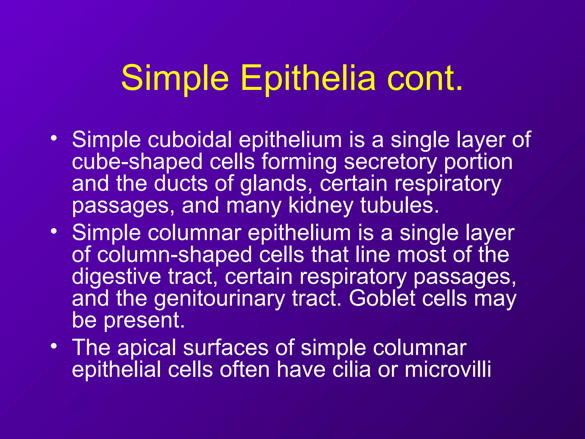


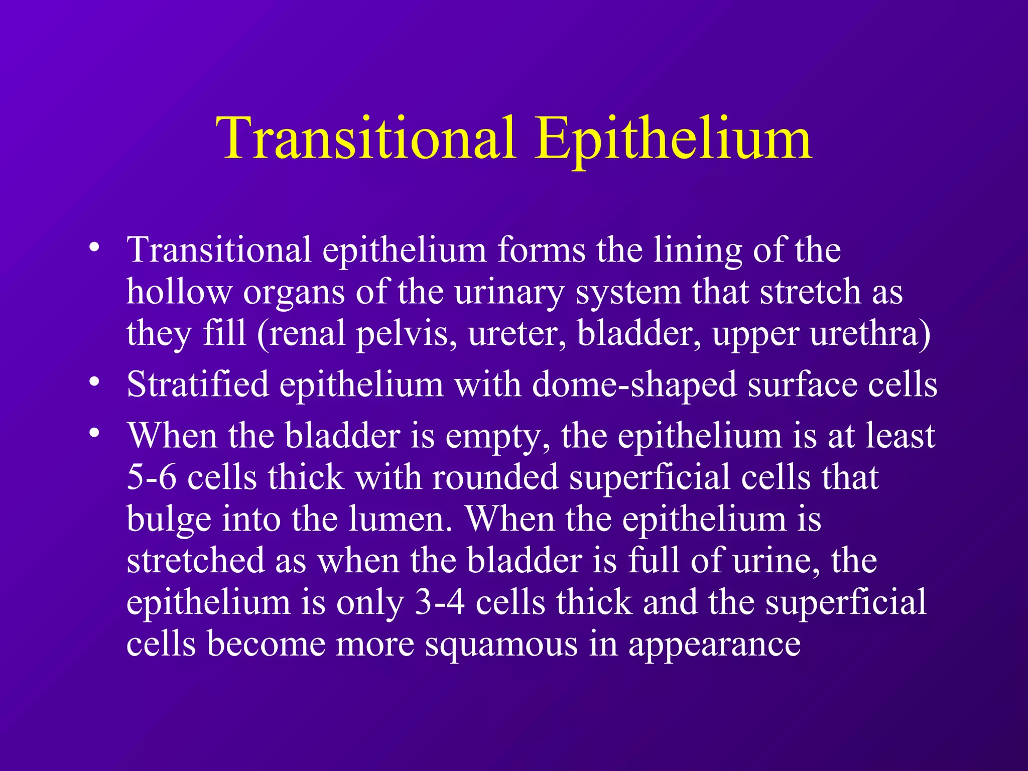
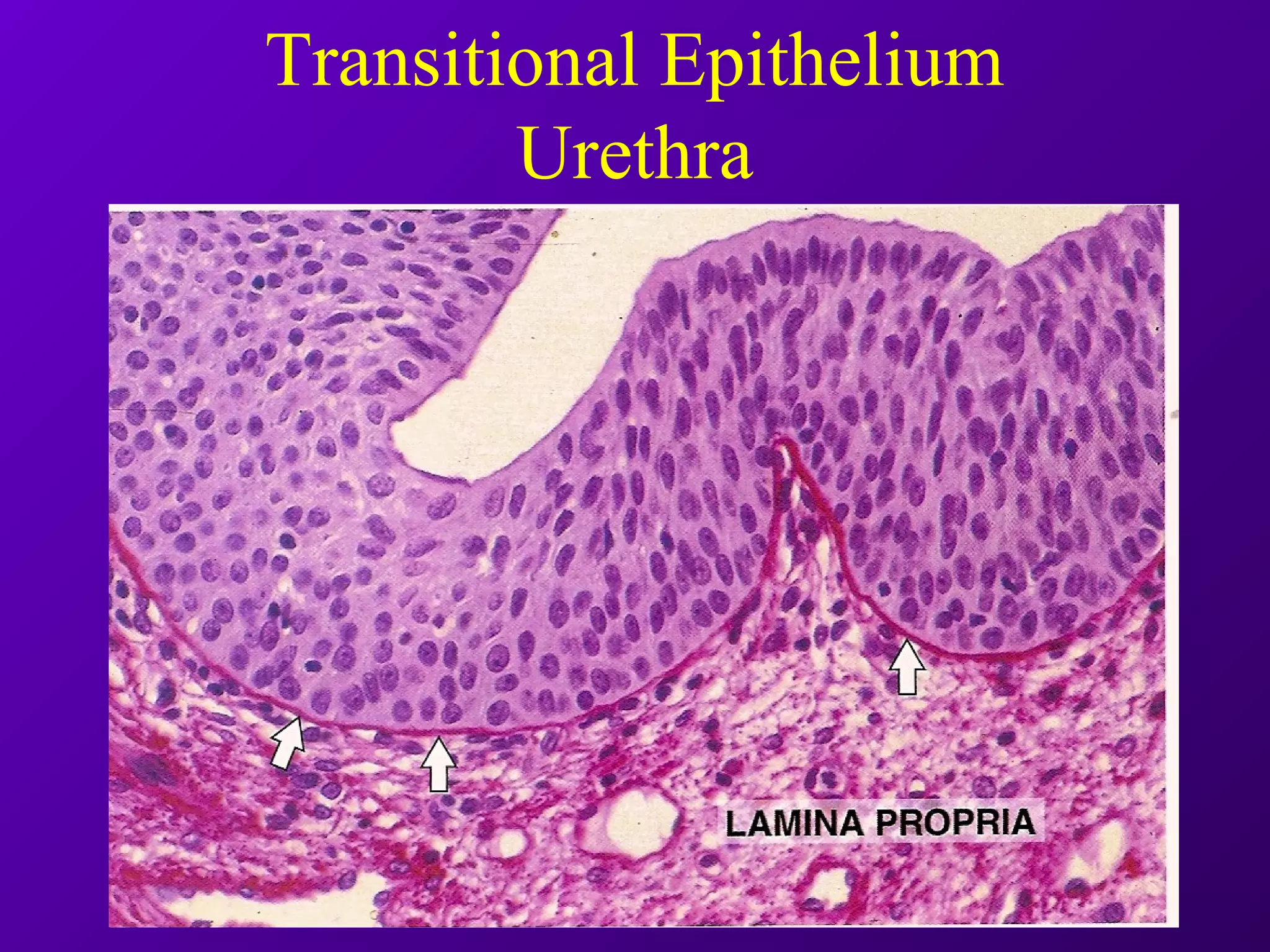


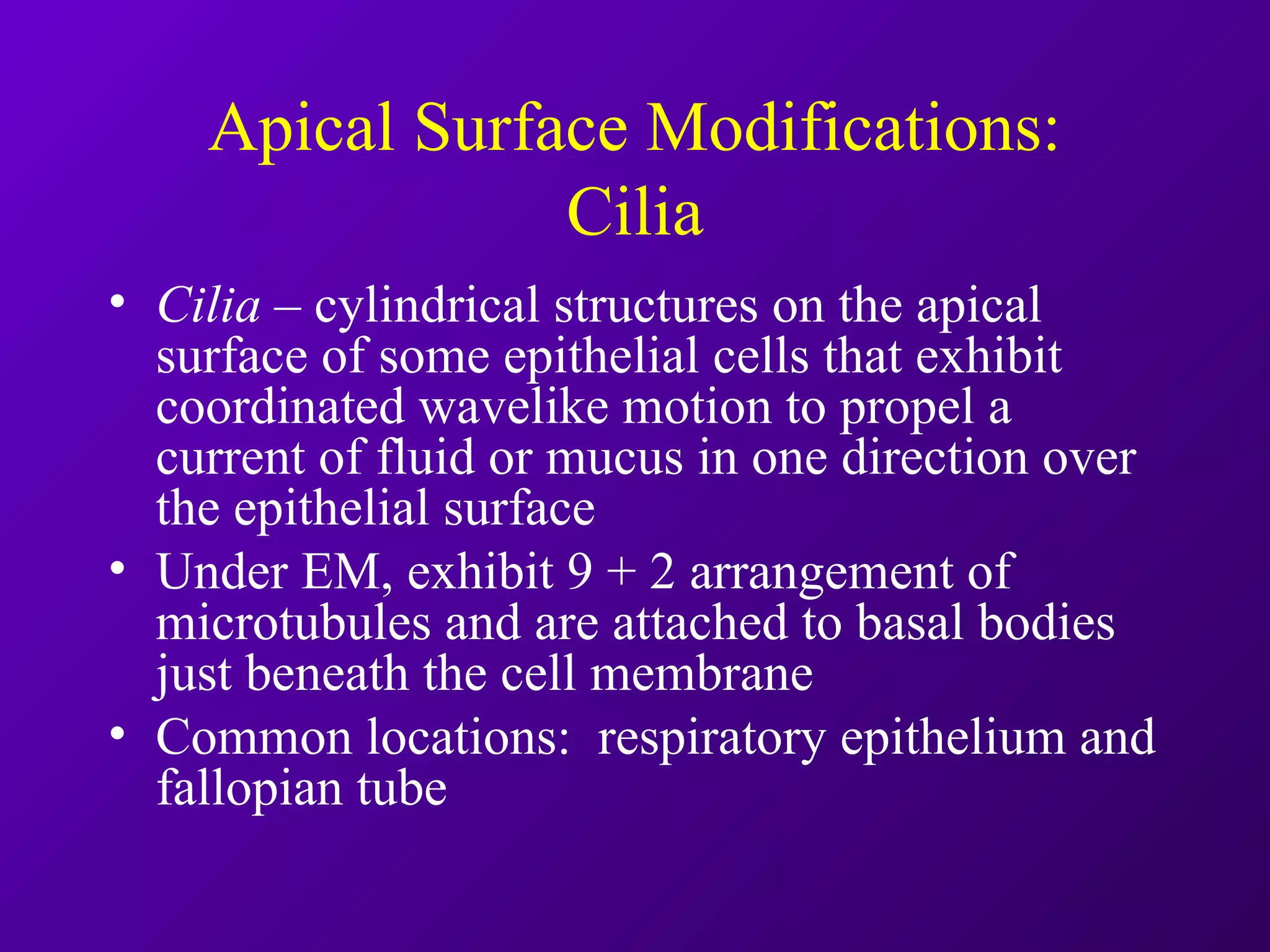


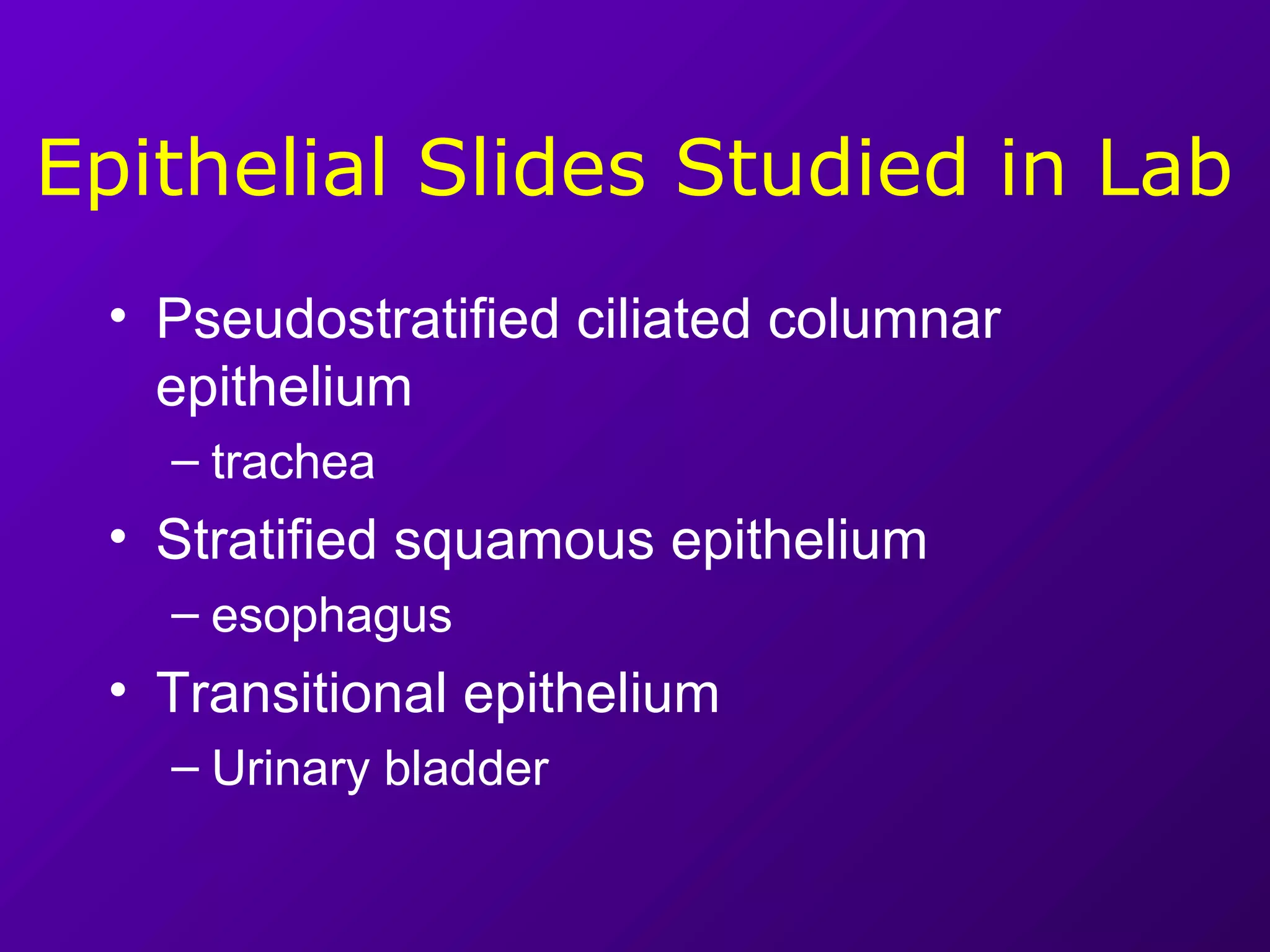


There are four primary types of tissues in the body: epithelial, connective, nervous, and muscular. Epithelial tissue forms sheets that cover surfaces and line cavities. There are several types of epithelia including simple squamous, simple cuboidal, simple columnar, stratified squamous, pseudostratified columnar and transitional epithelia. Each epithelium has a unique structure and location in the body suited to its functions like absorption, secretion, protection and selective permeability.


![2. epithelial-t[1]](https://cdn.slidesharecdn.com/ss_thumbnails/c55mbqopt3axovrntgld-signature-4c28f0f13a30c4ea316a9d58353990586de4897ab085203d01a9b7b7228e72f9-poli-180213061217-thumbnail.jpg?width=640&height=640&fit=bounds)


![Epithelium[1]](https://cdn.slidesharecdn.com/ss_thumbnails/epithelium1-200323141425-thumbnail.jpg?width=640&height=640&fit=bounds)


