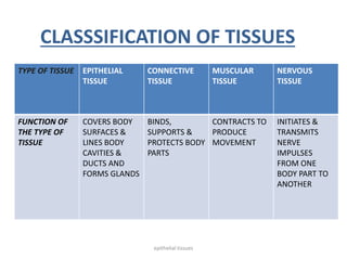
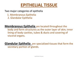
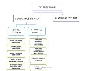
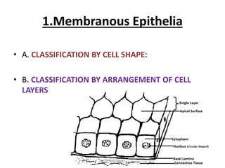
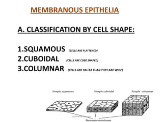
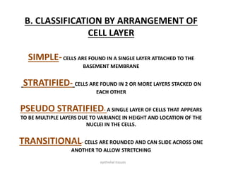
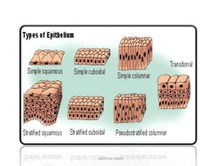
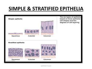
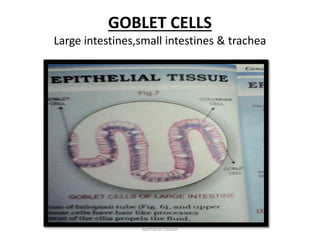
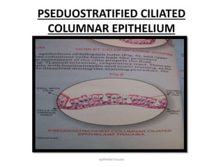
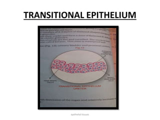
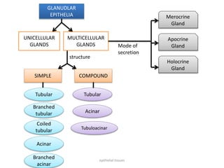
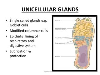
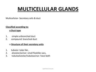
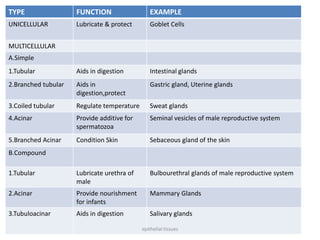
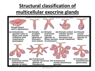
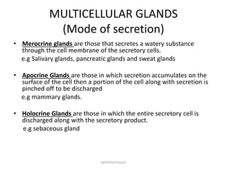
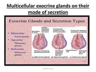
TISSUES
Dr.Tooba Rehan
Pharm-Depithelial tissues
• TISSUES:
Are aggregation of similar cells and cell products
that perform specific functions.
• HISTOLOGY:
Study of tissues is referred to as histology.
• PATHOLOGY:
Study of abnormal tissues in diseased organs.
epithelial tissues
CLASSSIFICATION OF TISSUES
TYPE OF TISSUE EPITHELIAL
TISSUE
CONNECTIVE
TISSUE
MUSCULAR
TISSUE
NERVOUS
TISSUE
FUNCTION OF
THE TYPE OF
TISSUE
COVERS BODY
SURFACES &
LINES BODY
CAVITIES &
DUCTS AND
FORMS GLANDS
BINDS,
SUPPORTS &
PROTECTS BODY
PARTS
CONTRACTS TO
PRODUCE
MOVEMENT
INITIATES &
TRANSMITS
NERVE
IMPULSES
FROM ONE
BODY PART TO
ANOTHER
epithelial tissues
EPITHELIAL TISSUE
Two major categories of epithelia
1. Membranous Epithelia
2. Glandular Epithelia
Membranous Epithelia are located throughout the
body and form structures as the outer layer of skin; inner
lining of body cavities, tubes & ducts and covering of
visceral organs.
Glandular Epithelia are specialized tissues that form the
secretary portion of glands.
epithelial tissues
EPITHELIAL TISSUES
MEMBRANOUS EPITHELIA GLANDULAR EPITHELIA
SIMPLE
EPITHELIA
STRATIFIED
EPITHELIA
SIMPLE SQUAMOUS
EPITHELIUM
SIMPLE CUBOIDAL
EPITHELIUM
SIMPLE COLUMNAR
EPITHELIUM
SIMPLE CILIATED
COLUMNAR EPITHELIUM
PSEUDOSTRATIFIED
CILIATED COLUMNAR
EPITHELIUM
STRATIFIED
SQUAMOUS
EPITHELIUM
(KERATINIZED)
STRATIFIED
SQUAMOUS
EPITHELIUM(NON
KERATINIZED)
STRATIFIED CUBOIDAL
EPITHELIUM
TRANSITIONAL
EPITHELIUM
epithelial tissues
1.Membranous Epithelia
• A. CLASSIFICATION BY CELL SHAPE:
• B. CLASSIFICATION BY ARRANGEMENT OF CELL
LAYERS
epithelial tissues
MEMBRANOUS EPITHELIA
A. CLASSIFICATION BY CELL SHAPE:
1.SQUAMOUS (CELLS ARE FLATTENED)
2.CUBOIDAL (CELLS ARE CUBE SHAPED)
3.COLUMNAR (CELLS ARE TALLER THAN THEY ARE WIDE)
epithelial tissues
B. CLASSIFICATION BY ARRANGEMENT OF
CELL LAYER
SIMPLE-CELLS ARE FOUND IN A SINGLE LAYER ATTACHED TO THE
BASEMENT MEMBRANE
STRATIFIED- CELLS ARE FOUND IN 2 OR MORE LAYERS STACKED ON
EACH OTHER
PSEUDO STRATIFIED- A SINGLE LAYER OF CELLS THAT APPEARS
TO BE MULTIPLE LAYERS DUE TO VARIANCE IN HEIGHT AND LOCATION OF THE
NUCLEI IN THE CELLS.
TRANSITIONAL- CELLS ARE ROUNDED AND CAN SLIDE ACROSS ONE
ANOTHER TO ALLOW STRETCHING
epithelial tissues
epithelial tissues
SIMPLE & STRATIFIED EPITHELIA
epithelial tissues
Simple Epithelia:
Single cell layer thick
Cells range from thin, flattened cell to tall,
columnar cell.
Diffusion, absorption, filtration & secretion
are principal functions
Some cells have cilia that create movement of
materials across the cell surface
Other have microvilli that increase surface
area for absorption.
epithelial tissues
SIMPLE SQUAMOUS EPITHELIA
 Flattened irregularly shaped cells
 Oval or spherical nucleus
 Thin cells with flattened nuclei
 Diffusion & filtration
 Pulmonary alveoli of lungs & portion of kidney
 Endothelium (blood & lymphatic vessels)
 Mesothelium(visceral organs & body cavities)
epithelial tissues
SIMPLE SQUAMOUS EPITHELIA
epithelial tissues
SIMPLE CUBOIDAL EPITHELIUM
• Single layer of tightly fitted cube shaped cells
• Cells have equal width & height
• Round central nuclei
• Excretory, secretory & absorptive function
• Surface of ovaries, tubules of kidneys & lines
the ducts of salivary glands & pancreas.
epithelial tissues
SIMPLE CUBOIDAL EPITHELIUM
epithelial tissues
SIMPLE COLUMNAR EPITHELIUM
• Tall columnar cells
• Height of cells varies depending on site &
function of tissues.
• Non ciliated
• Nucleus is basal & oval in shape.
• Protection, secretion & absorption.
• Goblets cells are scattered .
• Microvilli in gall bladder, small intestines.
• Lining inside wall of stomach to anal canal, gall
bladder, intestines cervical canal of uterus.
epithelial tissues
SIMPLE COLUMNAR EPITHELIUM
epithelial tissues
GOBLET CELLS
Large intestines,small intestines & trachea
epithelial tissues
SIMPLE CILIATED COLUMNAR
EPITHELUM
• Tall columnar cells
• Ciliated
• Epithelium of fallopian tubes & upper
respiratory passages.
• Cilia produce wave like movements.
• Transportive role through ciliary movement
epithelial tissues
SIMPLE CILIATED COLUMNAR
EPITHELUM
epithelial tissues
PSEDUOSTRATIFIED CILIATED
COLUMNAR EPITHELIUM
• Layered appearance (strata = layers).
• Not multi layer (pseudo = false).
• Nuclei located at different places.
• Goblet cells
• Ciliated / non ciliated
• Respiratory epithelium
• Protection, secretion & ciliary movement
epithelial tissues
PSEDUOSTRATIFIED CILIATED
COLUMNAR EPITHELIUM
epithelial tissues
Simple vs Stratified
Epithelia
epithelial tissues
STRATIFIED EPITHELIA
• Two or more layers of cells
• Poorly suited for absorption & secretion.
• Primarily protective function enhanced by
rapid cell division
• Classified according to shape of surface layer
of cells
• Cuboidal & columnar cells in contact with
basement membrane.
epithelial tissues
Stratified Squamous Epithelium
• Variable number of cell layers that are flattest at
the surface.
• Mitosis in deepest layers.
• Newly produces cells replace the cells that are
sloughed off.
• Keratinized & non keratinized
• First line of defense
• Protective (startification, rapid mitotic division &
keratinization)
epithelial tissues
Stratifed Squamous Epithelium
NON-KERATINIZED STRATIFIED
SQUAMOUS EPITHELIUM
1.No water proof protein keratin.
2.It has living squamous cells at the
surface. Stratum corneum absent
as non-kertinization or
cornification occurs.
3. It is formed in lining of cornea,
mouth, pharynx, oesophagus,,
vagina, cervix etc.
4. It provides mechanical protection
from injury.
KERATINIZED STRATIFIED
SQUAMOUS EPITHELIUM
1.The cells of few outer layers of
stratified squamous epithetium
replace their cytoplasm with a hard
water proof protein.
2. The layers of flat dead cells-
stratum corneum or horney layers
are present.
3. It forms epidermis of the skin.
4. It prevents loss of water and
mechanical injury.
epithelial tissues
Stratifed Squamous Epithelium
Non –Keratinized Keratinized
epithelial tissues
Layers of epidermis
epithelial tissues
STRATIFIED CUBOIDAL EPITHELIUM
• rare
• cube-shaped
• commonly cells make up two layers
• protection of larger ducts -cell layers
surround/protect gland ducts
• sweat glands in the skin
• mammary gland in the breast
• salivary glands in the mouth
epithelial tissues
STRATIFIED COLUMNAR EPITHELIUM
• column-shaped
• secretion and protection
• found near salivary glands - cell lining protects
the salivary duct, goblet cells found between the
cells
• found in vas deferens where it protects and aids
in secretion of glands
• intestinal lining - ciliated to help move nutrients
and increase absorption, also protects against
pathogens/bacteria
epithelial tissues
STRATIFIED COLUMNAR EPITHELIUM
epithelial tissues
TRANSITIONAL EPITHELIUM
• Similar to non keratinized startified squamous
epithelium except surface cells are large & round
• able to stretch
• Urinary system
• Surface cells round,cuboidal,umbrella shaped
• Intermediate zone (2-4 rows) are pear shaped cells
• Basal layer cuboidal cells.
• without distention (relaxed), the cells appear cuboidal-
shaped
• when distented (tense), the cells appear squamous-
shaped
epithelial tissues
Transitional epithelium is a stratified type that can be confused with stratified
squamous. The name transitional comes from the ability of this epithelium to
accommodate stretching in the urinary tract as fluid pressures vary. It is
found as a lining of the ureters, urethra, and bladder. The large, ovoid surface
cells are a key attribute of this epithelium type.
epithelial tissues
TRANSITIONAL EPITHELIUM
epithelial tissues
GLANDULAR EPITHELIA
• Glandular epithelial cells are specialized epithelial
cells that secrete bodily products, sometimes
called simply glands.
• Glands include two types: endocrine and
exocrine.
• Exocrine glands are often the only glands
associated with the term "glandular epithelium."
epithelial tissues
UNICELLULAR
GLANDS
MULTICELLULAR
GLANDS
GLANUDLAR
EPITHELIA
SIMPLE COMPOUND
Tubular
Branched
tubular
Coiled
tubular
Acinar
Branched
acinar
Tubular
Acinar
Tubuloacinar
Mode of
secretion
structure
Merocrine
Gland
Apocrine
Gland
Holocrine
Gland
epithelial tissues
UNICELLULAR GLANDS
• Single celled glands e.g.
Goblet cells
• Modified columnar cells
• Epithelial lining of
respiratory and
digestive system
• Lubrication &
protection
epithelial tissues
MULTICELLULAR GLANDS
Multicellular- Secretory cells & duct
Classified according to:
o Duct type
1. simple:unbranched duct
2. compound: branched duct
o Structure of their secretory units
1. tubular: tube like ,
2. alveolar/acinar: small flasklike sacs,
3. tubuloalveolar/tubuloacinar: have both
epithelial tissues
TYPE FUNCTION EXAMPLE
UNICELLULAR Lubricate & protect Goblet Cells
MULTICELLULAR
A.Simple
1.Tubular Aids in digestion Intestinal glands
2.Branched tubular Aids in
digestion,protect
Gastric gland, Uterine glands
3.Coiled tubular Regulate temperature Sweat glands
4.Acinar Provide additive for
spermatozoa
Seminal vesicles of male reproductive system
5.Branched Acinar Condition Skin Sebaceous gland of the skin
B.Compound
1.Tubular Lubricate urethra of
male
Bulbourethral glands of male reproductive system
2.Acinar Provide nourishment
for infants
Mammary Glands
3.Tubuloacinar Aids in digestion Salivary glands
epithelial tissues
Structural classification of
multicellular exocrine glands
epithelial tissues
MULTICELLULAR GLANDS
(Mode of secretion)
• Merocrine glands are those that secretes a watery substance
through the cell membrane of the secretory cells.
e.g Salivary glands, pancreatic glands and sweat glands
• Apocrine Glands are those in which secretion accumulates on the
surface of the cell then a portion of the cell along with secretion is
pinched off to be discharged
e.g mammary glands.
• Holocrine Glands are those in which the entire secretory cell is
discharged along with the secretory product.
e.g sebaceous gland
epithelial tissues
Multicellular exocrine glands on their
mode of secretion
epithelial tissues

Tissues

  • 1.
  • 2.
    • TISSUES: Are aggregationof similar cells and cell products that perform specific functions. • HISTOLOGY: Study of tissues is referred to as histology. • PATHOLOGY: Study of abnormal tissues in diseased organs. epithelial tissues
  • 3.
    CLASSSIFICATION OF TISSUES TYPEOF TISSUE EPITHELIAL TISSUE CONNECTIVE TISSUE MUSCULAR TISSUE NERVOUS TISSUE FUNCTION OF THE TYPE OF TISSUE COVERS BODY SURFACES & LINES BODY CAVITIES & DUCTS AND FORMS GLANDS BINDS, SUPPORTS & PROTECTS BODY PARTS CONTRACTS TO PRODUCE MOVEMENT INITIATES & TRANSMITS NERVE IMPULSES FROM ONE BODY PART TO ANOTHER epithelial tissues
  • 4.
    EPITHELIAL TISSUE Two majorcategories of epithelia 1. Membranous Epithelia 2. Glandular Epithelia Membranous Epithelia are located throughout the body and form structures as the outer layer of skin; inner lining of body cavities, tubes & ducts and covering of visceral organs. Glandular Epithelia are specialized tissues that form the secretary portion of glands. epithelial tissues
  • 5.
    EPITHELIAL TISSUES MEMBRANOUS EPITHELIAGLANDULAR EPITHELIA SIMPLE EPITHELIA STRATIFIED EPITHELIA SIMPLE SQUAMOUS EPITHELIUM SIMPLE CUBOIDAL EPITHELIUM SIMPLE COLUMNAR EPITHELIUM SIMPLE CILIATED COLUMNAR EPITHELIUM PSEUDOSTRATIFIED CILIATED COLUMNAR EPITHELIUM STRATIFIED SQUAMOUS EPITHELIUM (KERATINIZED) STRATIFIED SQUAMOUS EPITHELIUM(NON KERATINIZED) STRATIFIED CUBOIDAL EPITHELIUM TRANSITIONAL EPITHELIUM epithelial tissues
  • 6.
    1.Membranous Epithelia • A.CLASSIFICATION BY CELL SHAPE: • B. CLASSIFICATION BY ARRANGEMENT OF CELL LAYERS epithelial tissues
  • 7.
    MEMBRANOUS EPITHELIA A. CLASSIFICATIONBY CELL SHAPE: 1.SQUAMOUS (CELLS ARE FLATTENED) 2.CUBOIDAL (CELLS ARE CUBE SHAPED) 3.COLUMNAR (CELLS ARE TALLER THAN THEY ARE WIDE) epithelial tissues
  • 8.
    B. CLASSIFICATION BYARRANGEMENT OF CELL LAYER SIMPLE-CELLS ARE FOUND IN A SINGLE LAYER ATTACHED TO THE BASEMENT MEMBRANE STRATIFIED- CELLS ARE FOUND IN 2 OR MORE LAYERS STACKED ON EACH OTHER PSEUDO STRATIFIED- A SINGLE LAYER OF CELLS THAT APPEARS TO BE MULTIPLE LAYERS DUE TO VARIANCE IN HEIGHT AND LOCATION OF THE NUCLEI IN THE CELLS. TRANSITIONAL- CELLS ARE ROUNDED AND CAN SLIDE ACROSS ONE ANOTHER TO ALLOW STRETCHING epithelial tissues
  • 9.
  • 10.
    SIMPLE & STRATIFIEDEPITHELIA epithelial tissues
  • 11.
    Simple Epithelia: Single celllayer thick Cells range from thin, flattened cell to tall, columnar cell. Diffusion, absorption, filtration & secretion are principal functions Some cells have cilia that create movement of materials across the cell surface Other have microvilli that increase surface area for absorption. epithelial tissues
  • 12.
    SIMPLE SQUAMOUS EPITHELIA Flattened irregularly shaped cells  Oval or spherical nucleus  Thin cells with flattened nuclei  Diffusion & filtration  Pulmonary alveoli of lungs & portion of kidney  Endothelium (blood & lymphatic vessels)  Mesothelium(visceral organs & body cavities) epithelial tissues
  • 13.
  • 14.
    SIMPLE CUBOIDAL EPITHELIUM •Single layer of tightly fitted cube shaped cells • Cells have equal width & height • Round central nuclei • Excretory, secretory & absorptive function • Surface of ovaries, tubules of kidneys & lines the ducts of salivary glands & pancreas. epithelial tissues
  • 15.
  • 16.
    SIMPLE COLUMNAR EPITHELIUM •Tall columnar cells • Height of cells varies depending on site & function of tissues. • Non ciliated • Nucleus is basal & oval in shape. • Protection, secretion & absorption. • Goblets cells are scattered . • Microvilli in gall bladder, small intestines. • Lining inside wall of stomach to anal canal, gall bladder, intestines cervical canal of uterus. epithelial tissues
  • 17.
  • 18.
    GOBLET CELLS Large intestines,smallintestines & trachea epithelial tissues
  • 19.
    SIMPLE CILIATED COLUMNAR EPITHELUM •Tall columnar cells • Ciliated • Epithelium of fallopian tubes & upper respiratory passages. • Cilia produce wave like movements. • Transportive role through ciliary movement epithelial tissues
  • 20.
  • 21.
    PSEDUOSTRATIFIED CILIATED COLUMNAR EPITHELIUM •Layered appearance (strata = layers). • Not multi layer (pseudo = false). • Nuclei located at different places. • Goblet cells • Ciliated / non ciliated • Respiratory epithelium • Protection, secretion & ciliary movement epithelial tissues
  • 22.
  • 23.
  • 24.
    STRATIFIED EPITHELIA • Twoor more layers of cells • Poorly suited for absorption & secretion. • Primarily protective function enhanced by rapid cell division • Classified according to shape of surface layer of cells • Cuboidal & columnar cells in contact with basement membrane. epithelial tissues
  • 25.
    Stratified Squamous Epithelium •Variable number of cell layers that are flattest at the surface. • Mitosis in deepest layers. • Newly produces cells replace the cells that are sloughed off. • Keratinized & non keratinized • First line of defense • Protective (startification, rapid mitotic division & keratinization) epithelial tissues
  • 26.
    Stratifed Squamous Epithelium NON-KERATINIZEDSTRATIFIED SQUAMOUS EPITHELIUM 1.No water proof protein keratin. 2.It has living squamous cells at the surface. Stratum corneum absent as non-kertinization or cornification occurs. 3. It is formed in lining of cornea, mouth, pharynx, oesophagus,, vagina, cervix etc. 4. It provides mechanical protection from injury. KERATINIZED STRATIFIED SQUAMOUS EPITHELIUM 1.The cells of few outer layers of stratified squamous epithetium replace their cytoplasm with a hard water proof protein. 2. The layers of flat dead cells- stratum corneum or horney layers are present. 3. It forms epidermis of the skin. 4. It prevents loss of water and mechanical injury. epithelial tissues
  • 27.
    Stratifed Squamous Epithelium Non–Keratinized Keratinized epithelial tissues
  • 28.
  • 29.
    STRATIFIED CUBOIDAL EPITHELIUM •rare • cube-shaped • commonly cells make up two layers • protection of larger ducts -cell layers surround/protect gland ducts • sweat glands in the skin • mammary gland in the breast • salivary glands in the mouth epithelial tissues
  • 30.
    STRATIFIED COLUMNAR EPITHELIUM •column-shaped • secretion and protection • found near salivary glands - cell lining protects the salivary duct, goblet cells found between the cells • found in vas deferens where it protects and aids in secretion of glands • intestinal lining - ciliated to help move nutrients and increase absorption, also protects against pathogens/bacteria epithelial tissues
  • 31.
  • 32.
    TRANSITIONAL EPITHELIUM • Similarto non keratinized startified squamous epithelium except surface cells are large & round • able to stretch • Urinary system • Surface cells round,cuboidal,umbrella shaped • Intermediate zone (2-4 rows) are pear shaped cells • Basal layer cuboidal cells. • without distention (relaxed), the cells appear cuboidal- shaped • when distented (tense), the cells appear squamous- shaped epithelial tissues
  • 33.
    Transitional epithelium isa stratified type that can be confused with stratified squamous. The name transitional comes from the ability of this epithelium to accommodate stretching in the urinary tract as fluid pressures vary. It is found as a lining of the ureters, urethra, and bladder. The large, ovoid surface cells are a key attribute of this epithelium type. epithelial tissues
  • 34.
  • 35.
    GLANDULAR EPITHELIA • Glandularepithelial cells are specialized epithelial cells that secrete bodily products, sometimes called simply glands. • Glands include two types: endocrine and exocrine. • Exocrine glands are often the only glands associated with the term "glandular epithelium." epithelial tissues
  • 36.
  • 37.
    UNICELLULAR GLANDS • Singlecelled glands e.g. Goblet cells • Modified columnar cells • Epithelial lining of respiratory and digestive system • Lubrication & protection epithelial tissues
  • 38.
    MULTICELLULAR GLANDS Multicellular- Secretorycells & duct Classified according to: o Duct type 1. simple:unbranched duct 2. compound: branched duct o Structure of their secretory units 1. tubular: tube like , 2. alveolar/acinar: small flasklike sacs, 3. tubuloalveolar/tubuloacinar: have both epithelial tissues
  • 39.
    TYPE FUNCTION EXAMPLE UNICELLULARLubricate & protect Goblet Cells MULTICELLULAR A.Simple 1.Tubular Aids in digestion Intestinal glands 2.Branched tubular Aids in digestion,protect Gastric gland, Uterine glands 3.Coiled tubular Regulate temperature Sweat glands 4.Acinar Provide additive for spermatozoa Seminal vesicles of male reproductive system 5.Branched Acinar Condition Skin Sebaceous gland of the skin B.Compound 1.Tubular Lubricate urethra of male Bulbourethral glands of male reproductive system 2.Acinar Provide nourishment for infants Mammary Glands 3.Tubuloacinar Aids in digestion Salivary glands epithelial tissues
  • 40.
    Structural classification of multicellularexocrine glands epithelial tissues
  • 41.
    MULTICELLULAR GLANDS (Mode ofsecretion) • Merocrine glands are those that secretes a watery substance through the cell membrane of the secretory cells. e.g Salivary glands, pancreatic glands and sweat glands • Apocrine Glands are those in which secretion accumulates on the surface of the cell then a portion of the cell along with secretion is pinched off to be discharged e.g mammary glands. • Holocrine Glands are those in which the entire secretory cell is discharged along with the secretory product. e.g sebaceous gland epithelial tissues
  • 42.
    Multicellular exocrine glandson their mode of secretion epithelial tissues