Download to read offline
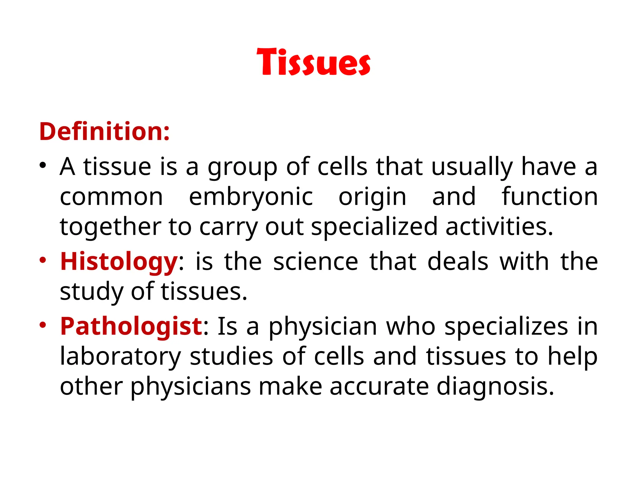
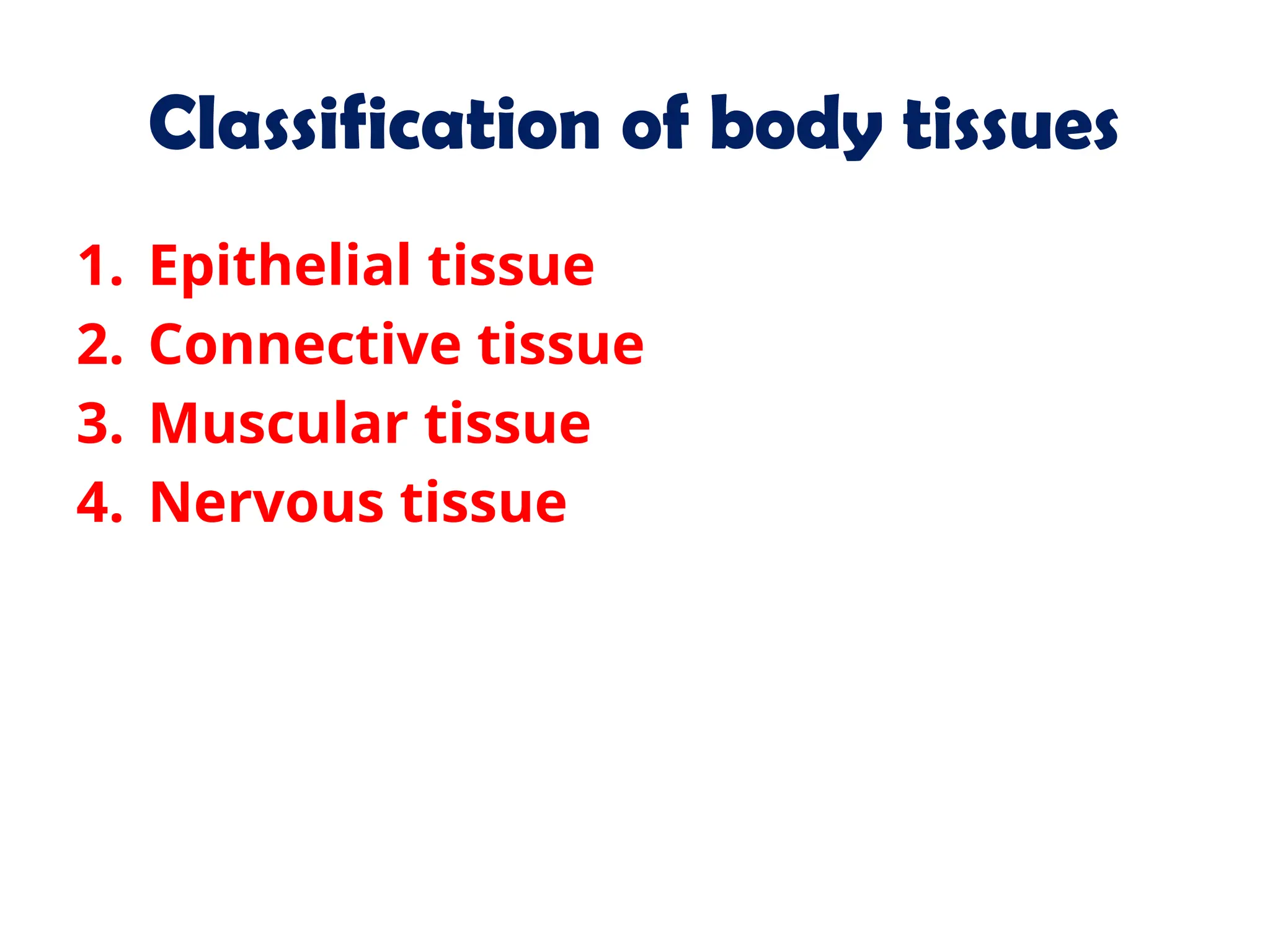
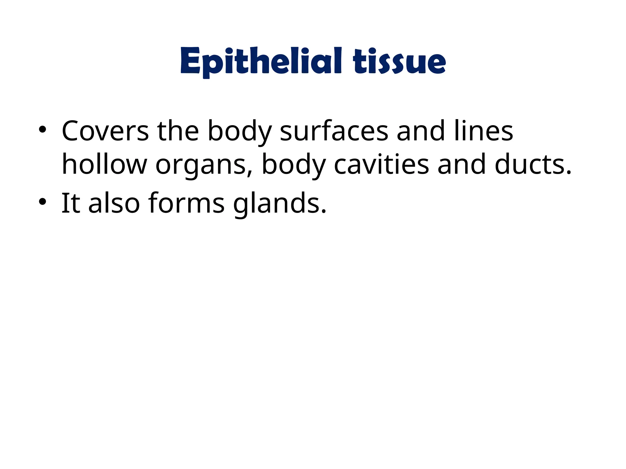
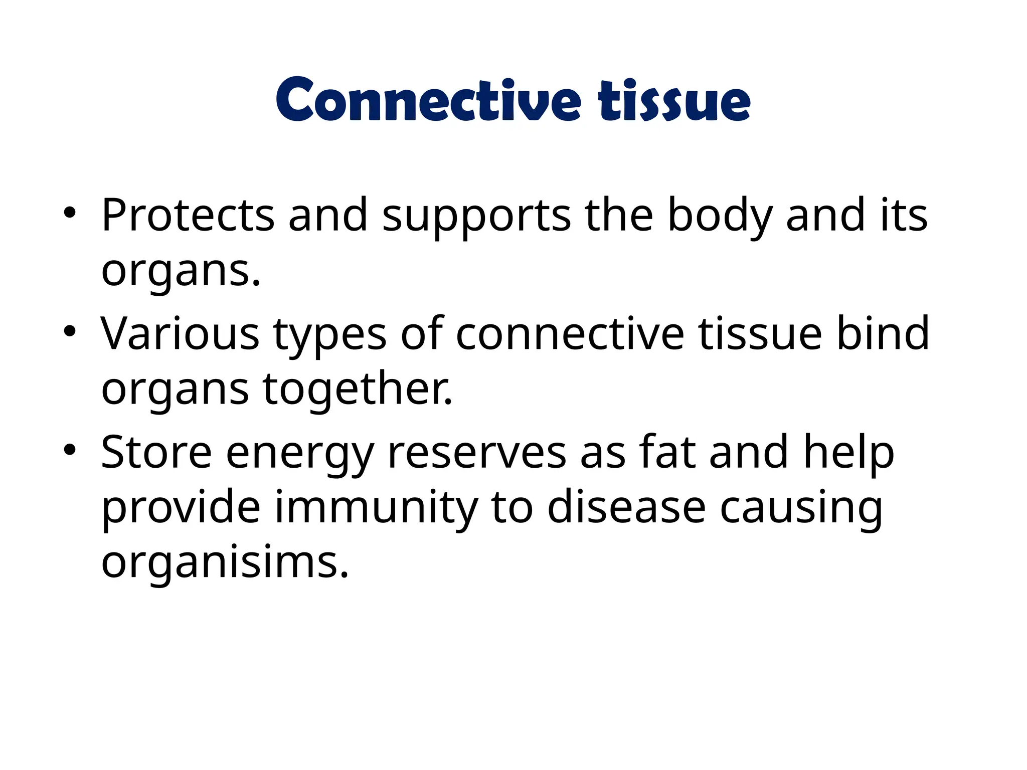
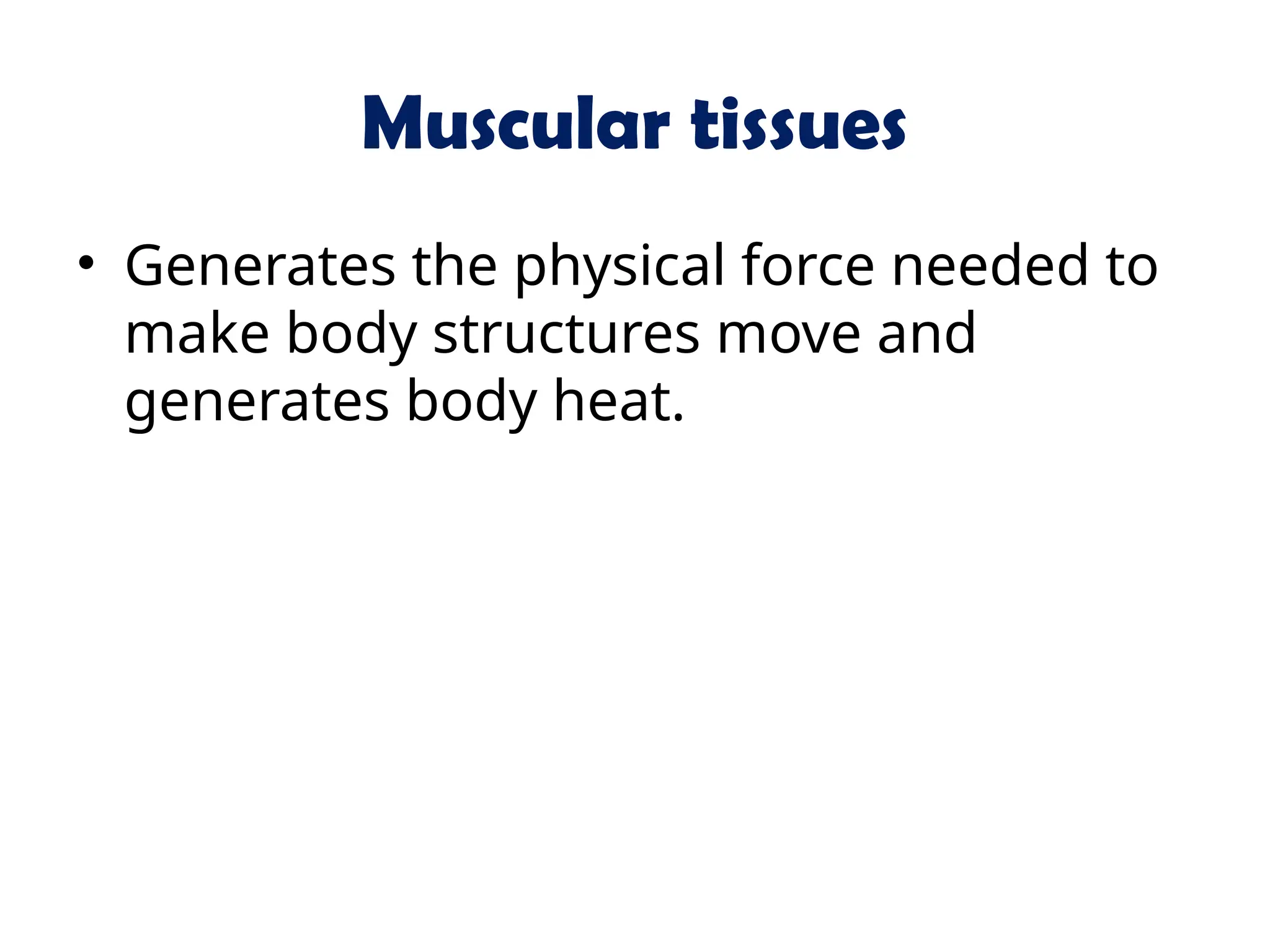
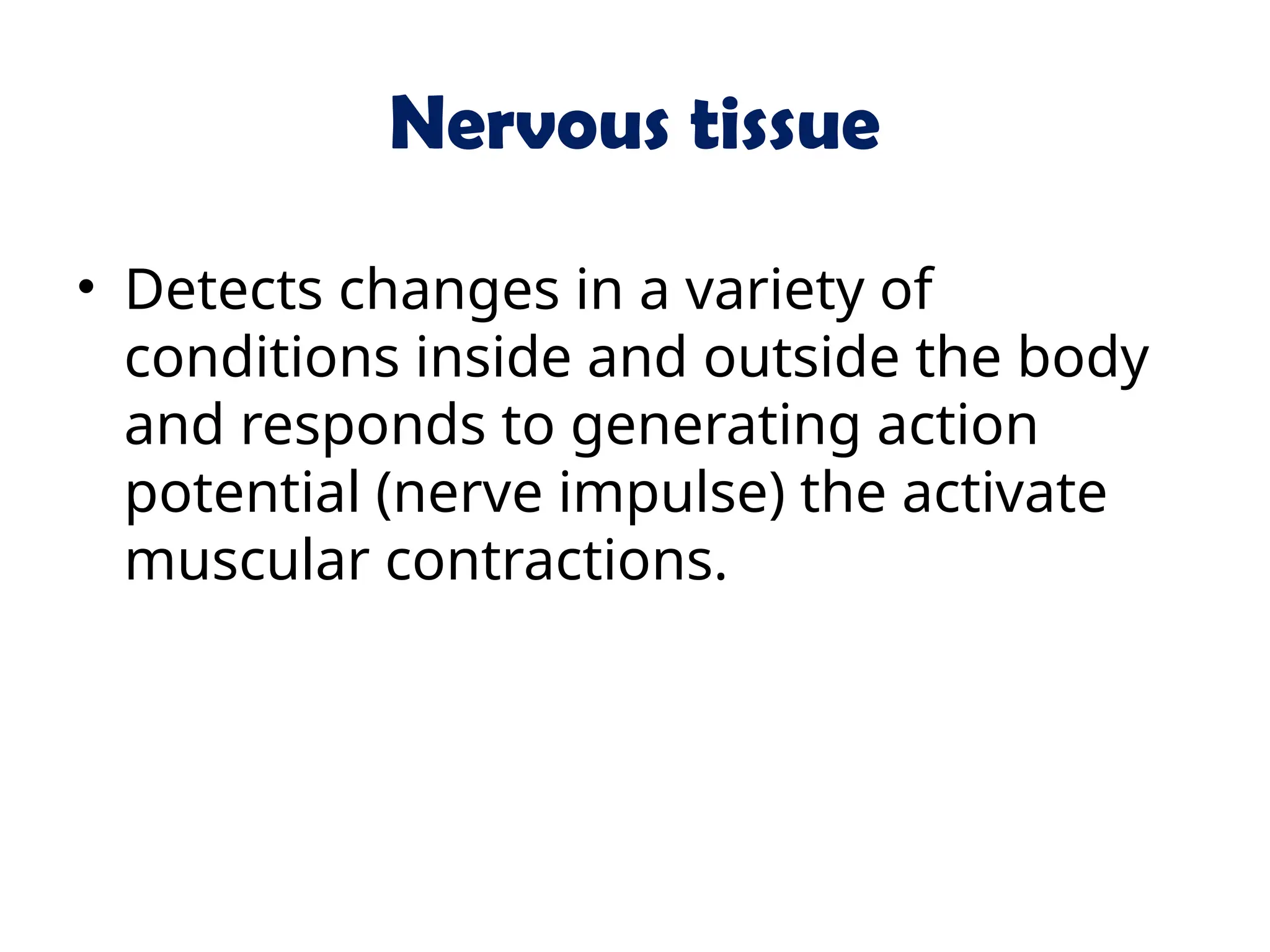
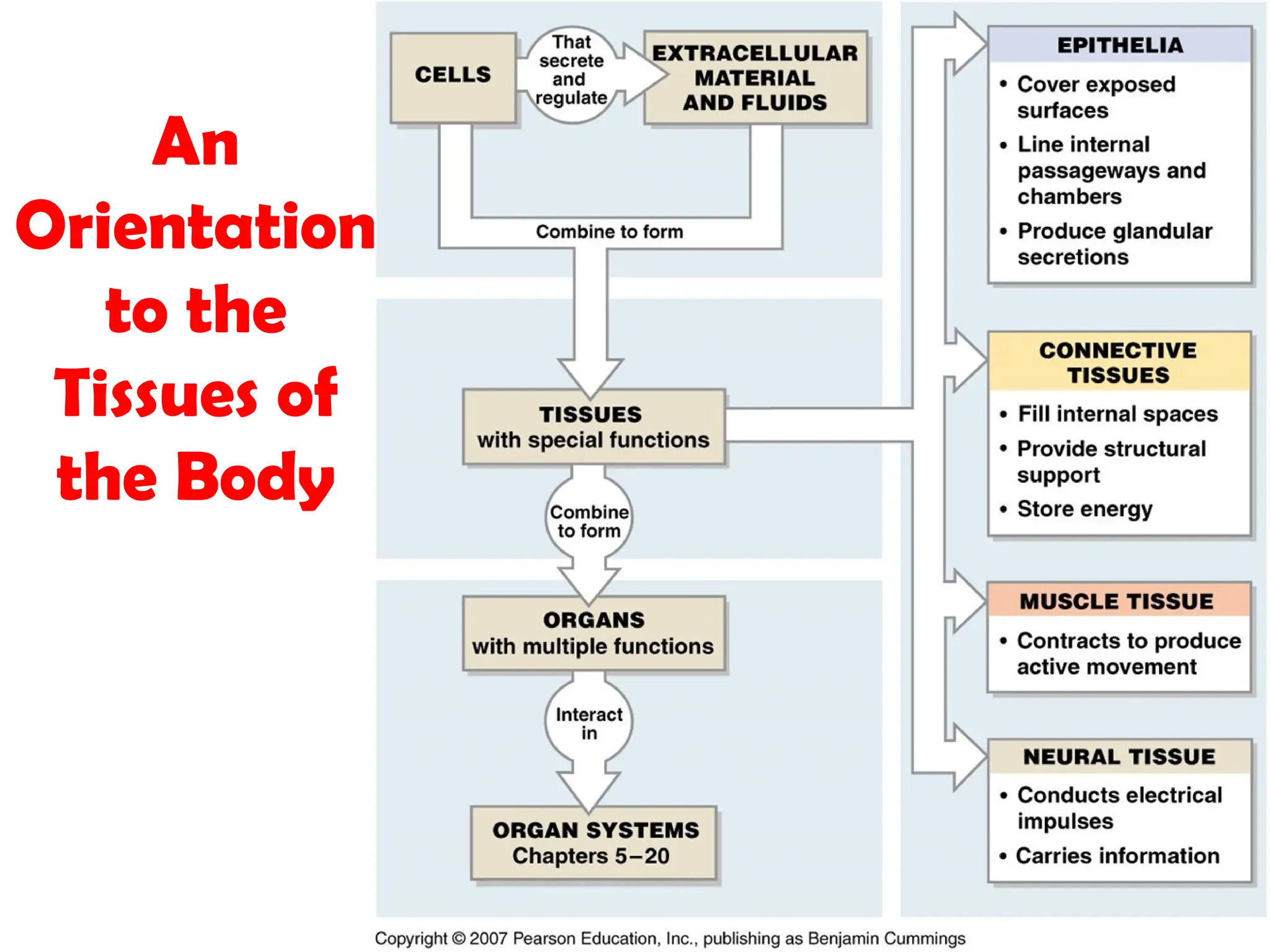
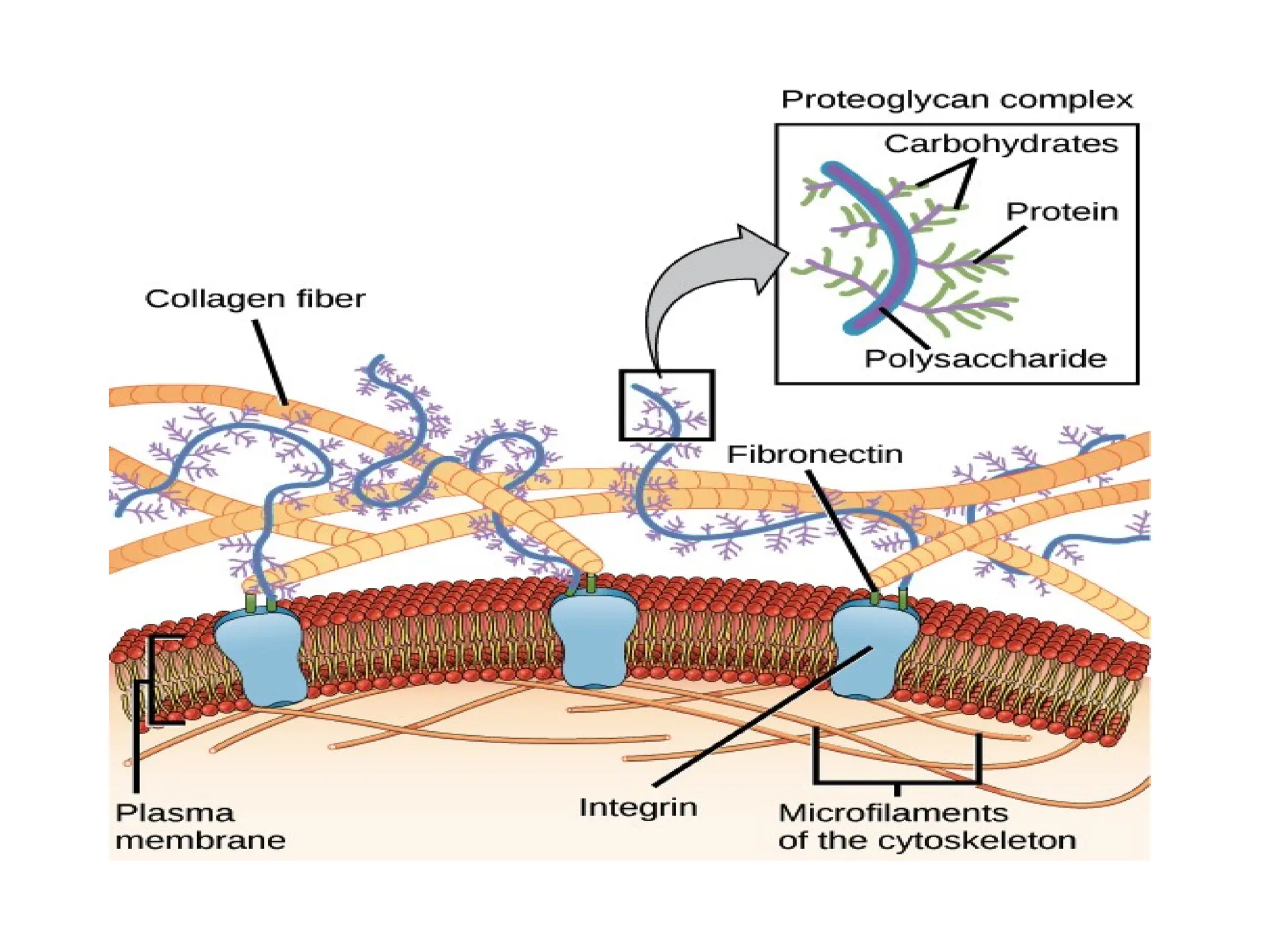
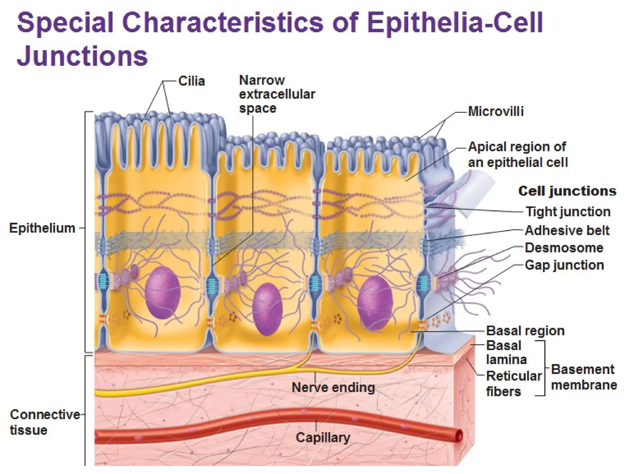
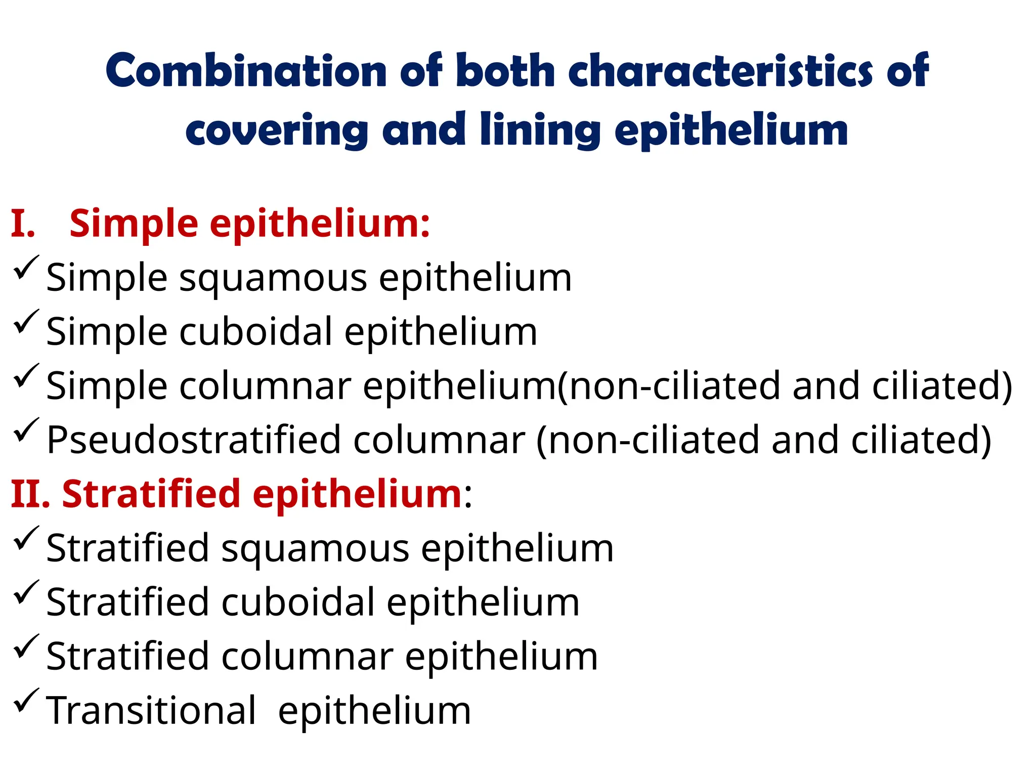
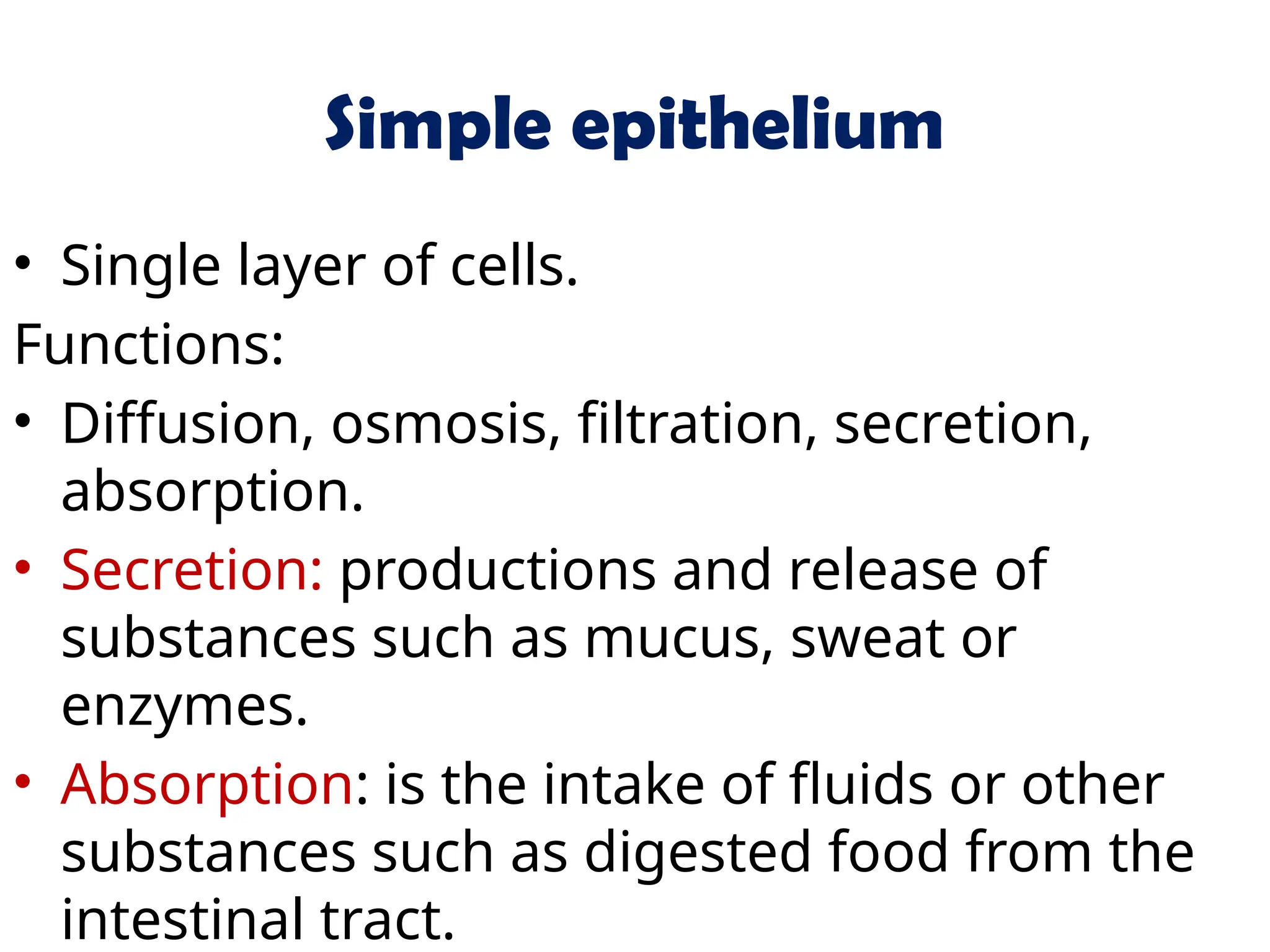
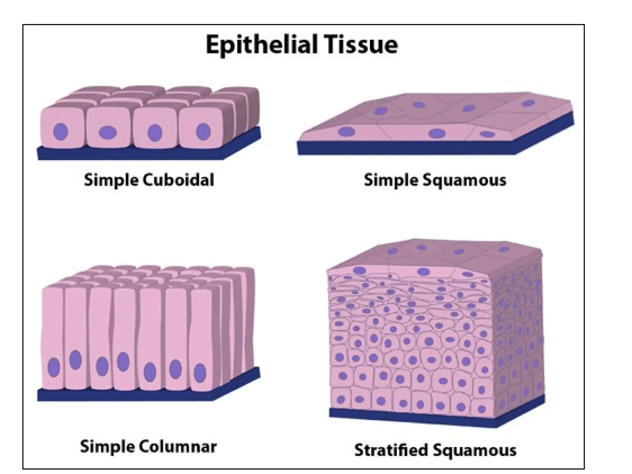
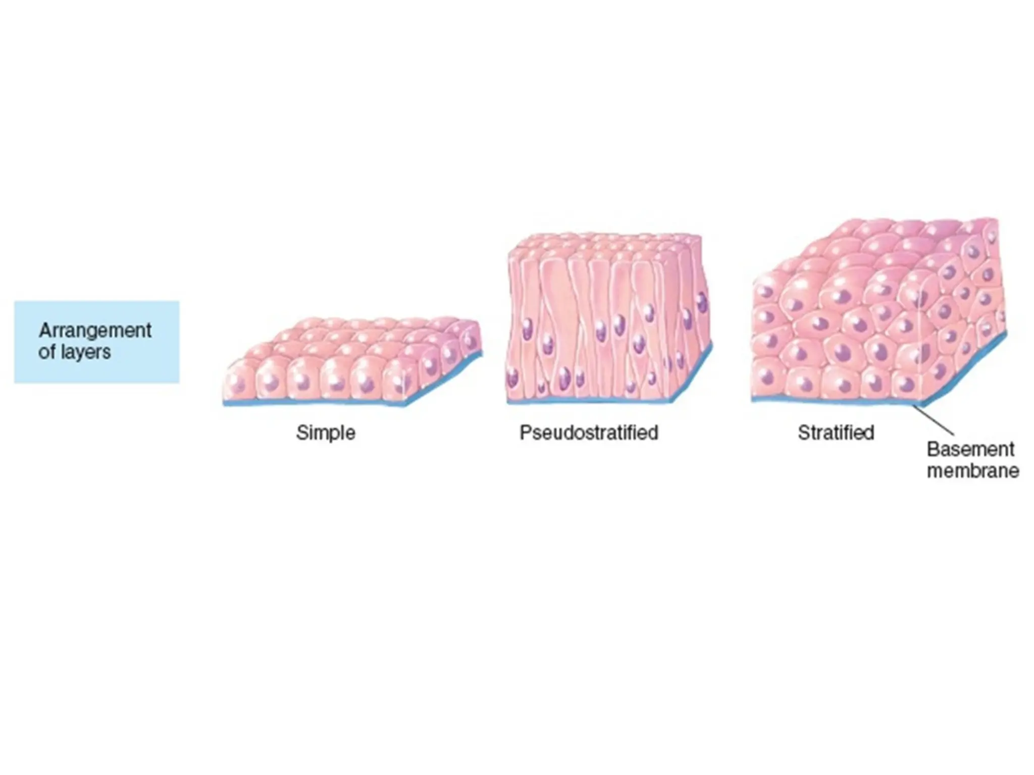
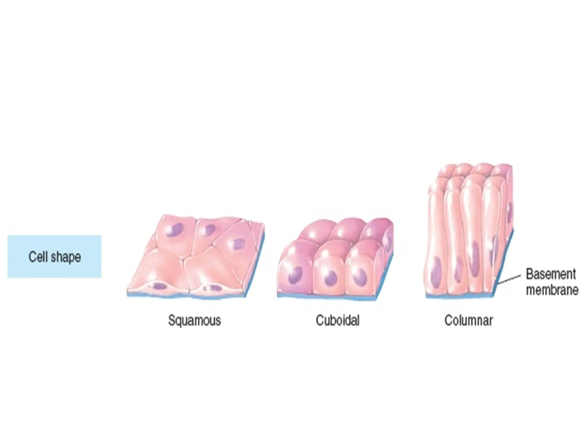
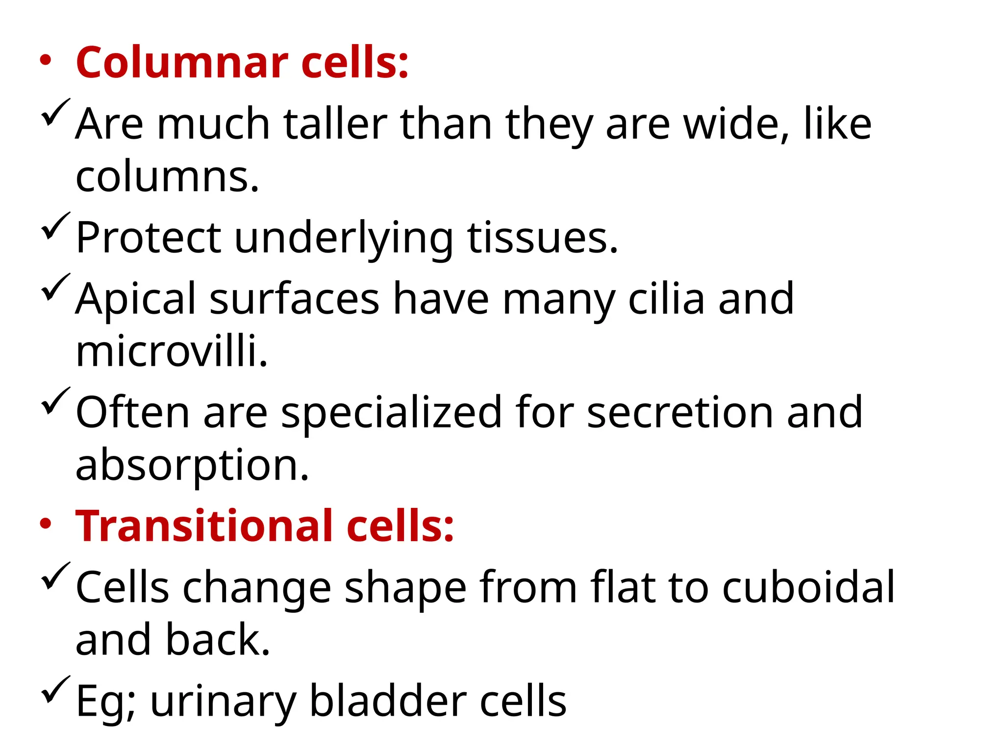
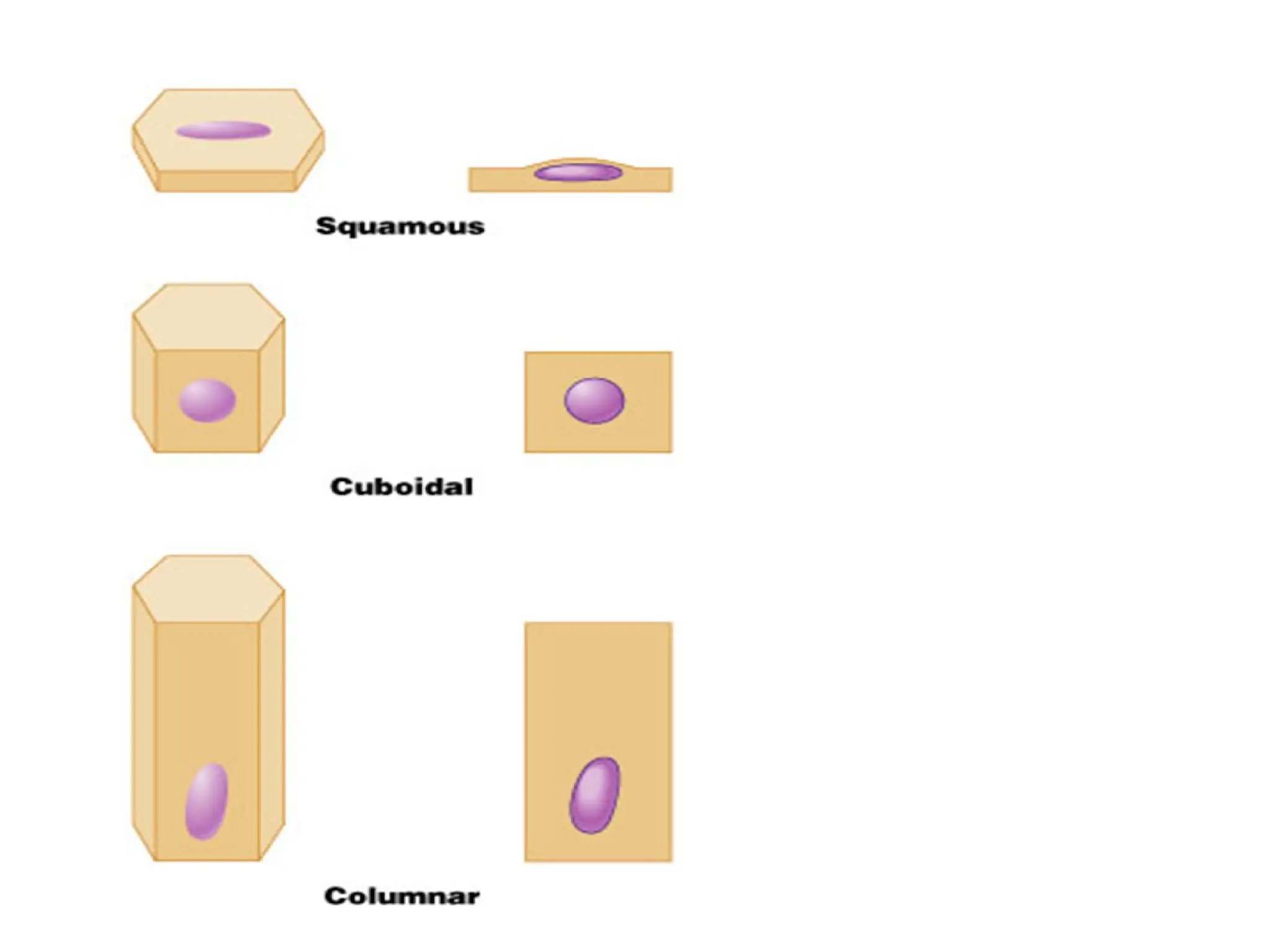
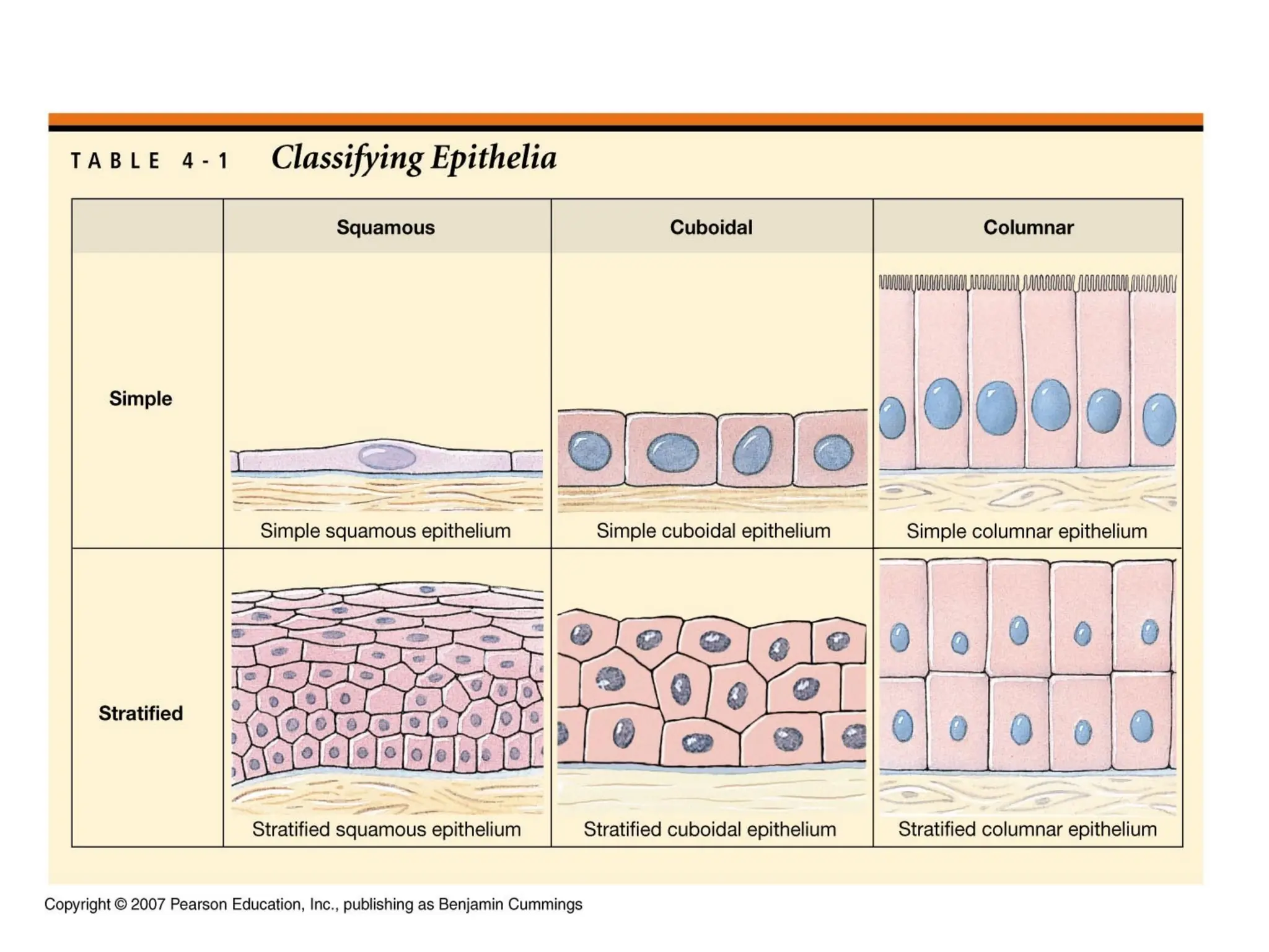
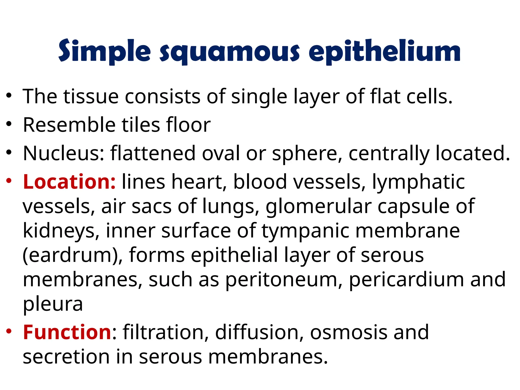
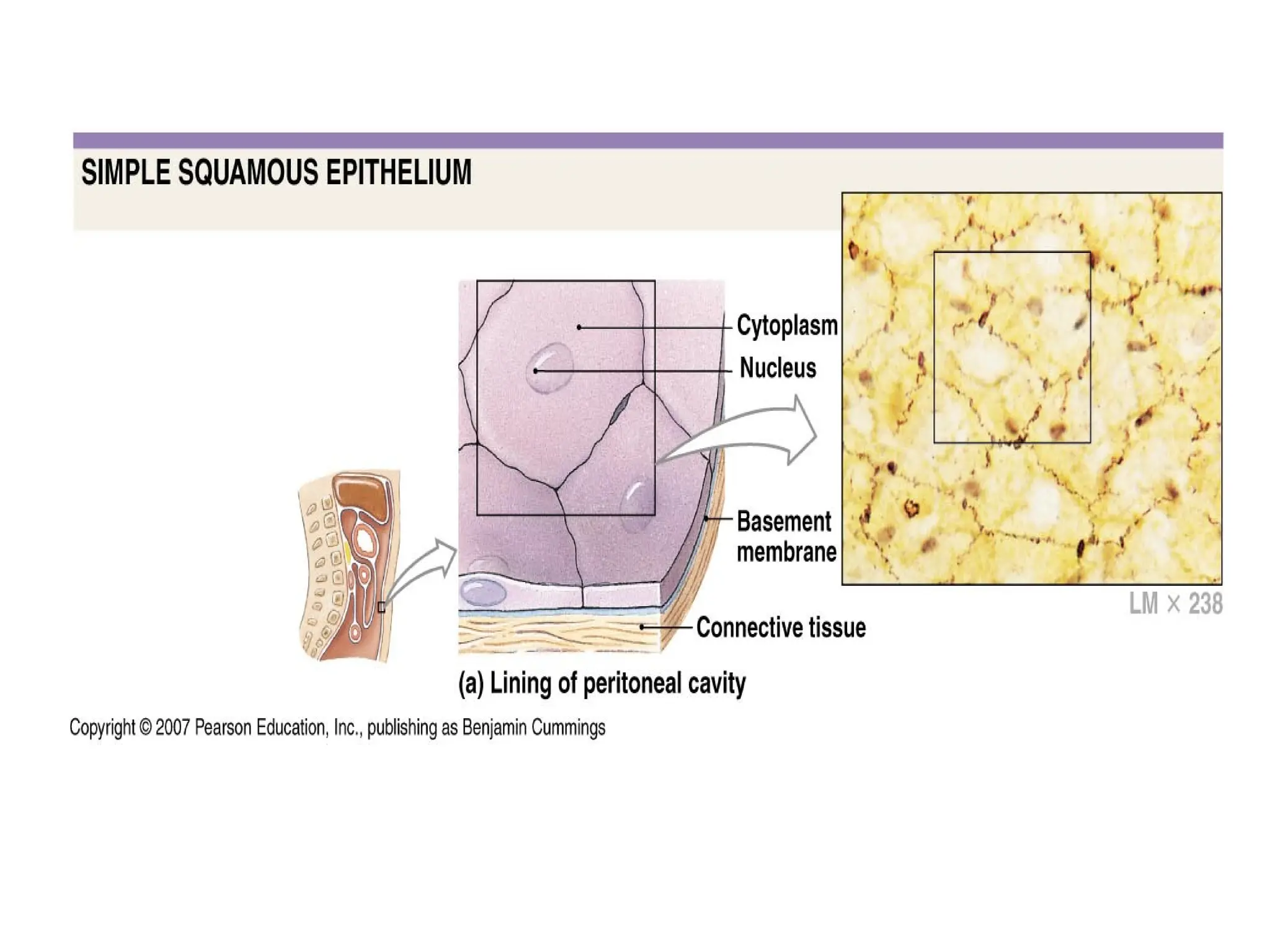
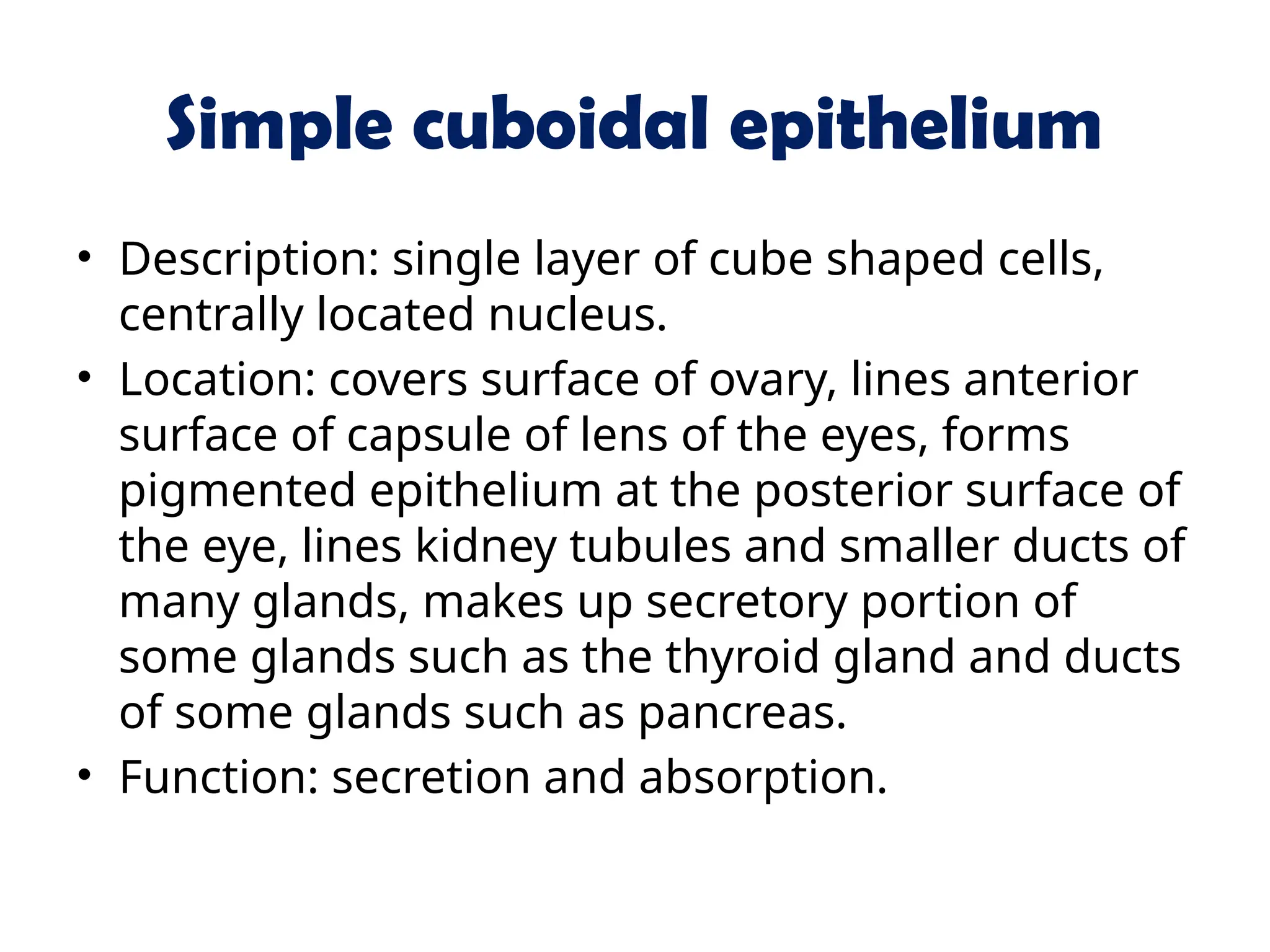
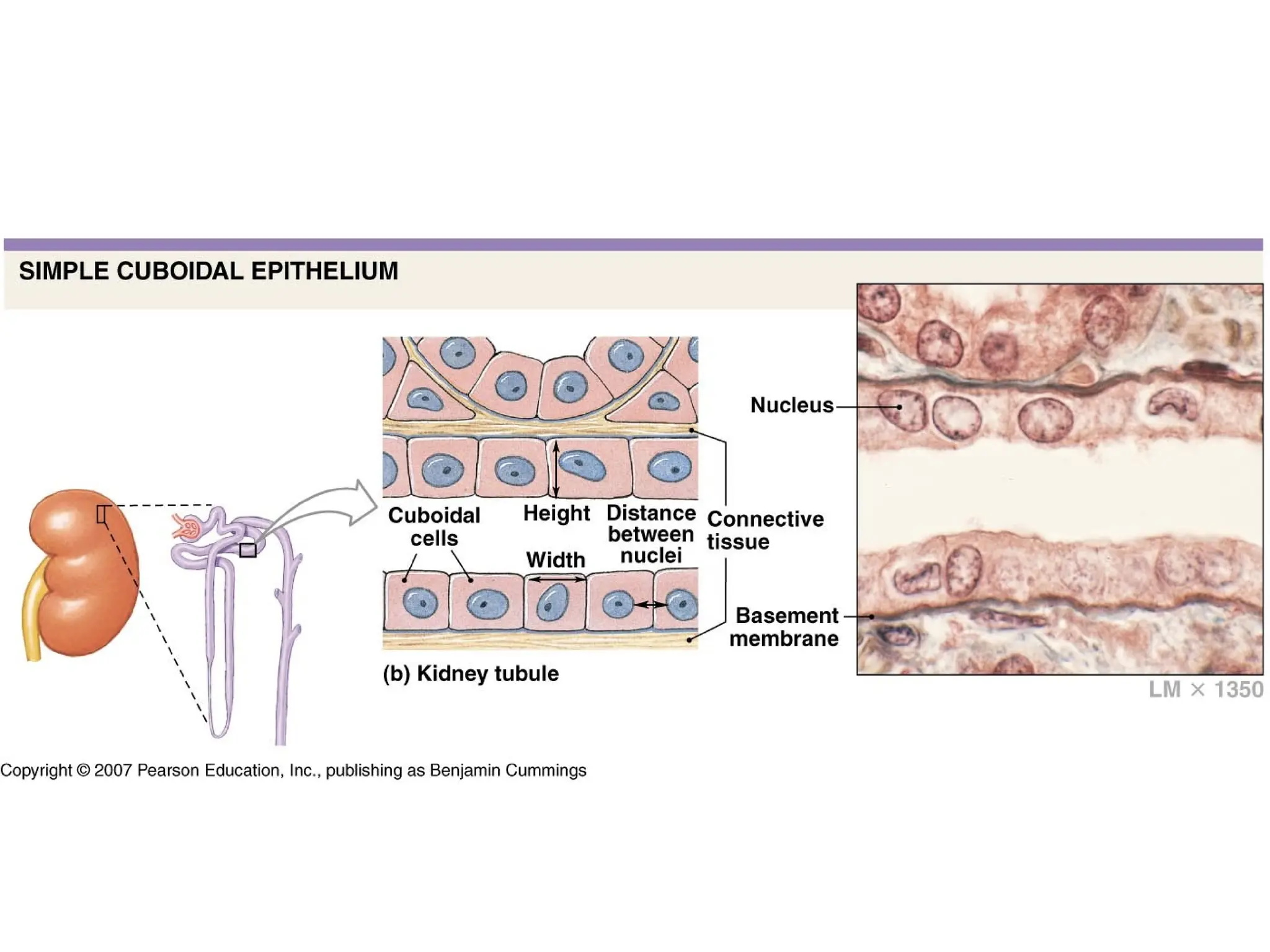
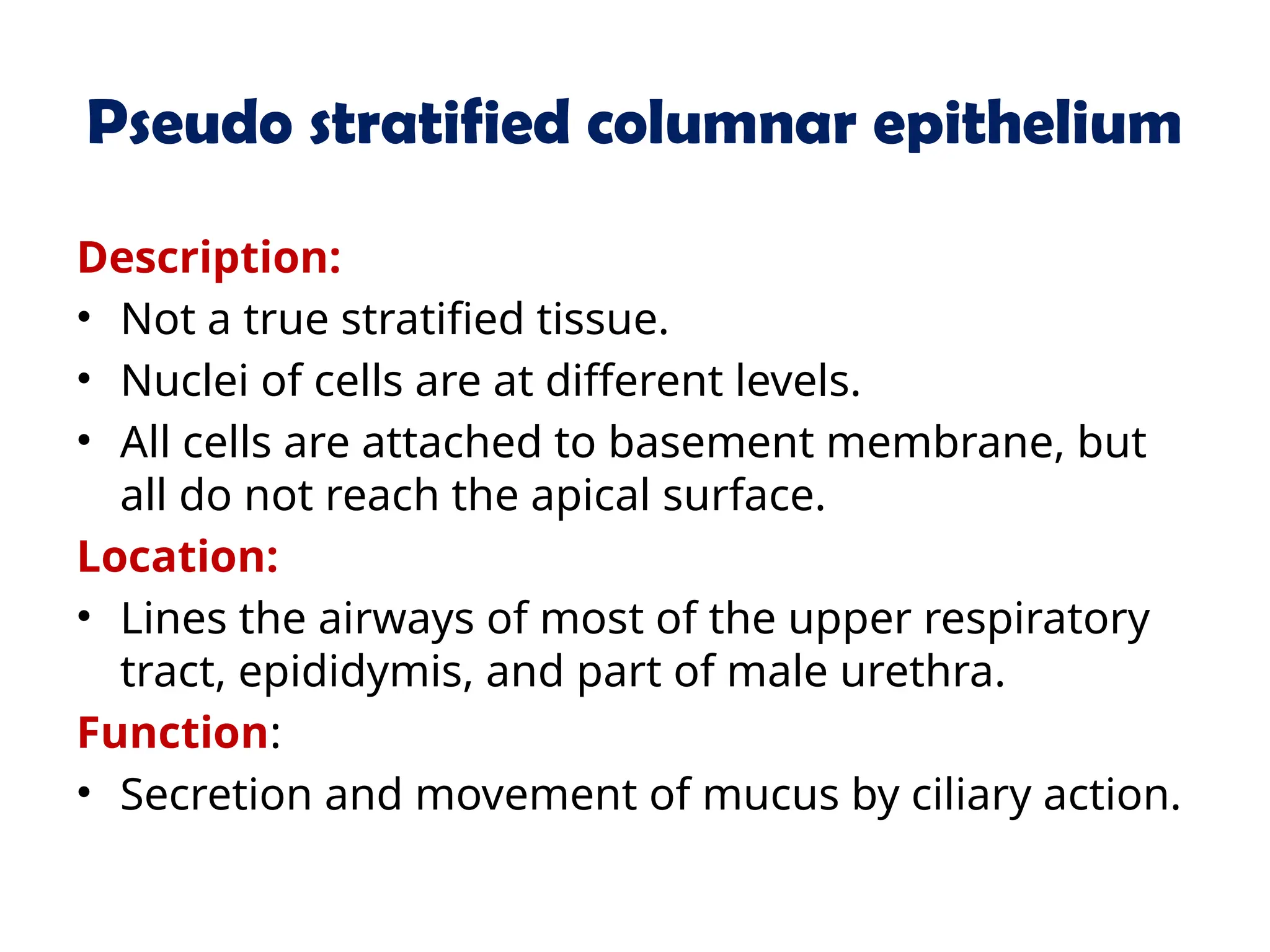
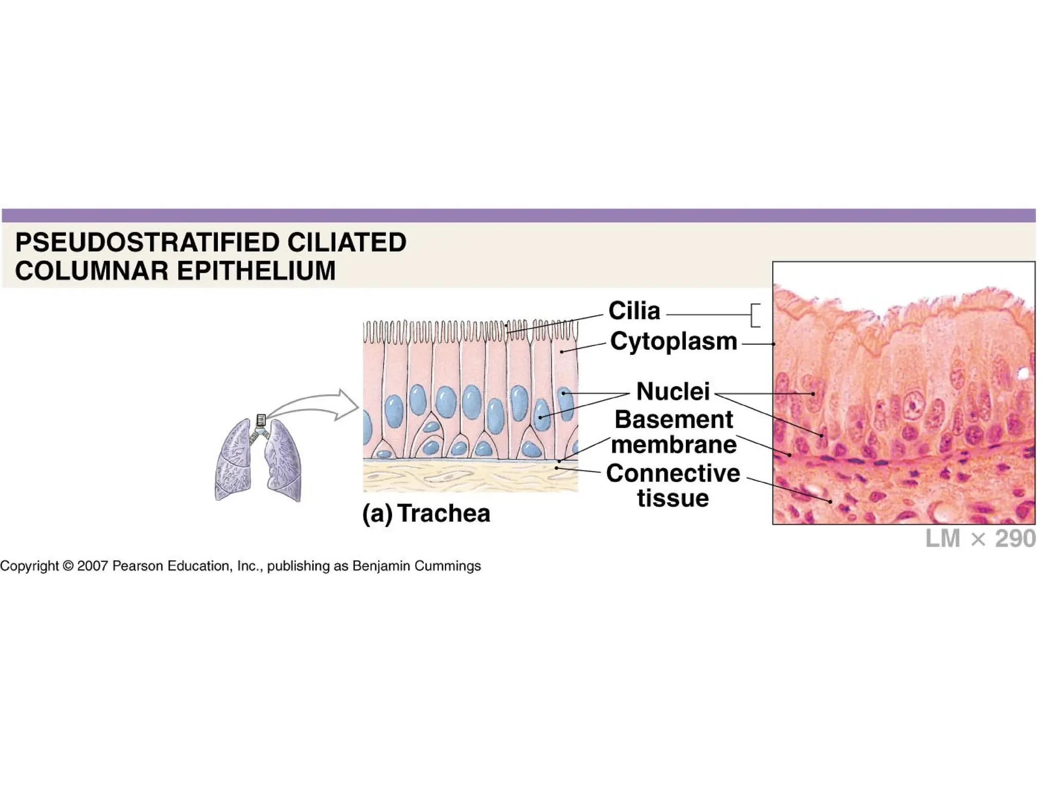
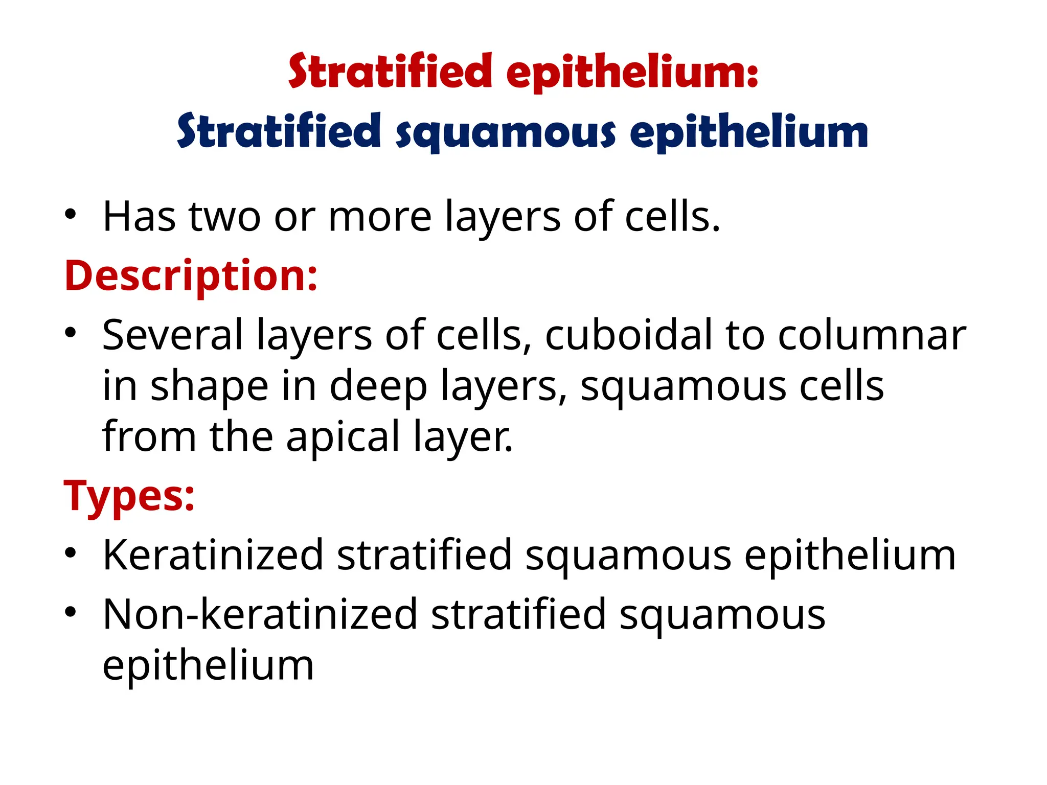
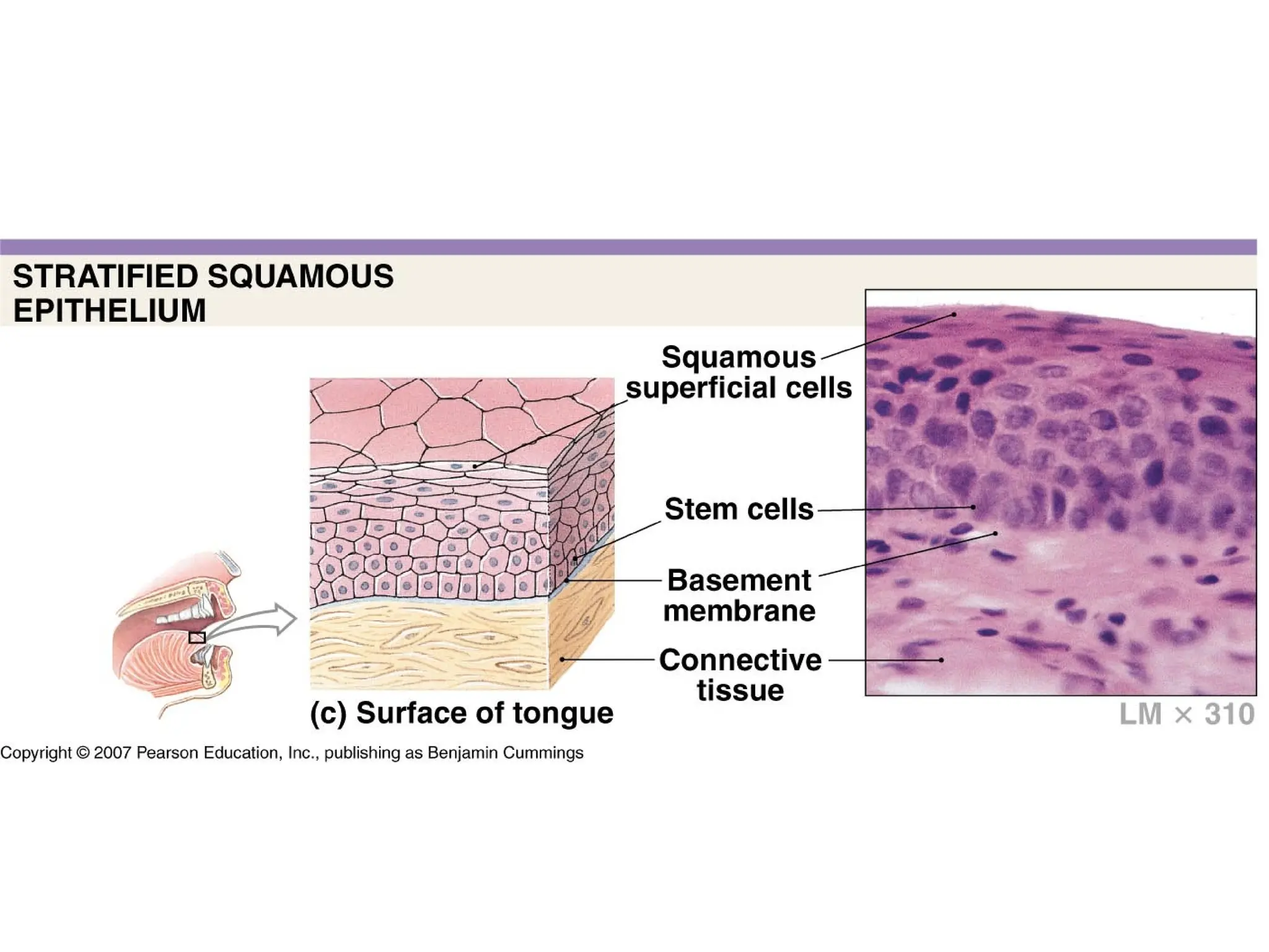
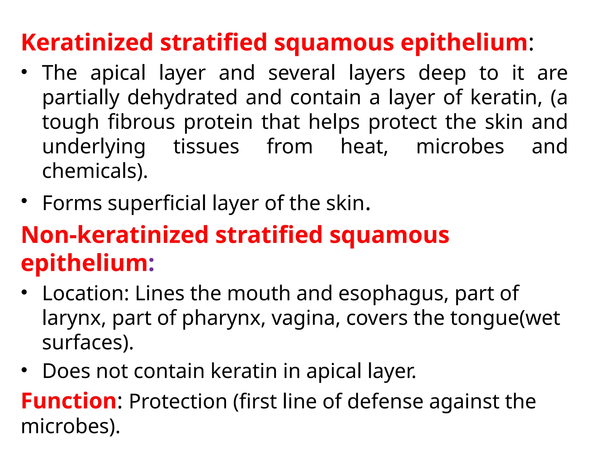
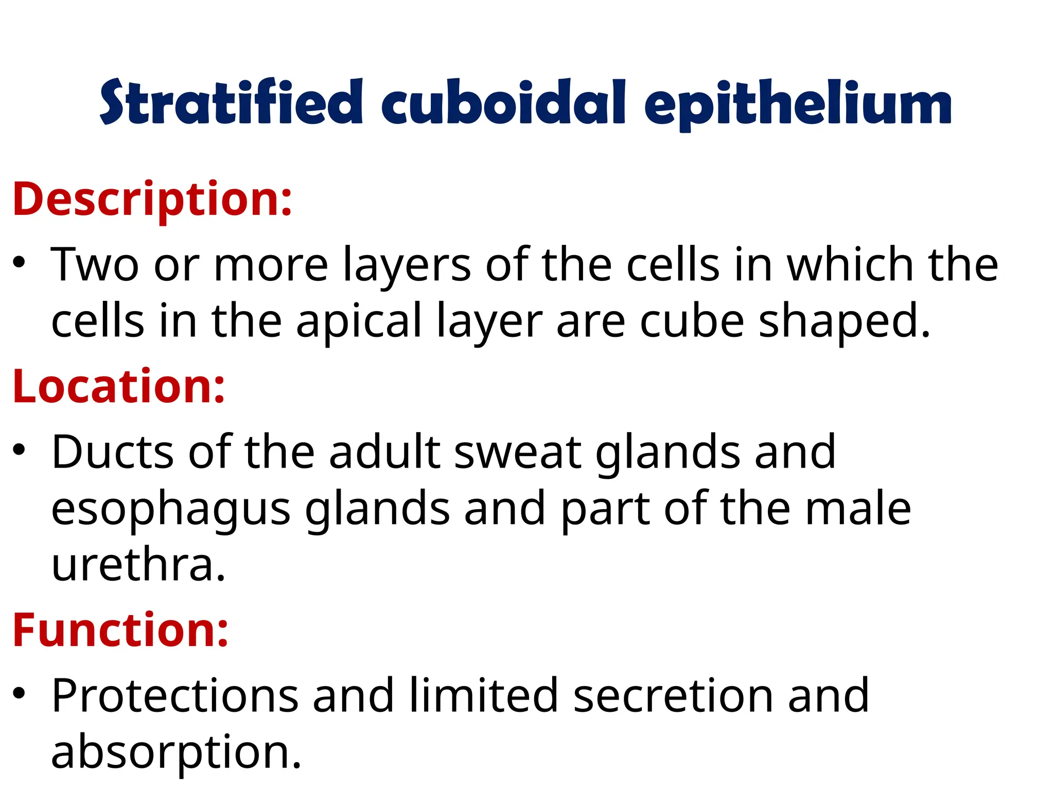
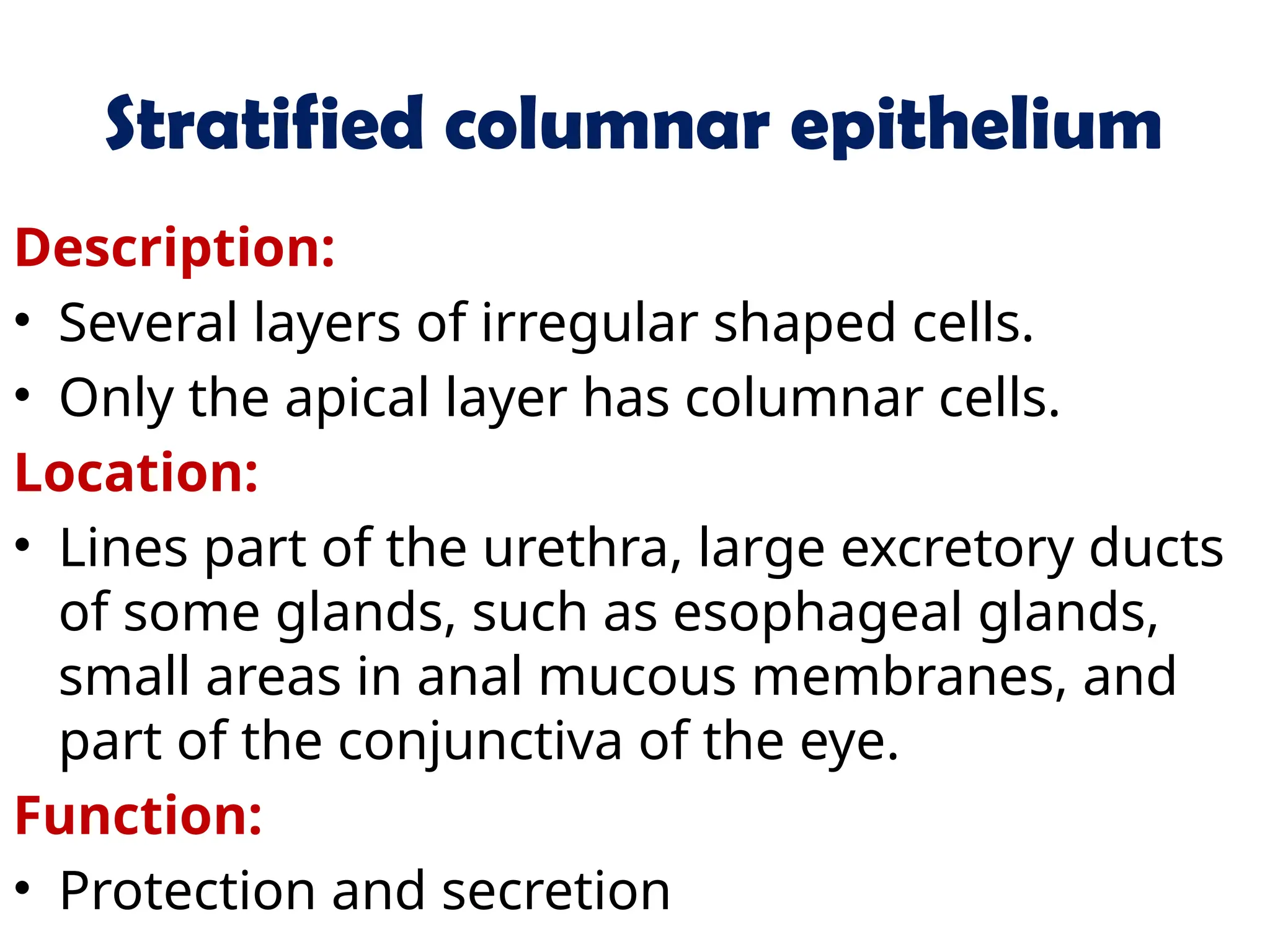
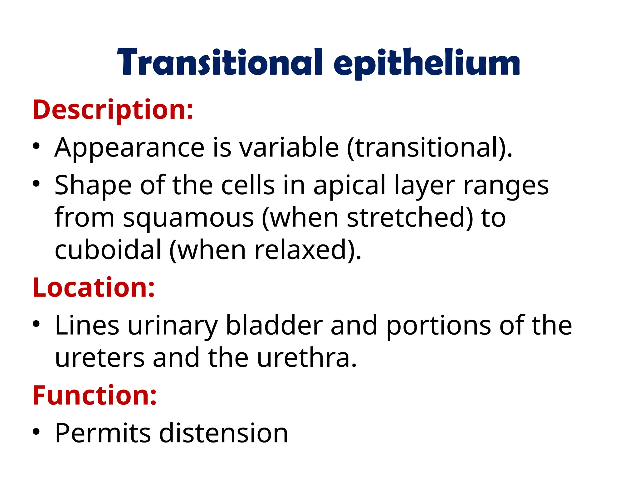
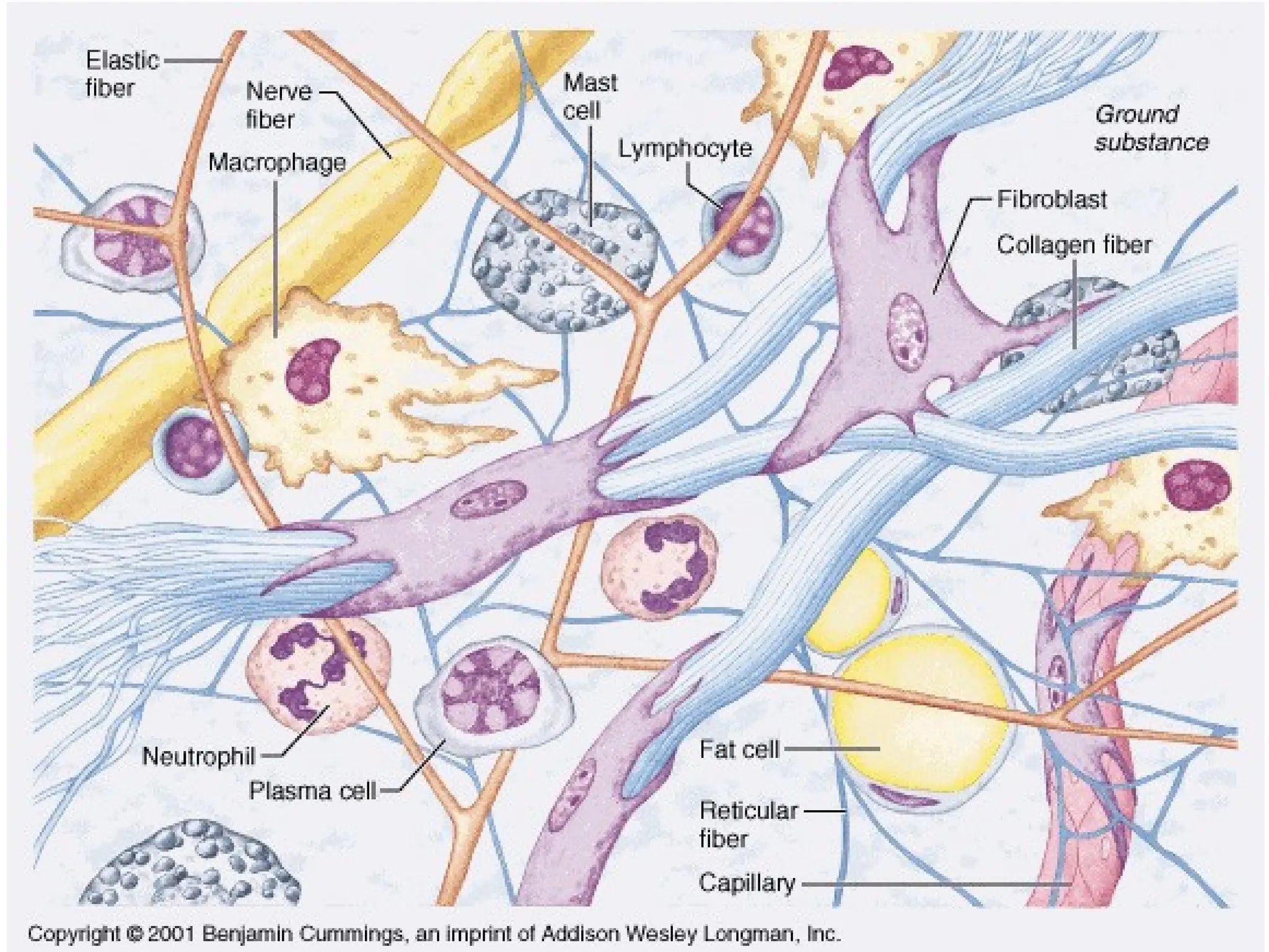
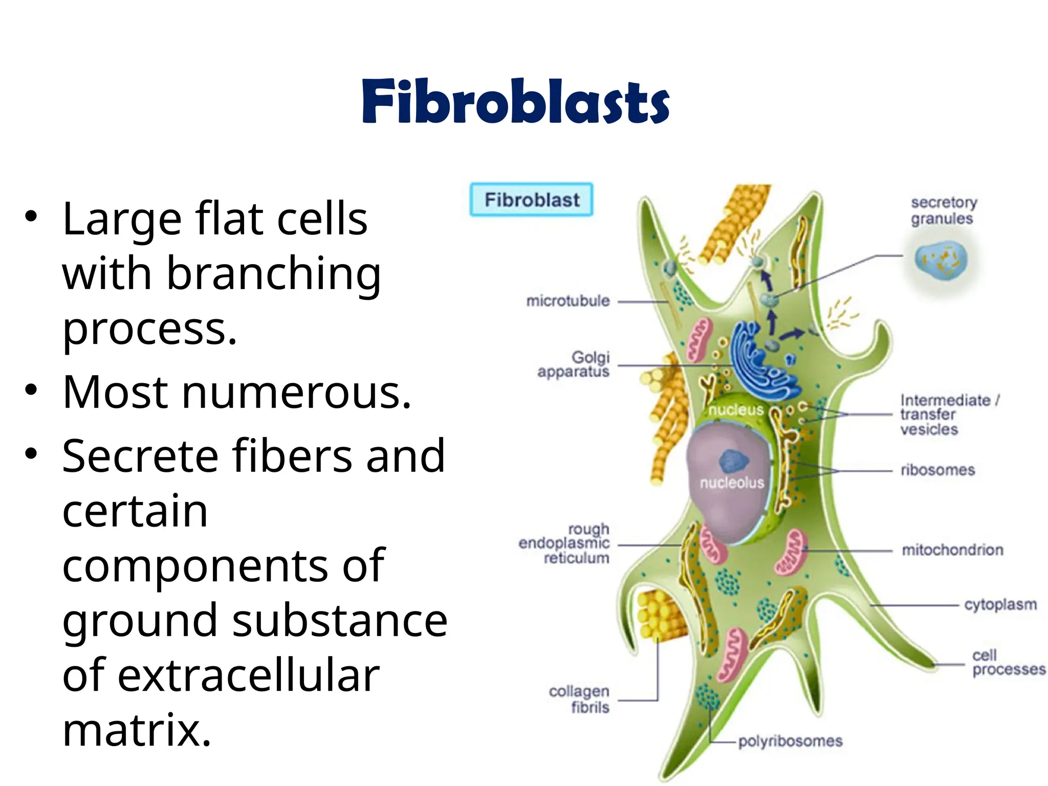
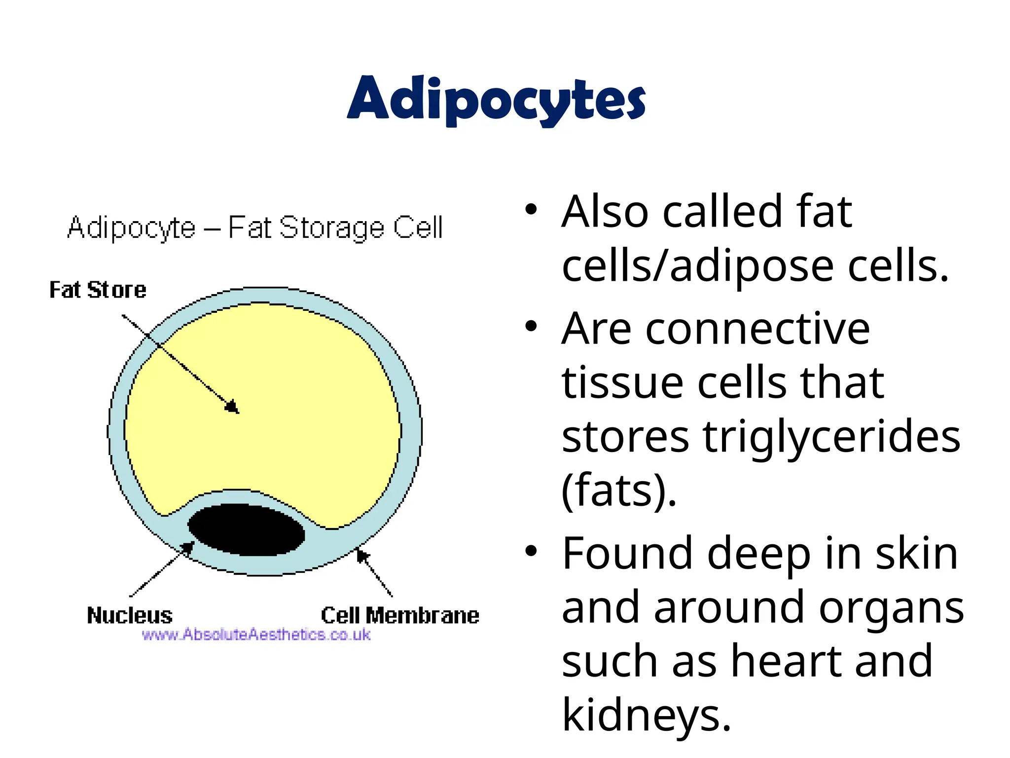
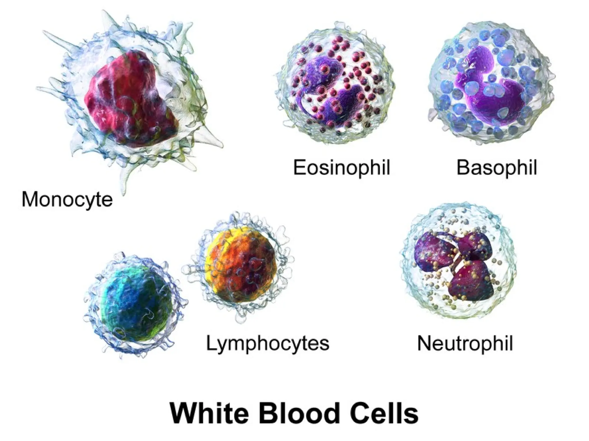
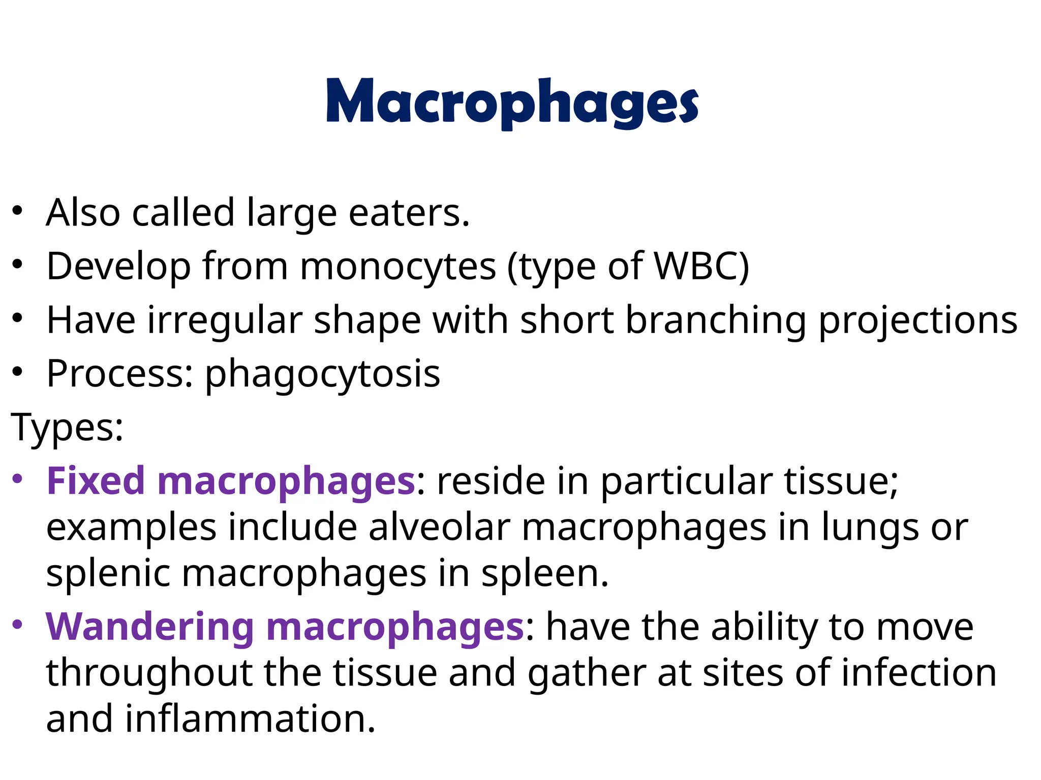
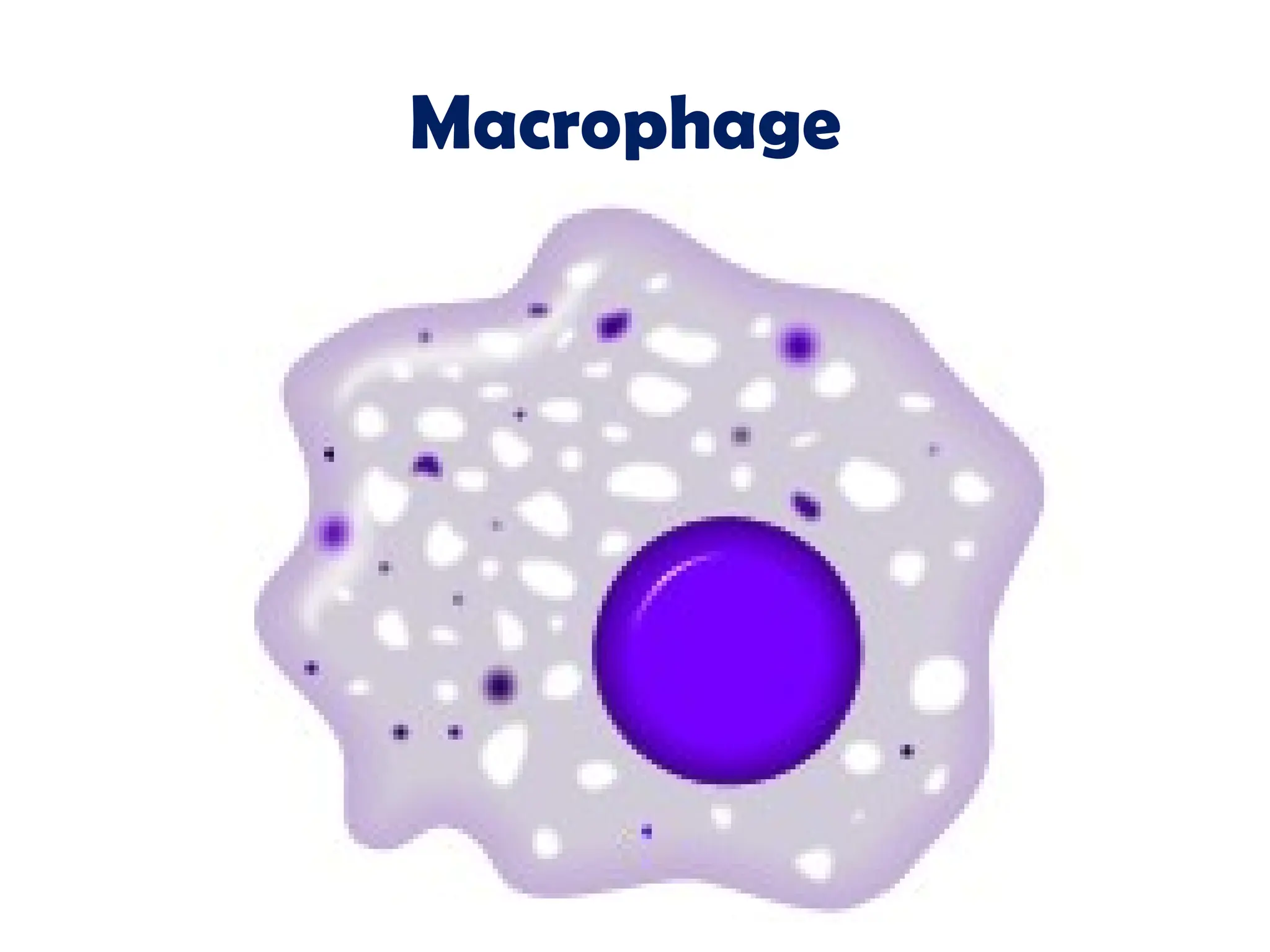
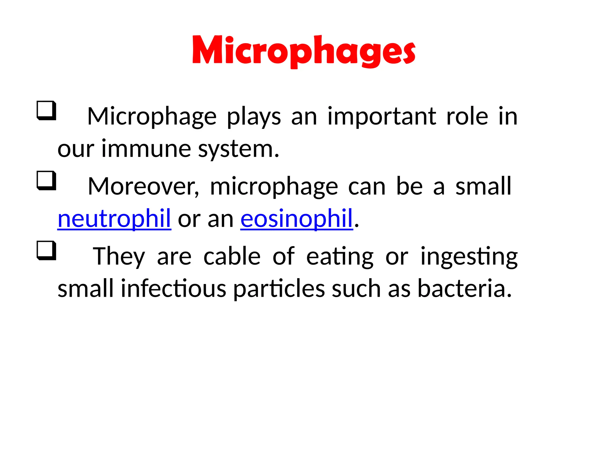
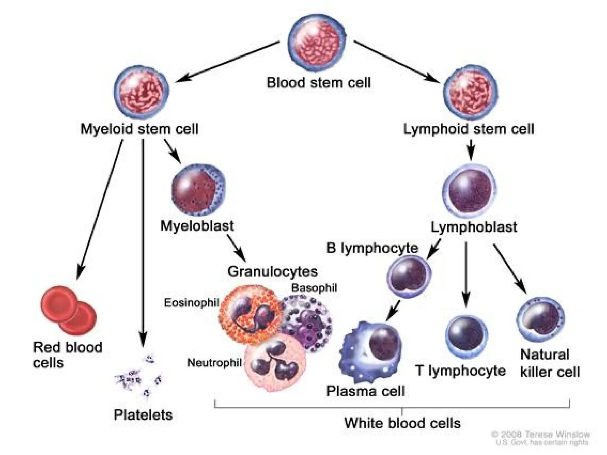
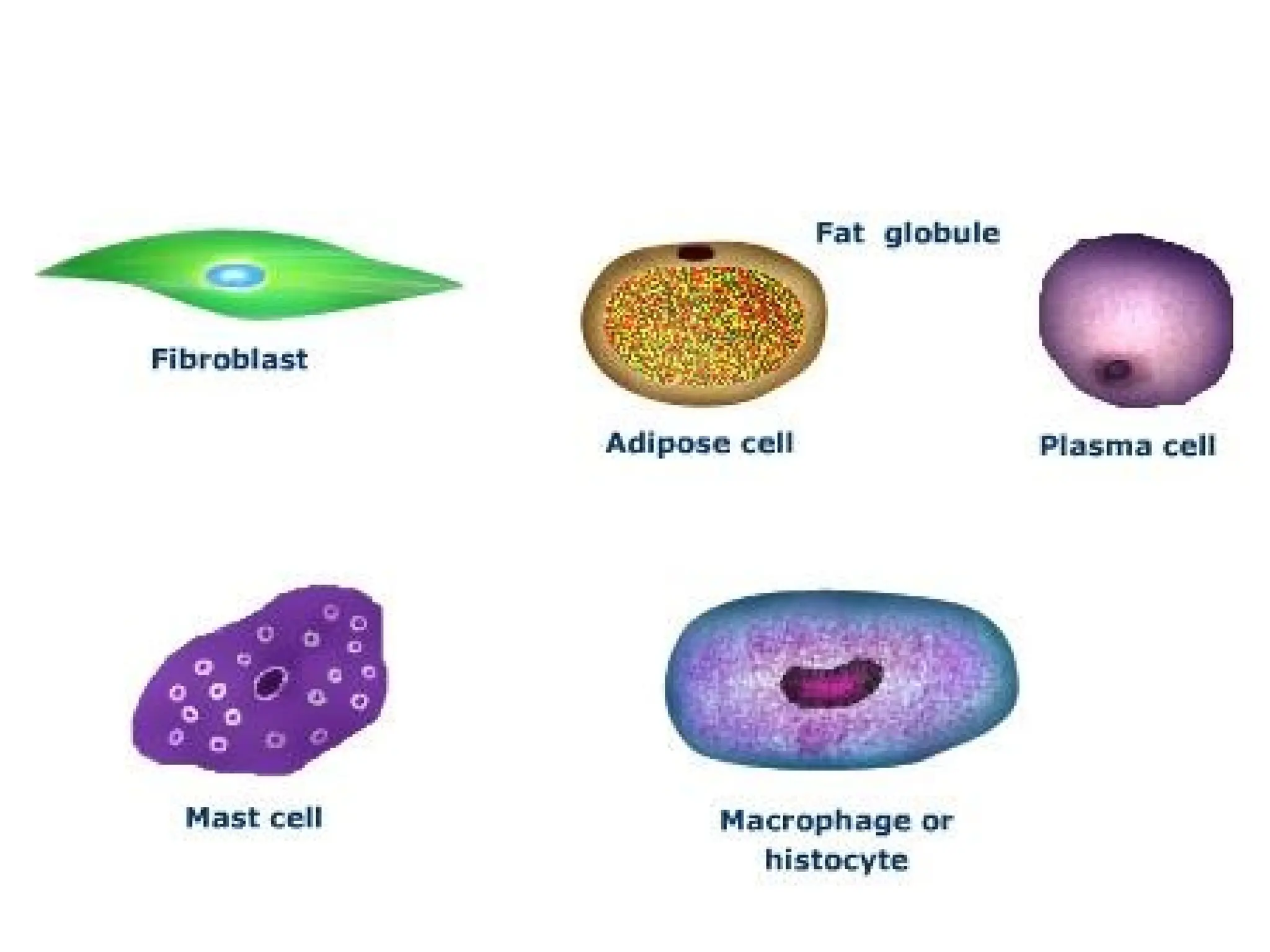
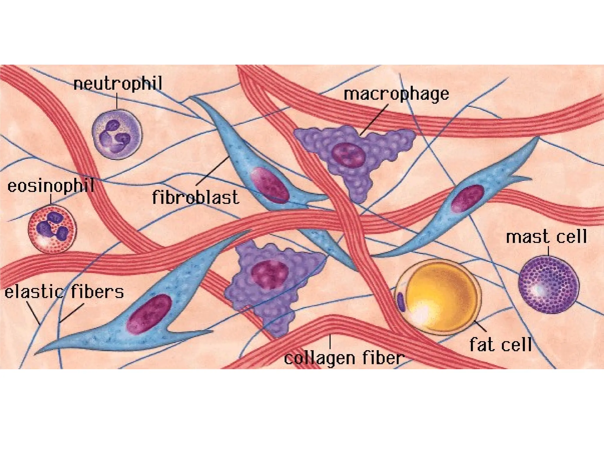
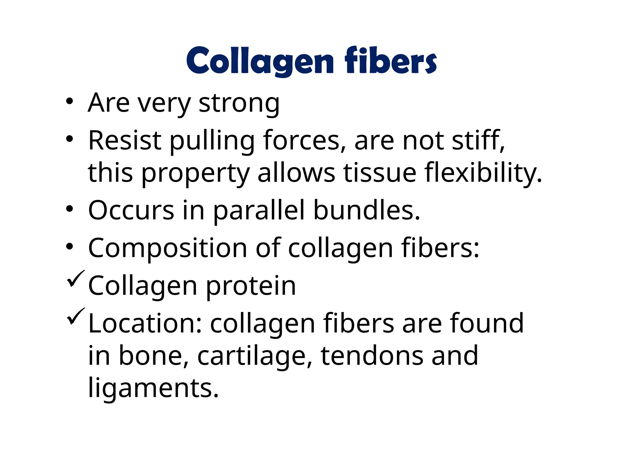
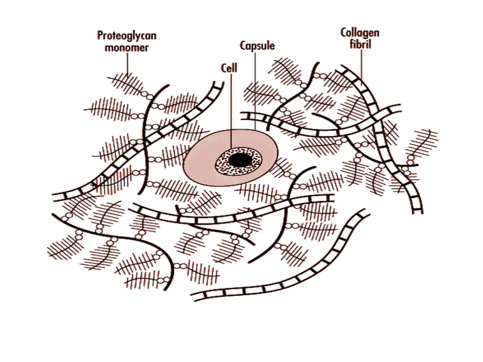
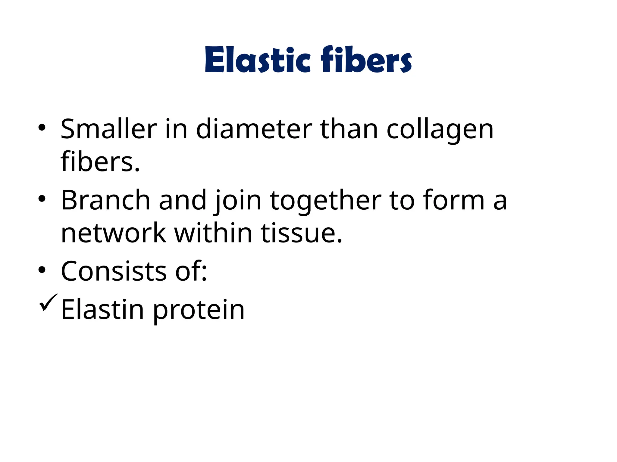
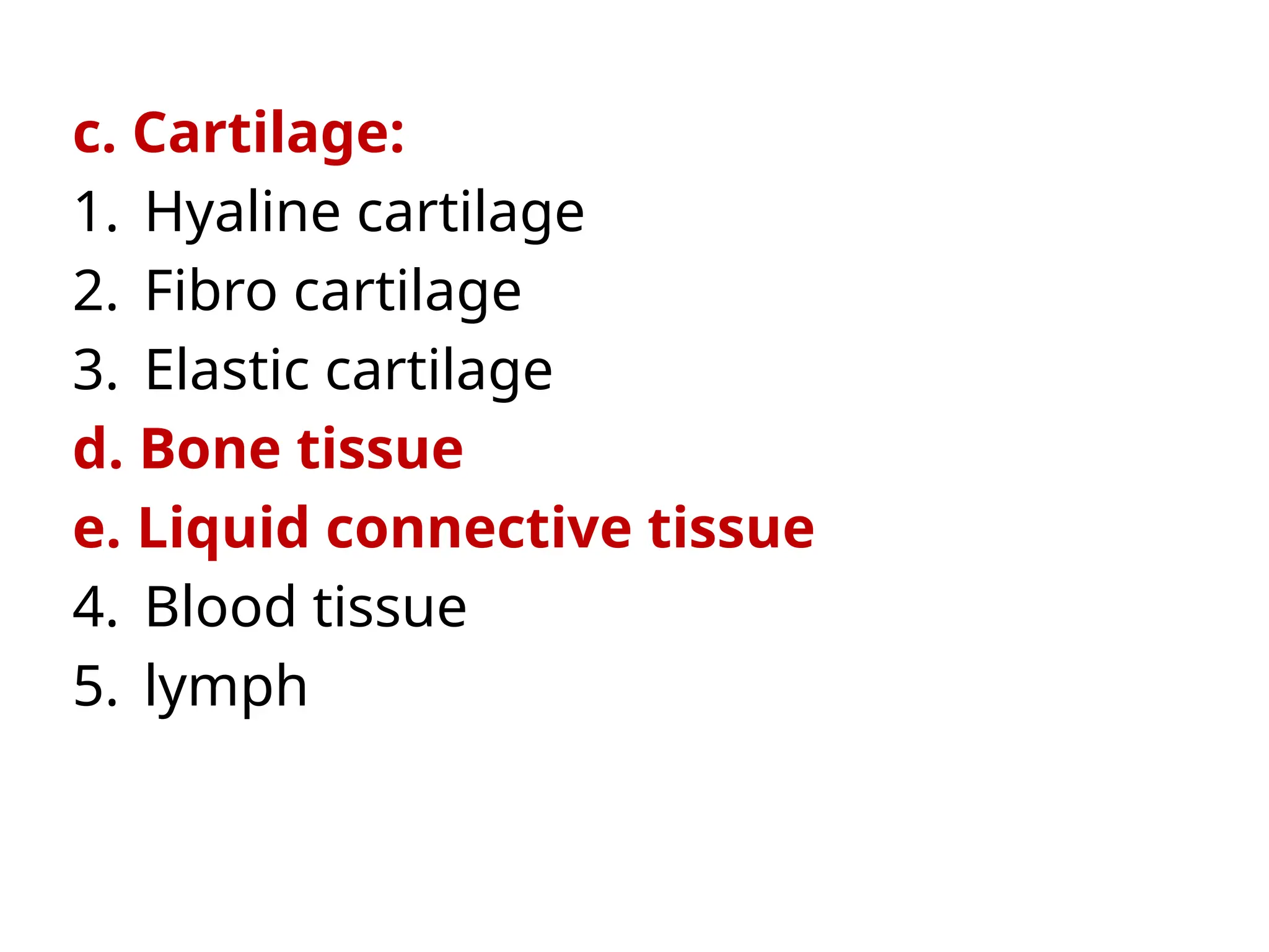
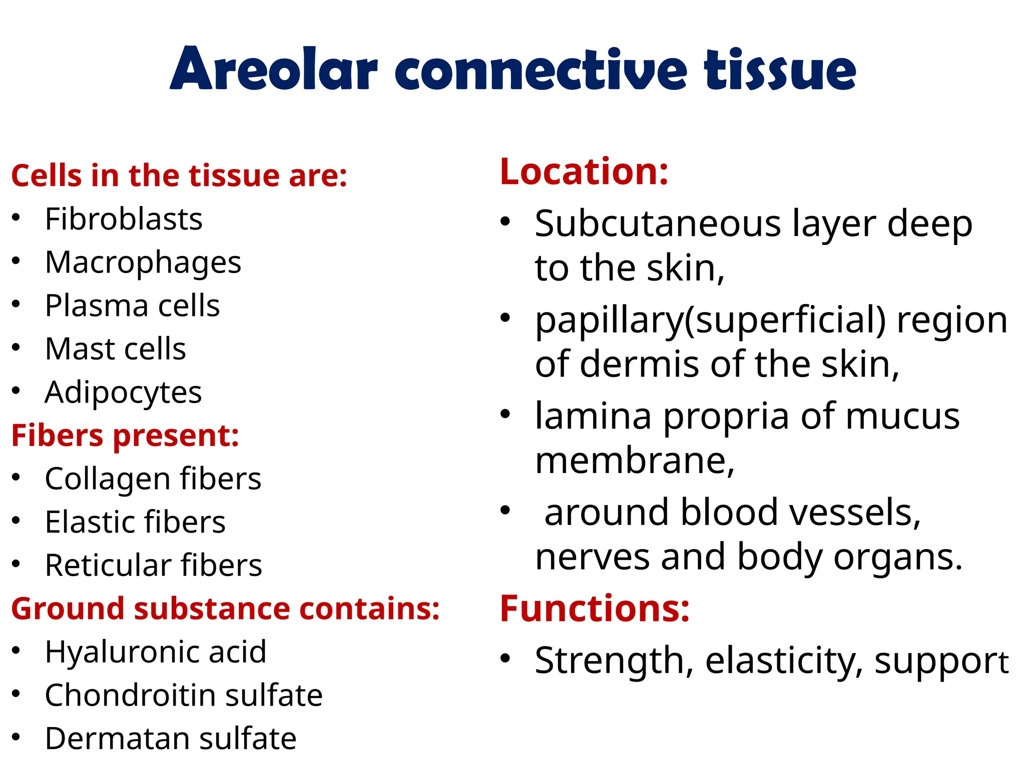
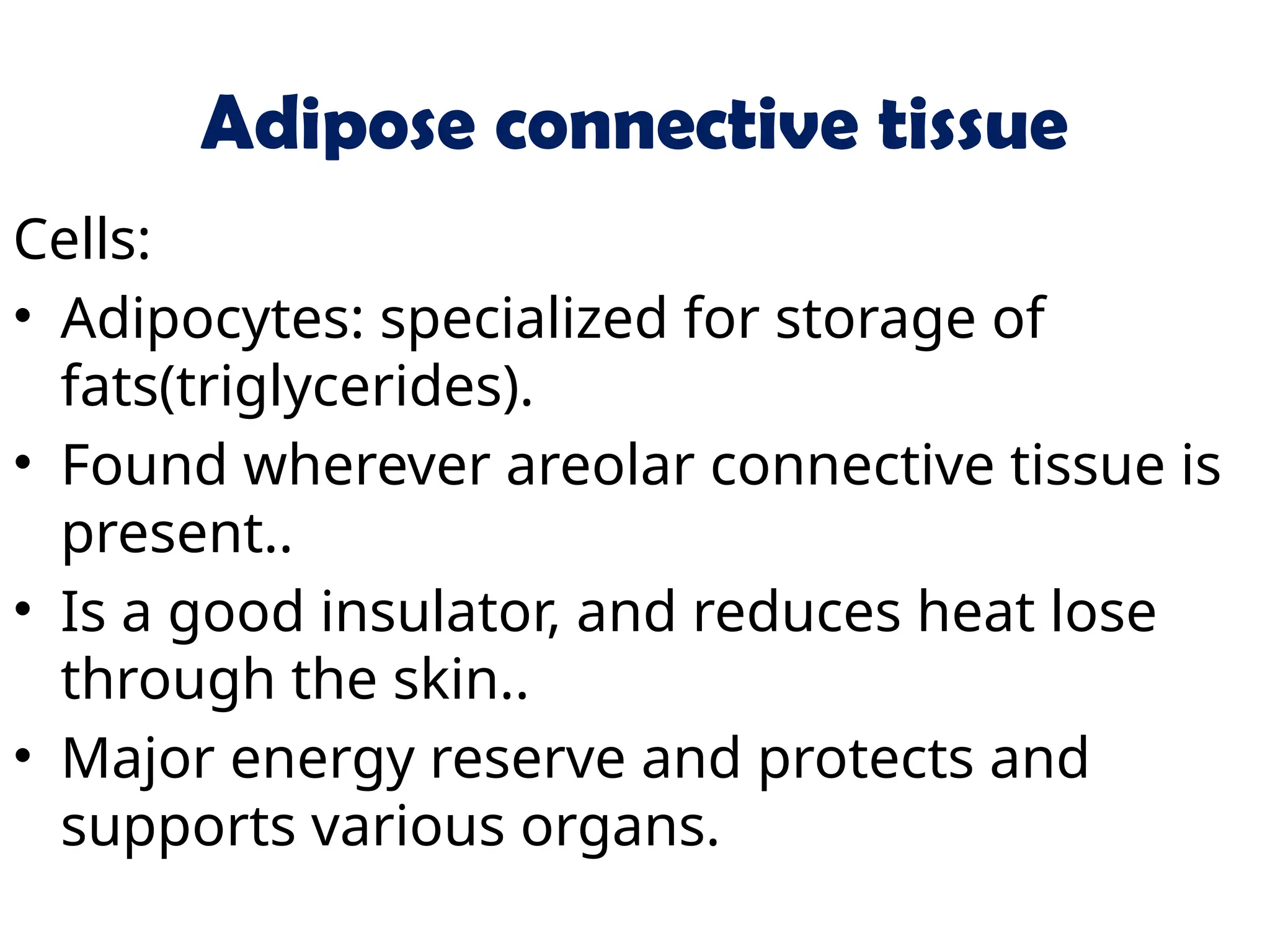


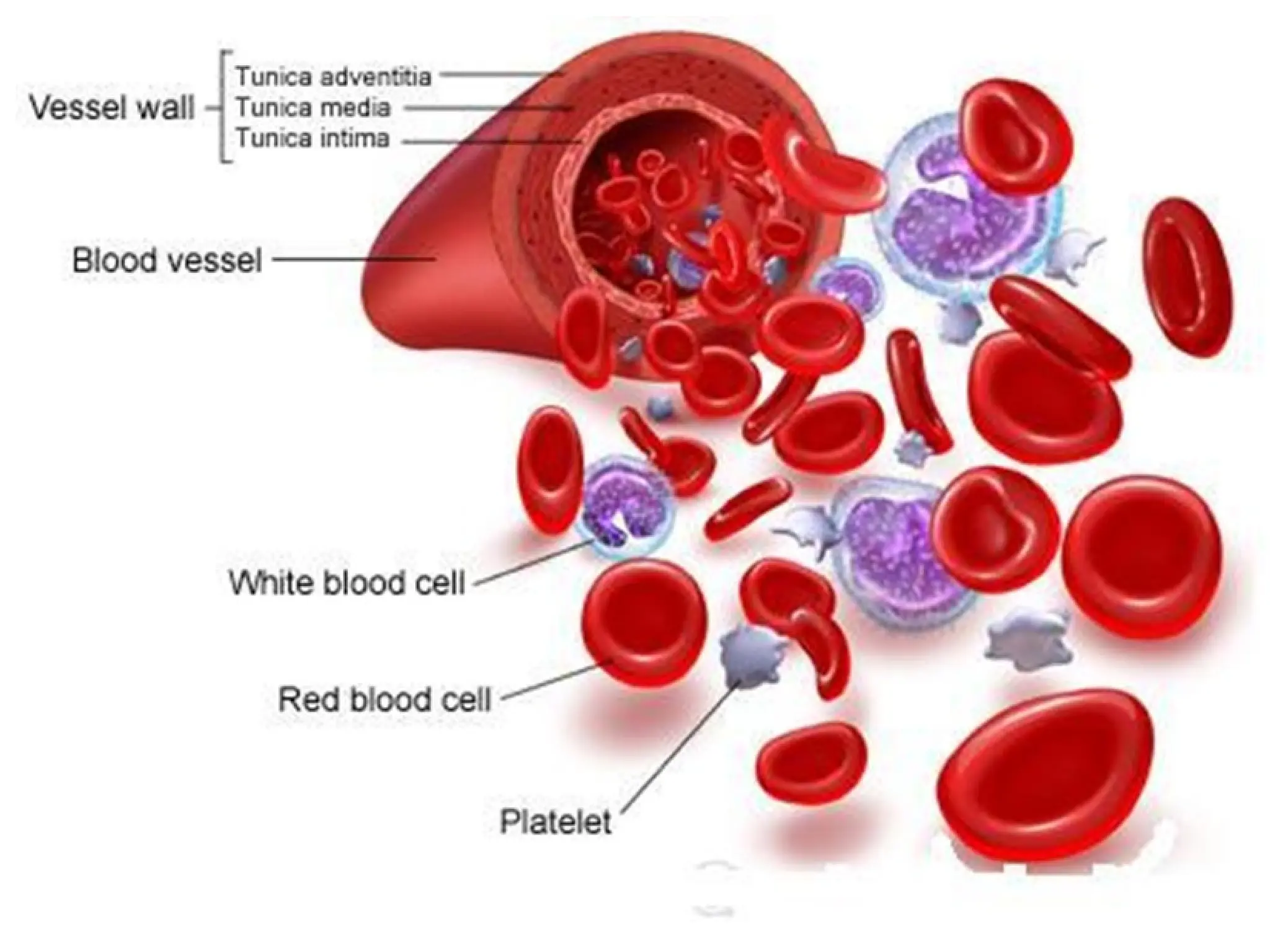
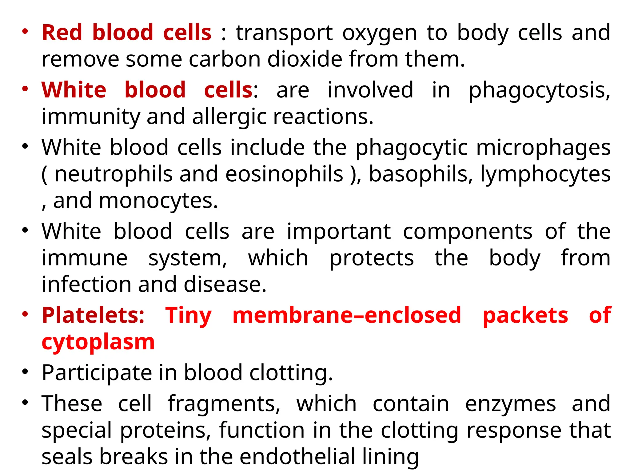
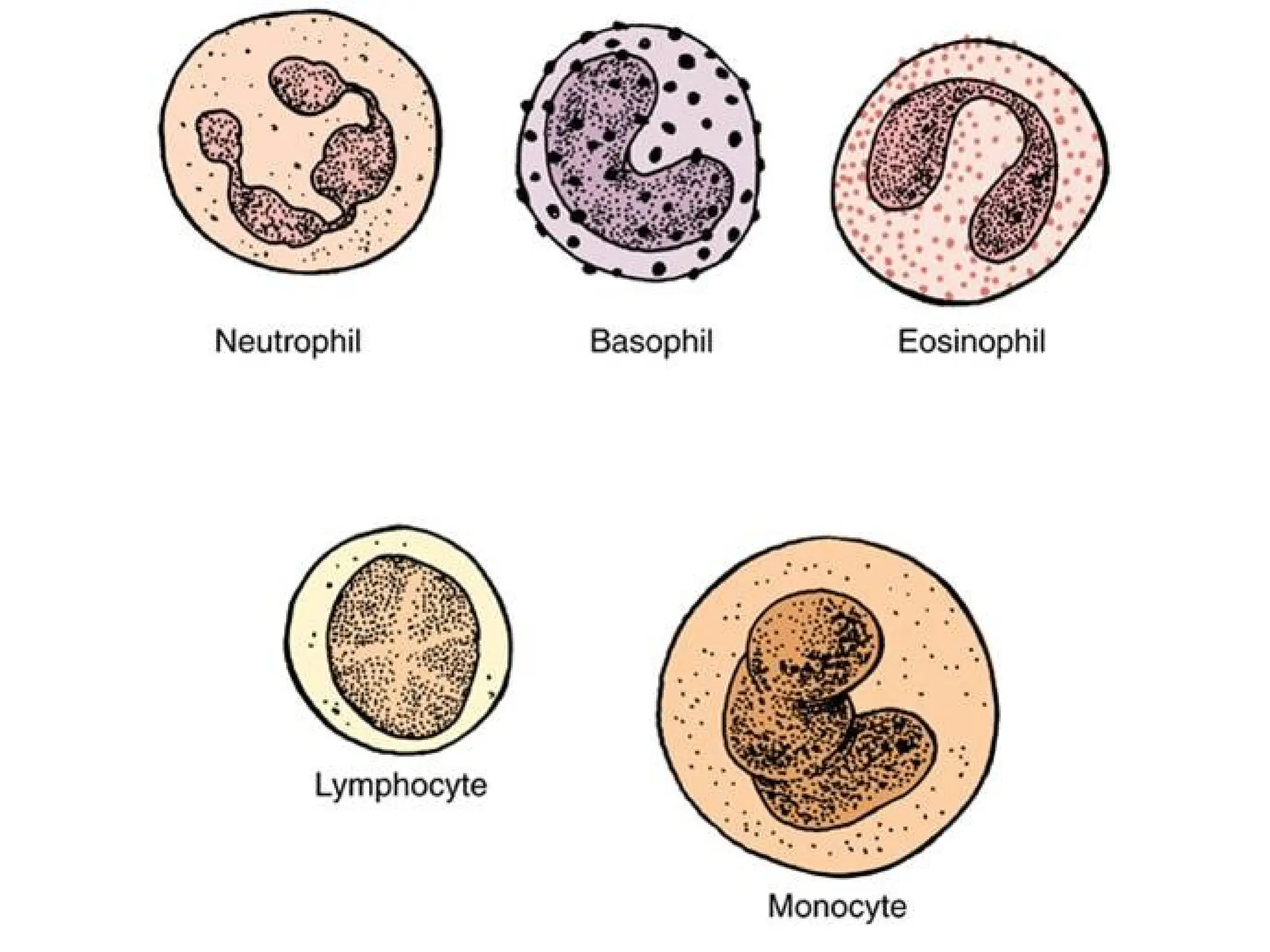
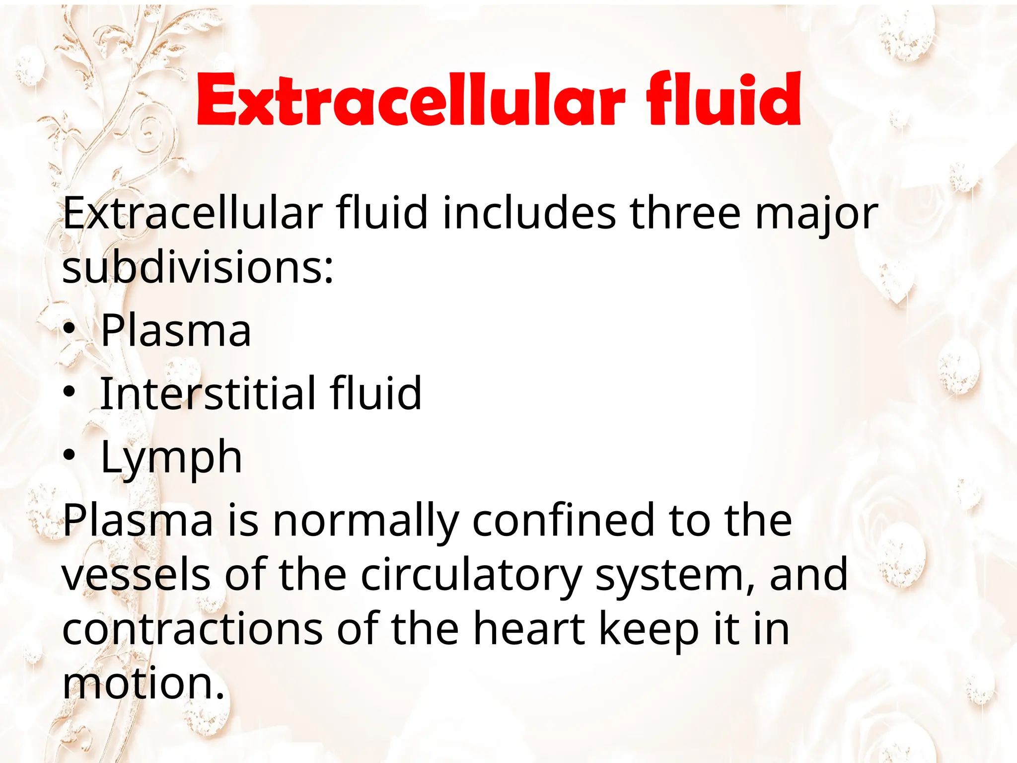
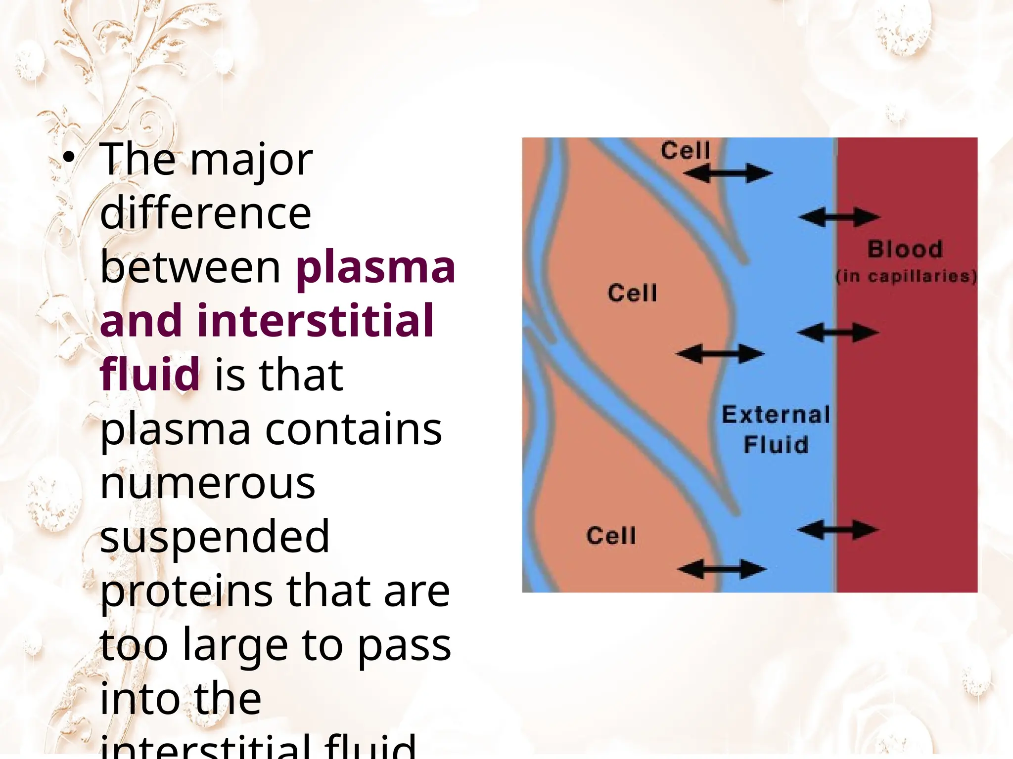
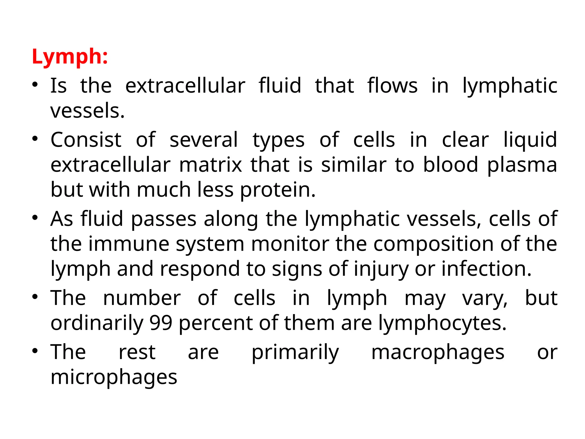
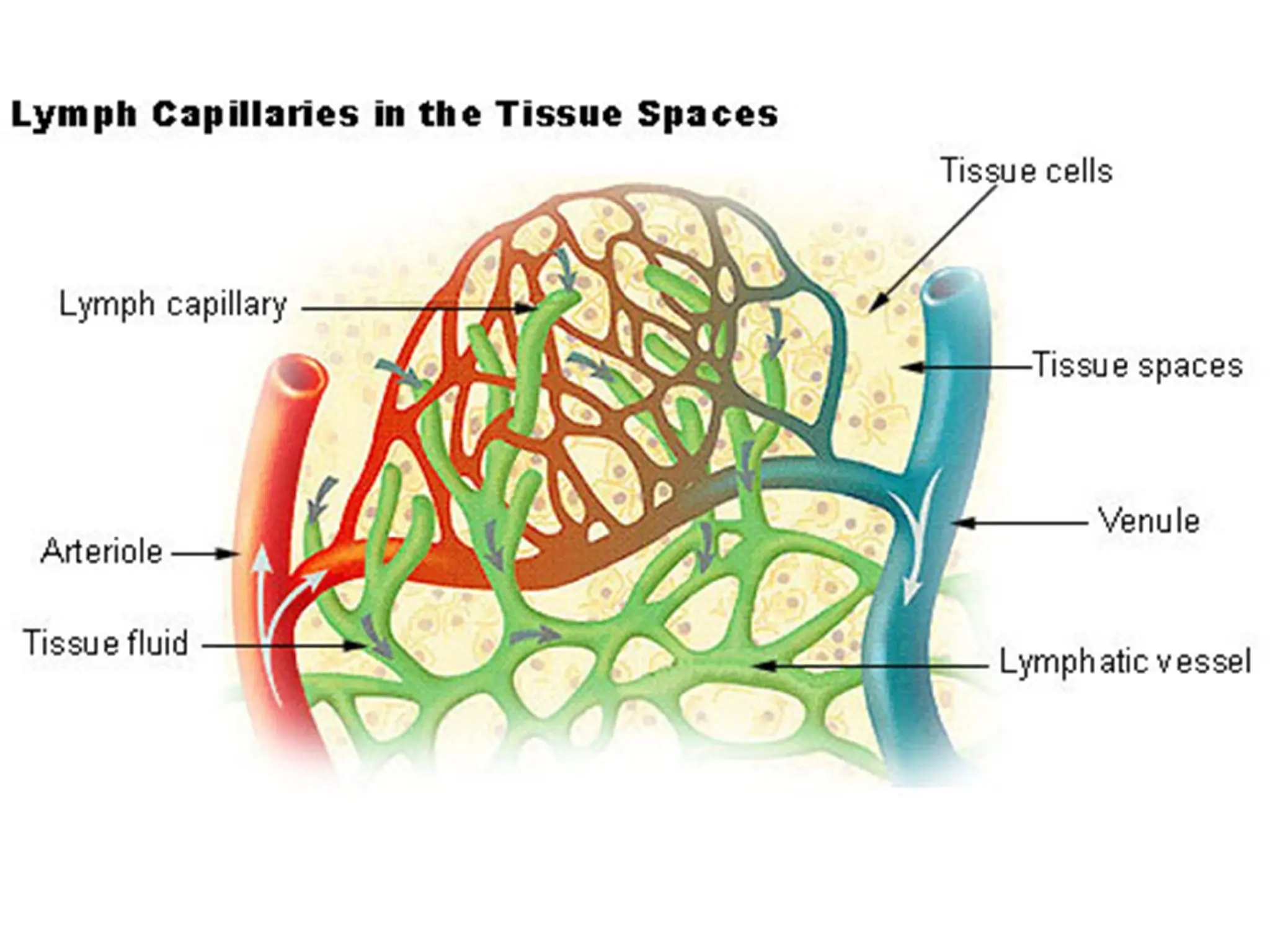
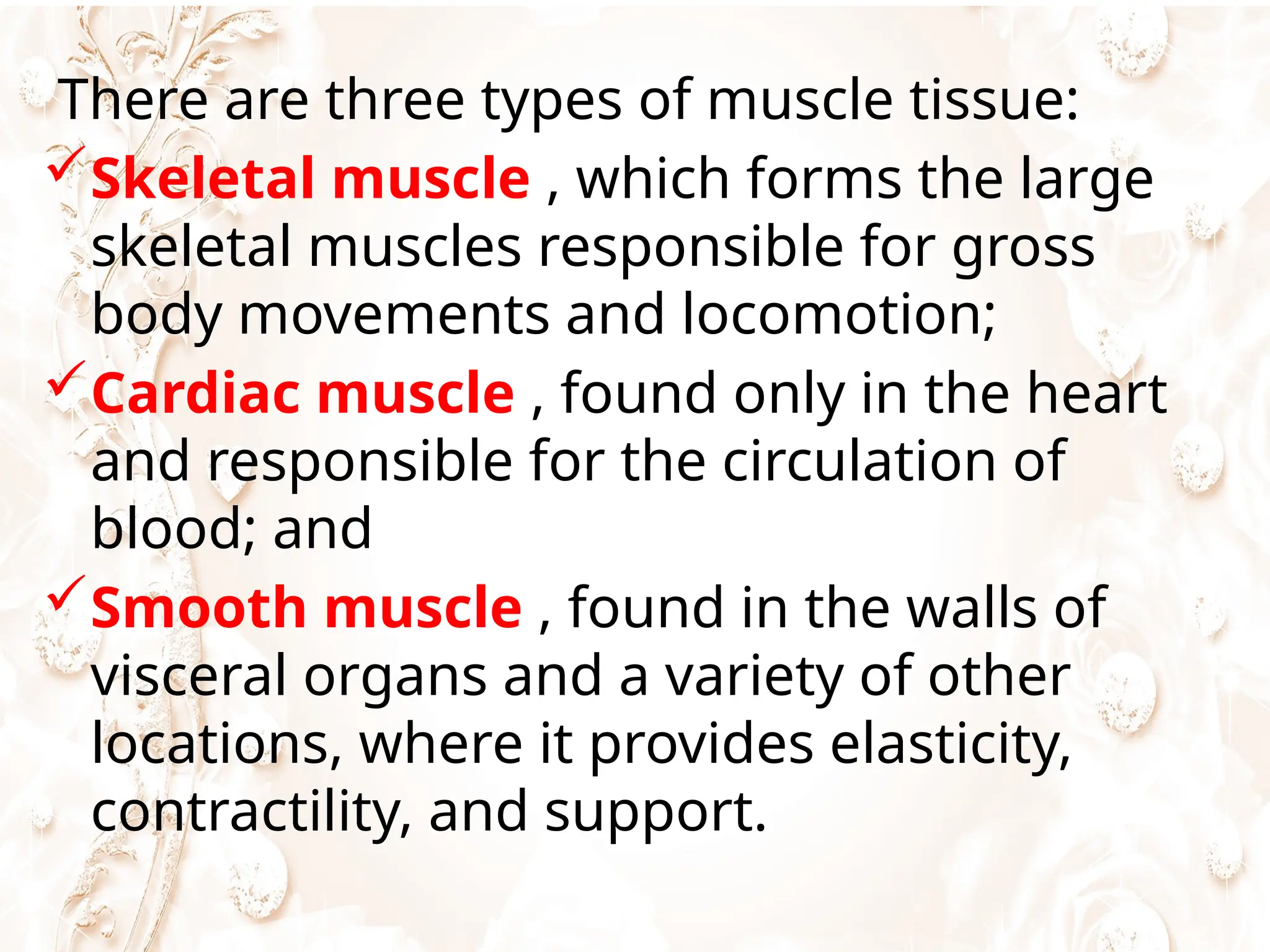
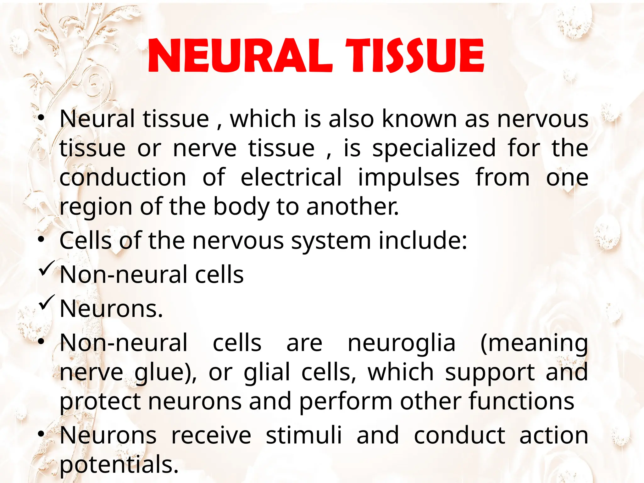
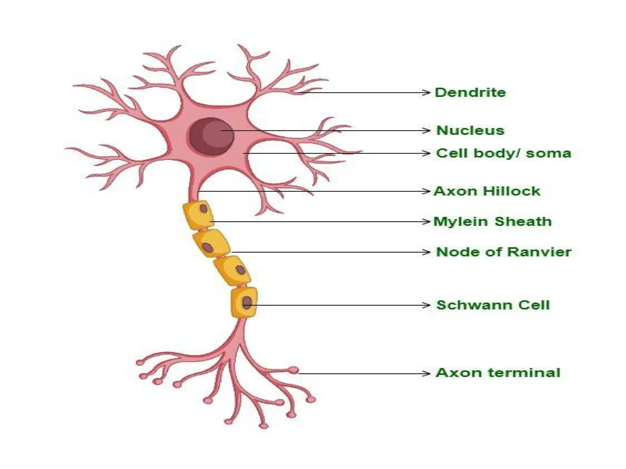
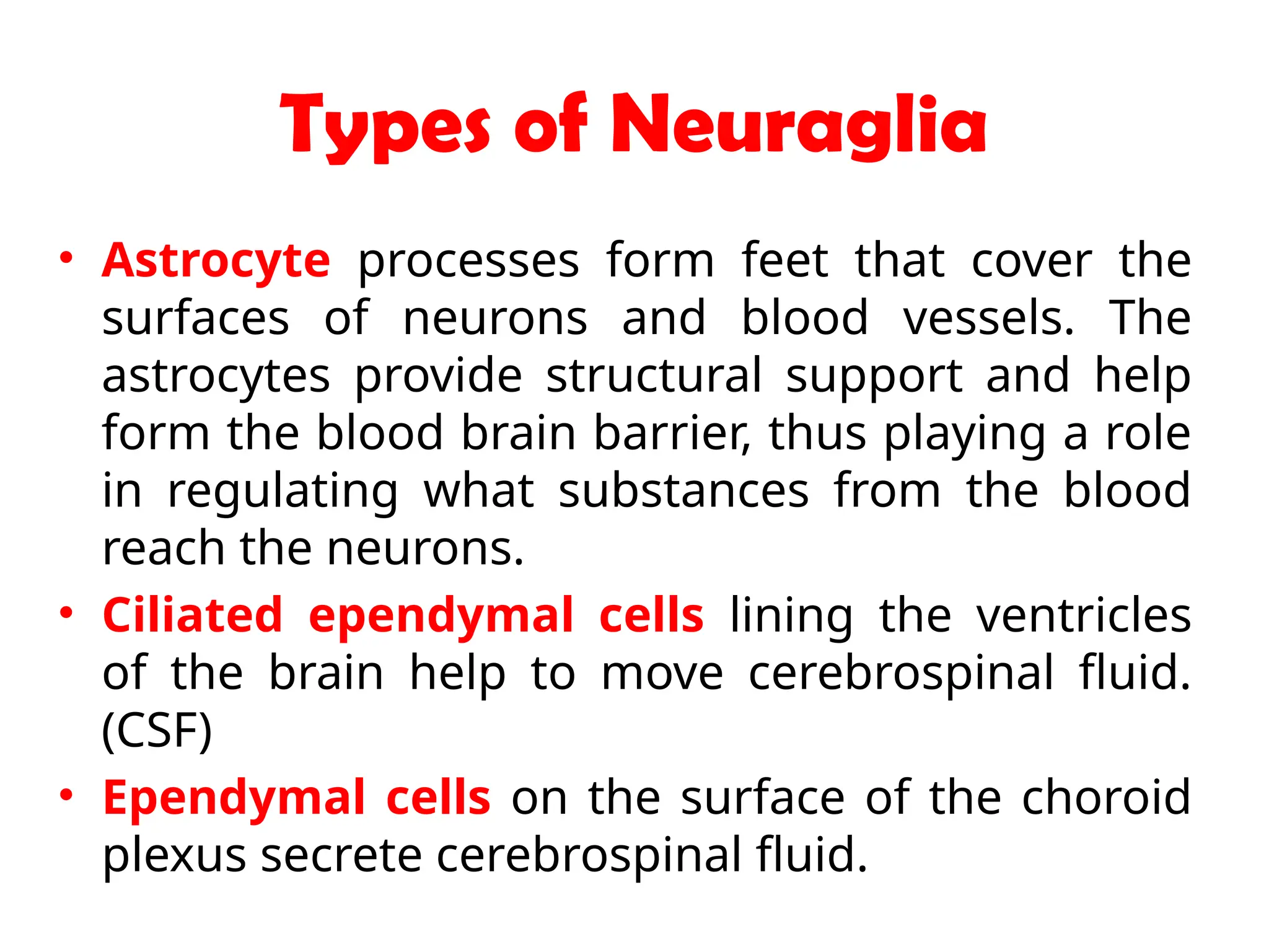


The document discusses tissue types in the body, encompassing their definitions, characteristics, and classifications, focusing on epithelial, connective, muscular, and nervous tissues. It describes the structures and functions of various epithelial tissues and the components of the extracellular matrix, along with connective tissue cells and their roles. Key details include the types of epithelial cells, their locations and functions, and the composition and characteristics of connective tissues.


