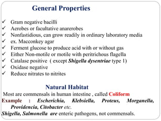
Enterobacteriaceae
Arun Kumar P
Dr. Roma A Chougale M.D
Introduction
Largest, most heterogeneous collection of medically
important Gram negative bacilli.
Normal habitat in Humans. These organisms are referred
to as “ Enteric Bacilli” or “Enterics”.
General Properties
 Gram negative bacilli
 Aerobes or facultative anarerobes
 Nonfastidious, can grow readily in ordinary laboratory media
 ex. Macconkey agar
 Ferment glucose to produce acid with or without gas
 Either Non-motile or motile with peritrichous flagella
 Catalase positive ( except Shigella dysentriae type 1)
 Oxidase negative
 Reduce nitrates to nitrites
Natural Habitat
Most are commensals in human intestine , called Coliform
Example : Escherichia, Klebsiella, Proteus, Morganella,
Providencia, Citobacter etc.
Shigella, Salmonella are enteric pathogens, not commensals.
Classification
Oldest method :
Classified based on fermentation of lactose on macconkey
agar.
Most widely used classification having great practical
application in laboratories to differentiate various members.
Lactose Fomenters Produce pink
color colonies
Escherichia,
Klebsiella
Non lactose Fomenters Pale or colorless
colonies
Salmonella,
Shigella, Proteus,
Morganella,
Providencia ,
Yersinia.
Late Lactose Fomenters Pink color
colonies – 2 days
of incubation
Shigella sonnei
Tribe Genus
Tribe I : Escherichiae Escherichia, Shigella.
Tribe II : Edwadrsielleae Edwardsiella
Tribe III : Salmonelleae Salmonella
Tribe IV : Citrobactereae Citrobacter
Tribe V : Klebsielleae Klebsiella, Enterobacter, Hafina,
Serratia, Pantoea
Tribe VI : Proteeae Proteus, Morganella, Providencia
Tribe VII : Yersinieae Yersinia
Tribe VII : Erwinieae Erwinia
Newer Method :
 Availability of Molecular methods, Taxonomy is greatly changed
 Tribe classification was proposed by Ewing. It has great impact
in laboratory for easy identification.
1. Escherichia
Escherichia coli
It was first described by Odar Escherichia in 1885
Genus consist of 5 species – E. coli, E. fergusonii, E. hermannii,
E. vulneris, E. blattae.
Of these E.coli – most common and clinical important
Most common aerobe to be harbored in the gut .
After excreted in feces, it remains viable only for some days in
environment, hence it is taken as fecal indicator.
Morphology :
 Gram negative Bacilli
 Motile by peritrichate flagella, some strains – non motile.
 Capsule
 Non spore former.
Cultural Characteristics :
 Aerobe or facultative anaerobe
 Nutrient Agar: Large, Thick, Grayish, White, Moist,
Smooth, Opaque, or Partially Translucent discs.
 Blood Agar: Hemolytic
 Macconkey Agar : Pink in color ( LF colonies)
 EMB Agar : Golden yellow metallic sheen
Blood agar Macconkey Agar
EMB agar
Biochemical Reactions :
Ferments – Glucose, Lactose, Mannitol, Maltose.
Indole – positive
Methyl Red - positive
VP – Negative
Citrate- Negative ( IMViC ++-- )
Urease – Negative
H2S Production - positive
Gelatin - positive
TSI- A/A, G+
Indole MR VP
citrate
+Ve +Ve - Ve
-Ve
Virulence Factors :
1. Surface factors , 2. Toxins.
Surface Antigens :
A. Somatic ‘ O’Antigen - Lipopolysaccharide Antigen.
Responsible for endotoxic activity
Protect Bacteria from Phagocytosis
Bactericidal effect of complement.
B. Flagellar ‘H’ antigen - Heat labile,
makes bacteria motile.
C. Capsular ‘ K’ antigen - polysacchaide capsular antigen
inhibiting the phagocytosis
present only in few strains causing neonatal
meningitis, polynephritis, and speticemia.
Fimbrial antigen - helps in attachment and colonization
CFA- colonization factor antigen- Enterotoxigenic E.coli
Mannose resistant fimbriae ( P,M,S,F1C,Dr fimbriae)-
haemagglutinate with RBCs – Uropathogenic E.coli
Toxins
Exotoxins- E.coli produce two types of exotoxin
Hemolysin. Enterotoxin
Produced more commonly by
Virulent strain of E.coli.
Role in pathogenicity is not
clear.
Produced by Diarrheagenic strains.
3 types,
 Heat labile toxin(LT),
Heat stable toxin(ST)
Verotoxin (VT) also known as Shiga
–like toxin(SLT).
Heat Labile Toxin(LT)
Produced by Enterotoxigenic E.coli, Plasmid - coded
It resemble Cholera toxin in its structure and function but less potent
Mechanisms : 2 peptide fragments,(A and B).
Fragment B: binding fragment, 5 Subunits.
toxins binds to GM1 ganglioside receptors present on the intestinal
epithelium.
following which A fragment is internalized and cleaved into A1 and
A2peptides.
Fragment A:
Fragment A2 helps in tethering A and B subunits together.
Fragment A1 (Active ), activates adenyl cyclase in the enterocyte
Cyclic adenonsine 5’Monophosphate
Increased outflow of water and electrolytes in the gut lumen,
diarrohea
Detection:
In vivo In vitro
Ligated Rabbit ileal loop test-
Positive ( Fluid accumulation in
loop)- 18 hours.
Adult Rabbit skin Test
Tissue culture tests
Steroid Production in Y1 mouse
adernal cell culture.
Elongation in Chinese hamster ovary
cells precipitin test ( Eikens Test)
Positive.
Serological tests – ELISA, Latex
agglutination.
Heat Stable Toxin
Produced by Enterotoxigenic E.coli, Plasmid Coded
Mechanisms :
2 types ST-1 and ST-II
Bind to Guanylate cyclase C
Increased production of Cyclic
Guanosine monophosphate (cGMP).
Fluid accumulation in gut lumen
Diarrhea
ST-I: Small, Monomeric toxin,
Fluid accumulation in young piglets
Mechanisms is not yet known
ST-II
Detection
In vivo In Vitro
Ligated Rabbit ileal loop test- Positive –
6 hours.
Infant mouse intra gastric test- Positive -
4 hours
Tissue cultures test negative,
Eikens test negative
Verocytotoxin or Verotoxin (VT)
Produced by Enterohemorrhagic E.coli, Bacteriophage coded.
It is so named because it is cytotoxic to vero cell lines.
Also called Shiga like toxin.
Mechanisms : 2 fragments A and B.
Fragment B : Binds to a globotriosyl ceramide(Gb3) Receptor on intestinal
epithelium.
Fragment A : active fragment, cleaved into A1 and A2
A1 fragment binds to 28s ribosomal ribonucleic acid.
Distrupts the protein synthesis, Destruction of Intestinal villus
Decreased adsorption , increase in fluid secretion.
Clinical Manifestations
4 Main types of clinical syndrome caused by E.coli
1. Diarrhea
2. Urinary Tract Infection
3. Pyogenic infection
4. Speticemia
Other Infections – Pneumonia- specially in hospitalised patients-ventilator
associated pneumonia.
Meningitis- neonatal
Osteomyelitis, Endovascular infection
Wound and soft tissue infetion
Urinary tract infection
Uropathogenic E.coli (UPEC), accounting for 70-75% of all cases of UTI
serotypes O1,O2,O4,O6,O7 and O75
Route of spread
Ascending route - periuretheral area to urinary bladder
Descending route- hematogenous spread into kidney
Types :
Lower UTI
upper UTI
Predisposing factors :
Females- due to short urethra and close proximity to anus, have
more chance of getting lower UTI than males
Urinary obstruction – prostate enlargement or urinary stones leads
to urine stasis
Pregnancy- physiological obstruction in urinary tract due to
growing fetus may lead to prolonged stasis of urine.
Asymptomatic bacteriuria is common in pregnant women
characteristics Lower UTI Upper UTI
Site involved/
syndromes
Bladder -cystitis
Urethra- urethritis
Kidney and ureter-
polynephritis
symptoms Local
manifestations-
Dysuria, urgency,
frequency
Local and systemic
manifestations
(vomiting, fever,
abdominal pain)
occurence common Less common
Virulence factor of
E.coli
Fimbriae Capsular K antigen
Diarrheagenic E.coli
Enteropathogenic E.coli (EPEC)
Enterotoxigenic E.coli (ETEC)
Enteroinvasive E.coli (EIEC)
Enterohemorrhagic E.coli (EHEC)
Enteroagrgregative E.coli (EAEC)
Diffusely- adherent E.coli (DAEC)
Enteropathogenic E.coli (EPEC)
Frequently causes infantile diarrhea, ocassionally cause sporadic diarrhea in
adults
spread : person – person, non toxigenic and non invasive
Mechasims:
adhesion to intestinal mucosa, mediated by plasmid coded bundle forming pili,
which form cup like projections called pedestals
Adhesion and effacing lesions(A/E lesions)
These lesions produced on the intestinal epithelium( coded by chromosomal LEE
gene)
Distruption of brush border epithelium
Increased secretion and watery diarrhea
Enterotoxigenic E.coli
Common causes of traveller’s diarrhea (25-75% of cases)
Causes acute watery diarrhea in infants and adults
Serotypes : O6, O8, O15, O25, 027, O153, O159 etc
Toxigenic but not invasive
Enteroinvasive E.coli
Serotypes : O28, O112, O114, 0124, O136, O152
Pathogenesis: not toxigenic, not invasive
Epithelial cell invasion is mediated by plasmid coded antigen
called Virulence marker antigen (VMA)
Manifestation: Ulceration of bowel
Dysentery (diarrhea with mucoid with blood,
called bacillary dysentery resembling shigellosis)
Enterohemorrhagic E.coli
Serotypes : O157: H7 (most common serotypes)
Others : O26:H11, O6, O55, O91, O103, O111, O113
Mode of transmission : contaminated food (lettuce, spinach, sprouts,
undercooked ground beef)
Infective dose : low infective dose <102 bacilli
Manifestations:
capillary microangiopathy
Hemorrhagic colitis- bloody diarrhea, abdominal pain and fecal leukocytes
Hemorrhagic Uremic Syndrome (HUS)- injury to small vessels of the kidney
and brain.
Bloody diarrhea, thrombocytopenia, renal failure and encephalopathy, common
in children.
Diagnosis :
Sorbitol Macconkey Agar: produce pale colonies , does not
ferment sorbitol
Rainbow Agar : O157 strains appear black colonies.
Toxin Detection :
cytotoxicity in vero cell lines
fecal toxin detection by ELISA
PCR: differentiate Stx-1 and Stx-2
Enteroaggregative E.coli
It adheres to Hep-2 cells in a distinct pattern,
Layering of bacteria aggregated in a stacked-brick fashion
Pathogenesis:
Intestinal colonization – aggregative adhesion fimbriae I (regulated
by aggR gene)
Produces EAST 1 Toxin (Entero- Aggregative Heat Stable
Enterotoxin 1)
Manifestation : persistent and acute diarrhea, in developing
countries.
Laboratory diagnosis
Sample collected
Specimen collected Disease
Pus, exudate and wound swab Cellulitis or wound infection
Urine Urinary tract infection
stool Diarrhea
CSF Meningitis
Peritoneal exudate Peritonitis
Sputum Pneumonia
Tracheal aspirate Ventilator Associated Pneumonia
Blood Bactereimia
Direct Microscopy – gram staining
Culture- Blood agar
Macconkey agar
EMB agar
Biochemical reaction
In urine,
Clean catch midstream urine
Suprapubic aspiration
Catheterized urine
Direct Examination
Wet mount examination- pus cells in urine, pyuria > 8 pus cells/
mm3 is significant
Leukocyte esterase test- detect leukocyte esterases secreted by pus
cells present in urine
Nitrate reduction test
Quantitative culture –
a. Semiquantitive method by standard loop technique
b. Pour plate method
105 Colony Forming Units (CFU)/mL – significant
< 104 CFU/ mL- commensal bacteria
Significant in following conditions
1. Patient on antibiotic or on diuretic treatment
2. infection with gram positive organisms (S.aureus and Candida)
3. Pyleonephritis and acute uretheral syndrome
4. Sample taken by suprapubic aspiration
Quantitative culture
Enterobacteriaceae

Enterobacteriaceae

  • 1.
  • 2.
    Introduction Largest, most heterogeneouscollection of medically important Gram negative bacilli. Normal habitat in Humans. These organisms are referred to as “ Enteric Bacilli” or “Enterics”.
  • 3.
    General Properties  Gramnegative bacilli  Aerobes or facultative anarerobes  Nonfastidious, can grow readily in ordinary laboratory media  ex. Macconkey agar  Ferment glucose to produce acid with or without gas  Either Non-motile or motile with peritrichous flagella  Catalase positive ( except Shigella dysentriae type 1)  Oxidase negative  Reduce nitrates to nitrites Natural Habitat Most are commensals in human intestine , called Coliform Example : Escherichia, Klebsiella, Proteus, Morganella, Providencia, Citobacter etc. Shigella, Salmonella are enteric pathogens, not commensals.
  • 4.
    Classification Oldest method : Classifiedbased on fermentation of lactose on macconkey agar. Most widely used classification having great practical application in laboratories to differentiate various members. Lactose Fomenters Produce pink color colonies Escherichia, Klebsiella Non lactose Fomenters Pale or colorless colonies Salmonella, Shigella, Proteus, Morganella, Providencia , Yersinia. Late Lactose Fomenters Pink color colonies – 2 days of incubation Shigella sonnei
  • 5.
    Tribe Genus Tribe I: Escherichiae Escherichia, Shigella. Tribe II : Edwadrsielleae Edwardsiella Tribe III : Salmonelleae Salmonella Tribe IV : Citrobactereae Citrobacter Tribe V : Klebsielleae Klebsiella, Enterobacter, Hafina, Serratia, Pantoea Tribe VI : Proteeae Proteus, Morganella, Providencia Tribe VII : Yersinieae Yersinia Tribe VII : Erwinieae Erwinia Newer Method :  Availability of Molecular methods, Taxonomy is greatly changed  Tribe classification was proposed by Ewing. It has great impact in laboratory for easy identification.
  • 6.
    1. Escherichia Escherichia coli Itwas first described by Odar Escherichia in 1885 Genus consist of 5 species – E. coli, E. fergusonii, E. hermannii, E. vulneris, E. blattae. Of these E.coli – most common and clinical important Most common aerobe to be harbored in the gut . After excreted in feces, it remains viable only for some days in environment, hence it is taken as fecal indicator.
  • 7.
    Morphology :  Gramnegative Bacilli  Motile by peritrichate flagella, some strains – non motile.  Capsule  Non spore former. Cultural Characteristics :  Aerobe or facultative anaerobe  Nutrient Agar: Large, Thick, Grayish, White, Moist, Smooth, Opaque, or Partially Translucent discs.  Blood Agar: Hemolytic  Macconkey Agar : Pink in color ( LF colonies)  EMB Agar : Golden yellow metallic sheen
  • 8.
    Blood agar MacconkeyAgar EMB agar
  • 9.
    Biochemical Reactions : Ferments– Glucose, Lactose, Mannitol, Maltose. Indole – positive Methyl Red - positive VP – Negative Citrate- Negative ( IMViC ++-- ) Urease – Negative H2S Production - positive Gelatin - positive TSI- A/A, G+ Indole MR VP citrate +Ve +Ve - Ve -Ve
  • 10.
    Virulence Factors : 1.Surface factors , 2. Toxins. Surface Antigens : A. Somatic ‘ O’Antigen - Lipopolysaccharide Antigen. Responsible for endotoxic activity Protect Bacteria from Phagocytosis Bactericidal effect of complement. B. Flagellar ‘H’ antigen - Heat labile, makes bacteria motile. C. Capsular ‘ K’ antigen - polysacchaide capsular antigen inhibiting the phagocytosis present only in few strains causing neonatal meningitis, polynephritis, and speticemia. Fimbrial antigen - helps in attachment and colonization CFA- colonization factor antigen- Enterotoxigenic E.coli Mannose resistant fimbriae ( P,M,S,F1C,Dr fimbriae)- haemagglutinate with RBCs – Uropathogenic E.coli
  • 11.
    Toxins Exotoxins- E.coli producetwo types of exotoxin Hemolysin. Enterotoxin Produced more commonly by Virulent strain of E.coli. Role in pathogenicity is not clear. Produced by Diarrheagenic strains. 3 types,  Heat labile toxin(LT), Heat stable toxin(ST) Verotoxin (VT) also known as Shiga –like toxin(SLT).
  • 12.
    Heat Labile Toxin(LT) Producedby Enterotoxigenic E.coli, Plasmid - coded It resemble Cholera toxin in its structure and function but less potent Mechanisms : 2 peptide fragments,(A and B). Fragment B: binding fragment, 5 Subunits. toxins binds to GM1 ganglioside receptors present on the intestinal epithelium. following which A fragment is internalized and cleaved into A1 and A2peptides. Fragment A: Fragment A2 helps in tethering A and B subunits together. Fragment A1 (Active ), activates adenyl cyclase in the enterocyte Cyclic adenonsine 5’Monophosphate Increased outflow of water and electrolytes in the gut lumen, diarrohea
  • 13.
    Detection: In vivo Invitro Ligated Rabbit ileal loop test- Positive ( Fluid accumulation in loop)- 18 hours. Adult Rabbit skin Test Tissue culture tests Steroid Production in Y1 mouse adernal cell culture. Elongation in Chinese hamster ovary cells precipitin test ( Eikens Test) Positive. Serological tests – ELISA, Latex agglutination.
  • 14.
    Heat Stable Toxin Producedby Enterotoxigenic E.coli, Plasmid Coded Mechanisms : 2 types ST-1 and ST-II Bind to Guanylate cyclase C Increased production of Cyclic Guanosine monophosphate (cGMP). Fluid accumulation in gut lumen Diarrhea ST-I: Small, Monomeric toxin, Fluid accumulation in young piglets Mechanisms is not yet known ST-II
  • 15.
    Detection In vivo InVitro Ligated Rabbit ileal loop test- Positive – 6 hours. Infant mouse intra gastric test- Positive - 4 hours Tissue cultures test negative, Eikens test negative
  • 16.
    Verocytotoxin or Verotoxin(VT) Produced by Enterohemorrhagic E.coli, Bacteriophage coded. It is so named because it is cytotoxic to vero cell lines. Also called Shiga like toxin. Mechanisms : 2 fragments A and B. Fragment B : Binds to a globotriosyl ceramide(Gb3) Receptor on intestinal epithelium. Fragment A : active fragment, cleaved into A1 and A2 A1 fragment binds to 28s ribosomal ribonucleic acid. Distrupts the protein synthesis, Destruction of Intestinal villus Decreased adsorption , increase in fluid secretion.
  • 17.
    Clinical Manifestations 4 Maintypes of clinical syndrome caused by E.coli 1. Diarrhea 2. Urinary Tract Infection 3. Pyogenic infection 4. Speticemia Other Infections – Pneumonia- specially in hospitalised patients-ventilator associated pneumonia. Meningitis- neonatal Osteomyelitis, Endovascular infection Wound and soft tissue infetion
  • 18.
    Urinary tract infection UropathogenicE.coli (UPEC), accounting for 70-75% of all cases of UTI serotypes O1,O2,O4,O6,O7 and O75 Route of spread Ascending route - periuretheral area to urinary bladder Descending route- hematogenous spread into kidney Types : Lower UTI upper UTI
  • 19.
    Predisposing factors : Females-due to short urethra and close proximity to anus, have more chance of getting lower UTI than males Urinary obstruction – prostate enlargement or urinary stones leads to urine stasis Pregnancy- physiological obstruction in urinary tract due to growing fetus may lead to prolonged stasis of urine. Asymptomatic bacteriuria is common in pregnant women
  • 20.
    characteristics Lower UTIUpper UTI Site involved/ syndromes Bladder -cystitis Urethra- urethritis Kidney and ureter- polynephritis symptoms Local manifestations- Dysuria, urgency, frequency Local and systemic manifestations (vomiting, fever, abdominal pain) occurence common Less common Virulence factor of E.coli Fimbriae Capsular K antigen
  • 21.
    Diarrheagenic E.coli Enteropathogenic E.coli(EPEC) Enterotoxigenic E.coli (ETEC) Enteroinvasive E.coli (EIEC) Enterohemorrhagic E.coli (EHEC) Enteroagrgregative E.coli (EAEC) Diffusely- adherent E.coli (DAEC)
  • 22.
    Enteropathogenic E.coli (EPEC) Frequentlycauses infantile diarrhea, ocassionally cause sporadic diarrhea in adults spread : person – person, non toxigenic and non invasive Mechasims: adhesion to intestinal mucosa, mediated by plasmid coded bundle forming pili, which form cup like projections called pedestals Adhesion and effacing lesions(A/E lesions) These lesions produced on the intestinal epithelium( coded by chromosomal LEE gene) Distruption of brush border epithelium Increased secretion and watery diarrhea
  • 23.
    Enterotoxigenic E.coli Common causesof traveller’s diarrhea (25-75% of cases) Causes acute watery diarrhea in infants and adults Serotypes : O6, O8, O15, O25, 027, O153, O159 etc Toxigenic but not invasive
  • 24.
    Enteroinvasive E.coli Serotypes :O28, O112, O114, 0124, O136, O152 Pathogenesis: not toxigenic, not invasive Epithelial cell invasion is mediated by plasmid coded antigen called Virulence marker antigen (VMA) Manifestation: Ulceration of bowel Dysentery (diarrhea with mucoid with blood, called bacillary dysentery resembling shigellosis)
  • 25.
    Enterohemorrhagic E.coli Serotypes :O157: H7 (most common serotypes) Others : O26:H11, O6, O55, O91, O103, O111, O113 Mode of transmission : contaminated food (lettuce, spinach, sprouts, undercooked ground beef) Infective dose : low infective dose <102 bacilli Manifestations: capillary microangiopathy Hemorrhagic colitis- bloody diarrhea, abdominal pain and fecal leukocytes Hemorrhagic Uremic Syndrome (HUS)- injury to small vessels of the kidney and brain. Bloody diarrhea, thrombocytopenia, renal failure and encephalopathy, common in children.
  • 26.
    Diagnosis : Sorbitol MacconkeyAgar: produce pale colonies , does not ferment sorbitol Rainbow Agar : O157 strains appear black colonies. Toxin Detection : cytotoxicity in vero cell lines fecal toxin detection by ELISA PCR: differentiate Stx-1 and Stx-2
  • 27.
    Enteroaggregative E.coli It adheresto Hep-2 cells in a distinct pattern, Layering of bacteria aggregated in a stacked-brick fashion Pathogenesis: Intestinal colonization – aggregative adhesion fimbriae I (regulated by aggR gene) Produces EAST 1 Toxin (Entero- Aggregative Heat Stable Enterotoxin 1) Manifestation : persistent and acute diarrhea, in developing countries.
  • 28.
    Laboratory diagnosis Sample collected Specimencollected Disease Pus, exudate and wound swab Cellulitis or wound infection Urine Urinary tract infection stool Diarrhea CSF Meningitis Peritoneal exudate Peritonitis Sputum Pneumonia Tracheal aspirate Ventilator Associated Pneumonia Blood Bactereimia
  • 29.
    Direct Microscopy –gram staining Culture- Blood agar Macconkey agar EMB agar Biochemical reaction
  • 30.
    In urine, Clean catchmidstream urine Suprapubic aspiration Catheterized urine Direct Examination Wet mount examination- pus cells in urine, pyuria > 8 pus cells/ mm3 is significant Leukocyte esterase test- detect leukocyte esterases secreted by pus cells present in urine Nitrate reduction test
  • 31.
    Quantitative culture – a.Semiquantitive method by standard loop technique b. Pour plate method 105 Colony Forming Units (CFU)/mL – significant < 104 CFU/ mL- commensal bacteria Significant in following conditions 1. Patient on antibiotic or on diuretic treatment 2. infection with gram positive organisms (S.aureus and Candida) 3. Pyleonephritis and acute uretheral syndrome 4. Sample taken by suprapubic aspiration
  • 32.

Editor's Notes

  • #17 Vero cell line – African green monkey kidney cell line.