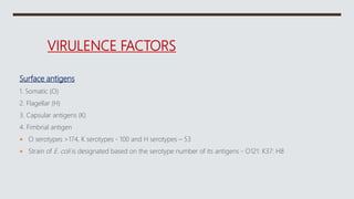
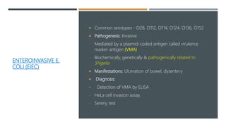
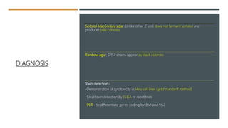
ESCHERICHIA
COLI
ENTEROBACTERIACE
AE
Family Characters (General Properties)
Gram-negative bacilli
™Aerobes and facultative anaerobes, Nonfastidious
Ferment glucose to produce acid with or without gas
™Reduce nitrate to nitrite
™Catalase positive, oxidase negative
Motile with peritrichous flagella, or nonmotile
Mostly commensals in human intestine - Coliform bacilli
CLASSIFICATION
Groups Lactose
fermentation
Colonies on
MacConkey agar
Examples
Lactose fermenters
(LF)-all are coliform
bacilli
Ferment lactose
producing acid
Produce pink colored
colonies,
(acid changes the
of neutral red indicator
to pink)
Escherichia,
Klebsiella
Citrobacter
Non lactose
fermenters (NLF)
Do not ferment
lactose
Produce pale or
colorless colonies
Salmonella, Shigella
Proteus,
Morganella,
Providencia and
Yersinia
Groups Lactose
fermentation
Colonies on
MacConkey agar
Examples
Late lactose
fermenters (LLF
or previously
called as
paracolon
Ferment lactose
after 2-8 days of
incubation
At 24 hrs
incubation-
produce pale or
colorless
colonies,
After 2 days-
produce pink
color colonies
Shigella sonnei
ESSENTIALS OF MEDICAL MICROBIOLOGY
EWING’S CLASSIFICATION
Tribe Genus
Tribe I-Escherichieae Escherichia
Shigella
Tribe II-Edwardsielleae Edwardsiella
Tribe III-Salmonelleae Salmonella
Tribe IV-Citrobactereae Citrobacter
Tribe Genus
Tribe V-Klebsielleae Klebsiella
Enterobacter, Hafnia
Serratia
Pantoea
Tribe VI-Proteeae Proteus
Morganella
Providencia
Tribe VII-Yersinieae Yersinia
Tribe VIII-Erwinieae Erwinia
ESCHERICHIA
COLI
Described first by Escherich in 1885
Most common aerobe harbored in gut of
and animals
Detection thermotolerant E. coli (survives at
44°C) in drinking water  recent contamination
with human or animal feces
Other species are less important as human
pathogens - E. fergusonii, E. hermannii and E.
vulneris
VIRULENCE FACTORS
Surface antigens
1. Somatic (O)
2. Flagellar (H)
3. Capsular antigens (K)
4. Fimbrial antigen
 O serotypes >174, K serotypes - 100 and H serotypes – 53
 Strain of E. coli is designated based on the serotype number of its antigens - O121: K37: H8
SOMATIC/ O
ANTIGEN
 Most important virulence factor  endotoxic
activity,
 Protects from phagocytosis and bactericidal
effect of complement
 Lipopolysaccharide (LPS) antigen, heat-stable
 Occasionally, it cross reacts with O antigens of
other species
 Early O serotypes - commensals of intestine
 Late O serotypes - diarrhea producing strains
 Flagellar or H antigen (H from Hauch, meaning film of breath)
- Heat labile
- Motility contributing to virulence
 Capsular or K antigen (K for Kapsel, German for capsule)-
- Polysaccharide capsular antigen present on the envelope or microcapsule
- Expressed by some strains only - neonatal meningitis, pyelonephritis and
- Encloses O antigen  inagglutinable by the O antiserum
- Inhibits Phagocytosis
FIMBRIAL ANTIGEN (PILUS) - ORGAN OF ADHESION
CFA (colonization factor antigen): enterotoxigenic E. Coli
Mannose resistant fimbriae (e.g. P, M, S, F1C and Dr fimbriae):
• Hemagglutinate with RBCs that is not inhibited by mannose
• Expressed by uropathogenic E. coli
P fimbriae
bind specifically to the P blood group antigens present on human RBCs and uroepithelial cells
TOXINS
 Enterotoxins: produced by diarrheagenic strains of E. coli
- Heat labile toxin, heat stable toxin and verocytotoxin
 Hemolysins: virulent strains of E. coli (especially pyelonephritis strains)
 ‰Cytotoxic necrotizing factor 1 (CNF1) and secreted autotransporter toxin (SAT): Cytotoxic
to bladder and kidney cells
 Siderophores (i.e. aerobactin)—Helps in iron uptake
HEAT-LABILE
TOXIN-LT
 Produced by: Enterotoxigenic E. Coli
 Plasmid coded, Resembles cholera toxin but less
potent
 Mechanism of action:
Subunit B:Binds to GM1 ganglioside receptors on
intestinal epithelium A fragment is internalized
and cleaved into A1 and A2 peptides
Fragment A:
- Fragment A2 helps in tethering A and B subunits
together
FRAGMENT A1 - ACTIVE FRAGMENT
diarrhea
increased
outflow of
water and
electrolytes
into the gut
lumen
intracellular
accumulation
of cAMP
upregulates
activity of
adenylate
cyclase
causes ADP
ribosylation
of G protein
Detection of LT:
Toxin detection: latex agglutination, ELISA
Molecular methods: PCR detecting gene coding for LT
HEAT STABLE TOXIN- ST
diarrhea
fluid
accumulat
ion in gut
lumen
increased
productio
n of
Cgmp
ST-I:
Binds to
the
guanylate
cyclase C
• Produced by: Enterotoxigenic E. Coli
• Plasmid-coded
• ST is of two types: ST-I and ST-II
-ST-II: causes fluid accumulation by an unknown mechanism
• Detection of ST: Same as for LT
VEROCYTOTOXIN OR SHIGA-LIKE TOXIN
 Produced by: Enterohemorrhagic E. Coli
 Bacteriophage-coded
 Cytotoxic to Vero cell lines,
 Also called Shiga-like toxin - resembles Shiga toxin in its structure &
function
Mechanism of action:
- Fragment B binds to a globotriosyl ceramide (Gb3) receptor on
intestinal epithelium
- Fragment A - Active fragment. Inhibits protein synthesis
Detection of VT:
• Serologically—Latex agglutination, ELISA
• Molecular methods—using specific DNA probe
• Cytotoxicity on Vero and HeLa cell lines
CLINICAL FEATURES
 one of the most common pathogen encountered clinically
 Urinary tract infection (UTI): uropathogenic E. coli (UPEC)
 Diarrhea: Six types of diarrheagenic E. Coli
1. Enteropathogenic E. coli (EPEC)
2. Enterotoxigenic E. coli (ETEC)
3. Enteroinvasive E. coli (EIEC)
4. Enterohemorrhagic E. coli (EHEC)
5. Enteroaggregative E. coli (EAEC)
6. Diffusely adherent E. coli (DAEC)
OTHER INFECTIONS
Abdominal infections: Commonest cause of primary and secondary bacterial peritonitis
Visceral abscesses - hepatic abscess
„Pneumonia in hospitalized patients— VAP
Meningitis (especially neonatal)
Wound and soft tissue infection - cellulitis and infection of wounds
Osteomyelitis, Endovascular infection and bacteremia
LABORATORY DIAGNOSIS – SPECIMEN COLLECTION
Specimens collected Disease
Pus, exudates and wound swab Cellulitis or pyogenic wound
Urine UTI
Stool Diarrhea
CSF Meningitis
Peritoneal exudate Peritonitis
Sputum Pneumonia
Tracheal aspirate Ventilator associated pneumonia
Blood Bacteremia
Direct Microscopy
Gram negative Bacilli arranged singly with pus cells
Culture:
Aerobe and facultative anaerobe, nonfastidious
-Blood agar:
Colonies are big, circular, grey, moist and occasionally β
hemolytic
-MacConkey agar: pink due to lactose fermentation
Liquid medium - uniform turbidity
Culture smear and motility testing
• Scattered gram-negative bacilli
• Hanging drop – Motile bacilli
- Catalase positive and oxidase negative
- Nitrate is reduced to nitrite
 ICUT tests:
- Indole test: Positive
- Citrate & Urease test: Negative
-Triple sugar iron agar) test: Shows
acid/acid, gas present, H2S absent
 ‰Sugar fermentation test:
ferments most of the sugars, such
as glucose, lactose, mannitol,
maltose (but not sucrose), with
production of acid and gas.
 MR (methyl red) test: Positive
 VP (Voges-Proskauer) test:
Negative
BIOCHEMICAL REACTIONS
Antimicrobial susceptibility testing:
• On Mueller-Hinton agar by using disk diffusion method
• Can rapidly develop resistance to multiple drugs
• Many strains produce β lactamases such as ESBL (extended
spectrum β lactamases) and MBL (metallo-β-lactamases)
URINARY TRACT INFECTION (UTI)
 Single most common pathogen, accounting for 70–75% of all
casesof UTI
 UPEC serotypes O1, O2, O4, O6, O7 and O75
 Route of spread:
1. Ascending route: From periurethral area
2. Descending route: Hematogenous seeding
UPPER V/S LOWER UTI
Lower UTI Upper UTI
Syndromes Cystitis and urethritis Pyelonephritis
Symptoms Local manifestations-
dysuria, urgency,
frequency,
Systemic manifestations-
Fever, vomiting,
pain
Route of spread Ascending route Both ascending (common)
and descending route
Lower UTI Upper UTI
Occurrence More common Less common
Virulence factors Fimbriae (e.g. P
fimbriae)
Capsular K antigen
Common virulence factors responsible are-
 Cytotoxins (CNF 1-cytotoxic necrotising factor 1 and SAT-Secreted
auto transporter toxin)
 Hemolysins
PREDISPOSING FACTORS THAT
PROMOTE UTI
 Females: short urethra and close
proximity to anus
 Presence of urinary catheters
 ™Urinary obstruction: prostate
enlargement or urinary stones
urine stasis
 Pregnancy:
- Physiological obstruction in urinary
tract due to growing fetus
- Asymptomatic bacteriuria is common
LABORATORY DIAGNOSIS OF UPEC
 Specimen Collection
-Clean voided midstream urine: Commonest specimen - collected after properly cleaning urethral meatus
or glans
-Suprapubic aspiration of urine from the bladder: most ideal specimen - for patients in coma or infants
-Catheterized patients - from the catheter tube (after clamping and disinfecting); but not from the bag
 Transport
-Processed immediately. Expected delay- refrigerator or adding boric acid, glycerol or formate
 Direct Examination & Screening Tests
• Wet™mount - more than 8 pus cells/mm3 or 4 lakh pus cells excreted in urine/hour is
significant
• Leukocyte esterase test: Rapid and cheaper method
• Nitrate reduction test (Griess test)
• Gram staining of urine is not a reliable indicator as—
-Bacterial count in urine is low
-Pus cells rapidly deteriorate in urine
-Limited to pyelonephritis and invasive UTI - count of ≥1 bacteria/oil immersion field is
significant
CULTURE
 Culture media: MacConkey agar and blood agar or CLED agar
 Kass concept of significant bacteriuria:
- A count of ≥105 colony forming units (CFU)/mL of urine is considered as significant 
Indicates infection „
- Count between 104 to 105 CFU/mL indicates doubtful significance - clinically correlated
Low count of ≤𝟏𝟎 𝟒CFU/mL –
• Commensal bacteria due to contamination during voiding.
• Low counts may be significant :
-Patient on antibiotic or diuretic treatment
-Infection with some gram-positive organisms such as S. aureus and Candida
-Pyelonephritis and acute urethral syndrome
-Sample taken by suprapubic aspiration
™ Quantitative culture:
-Semi-quantitative method - standardized loop technique
„ -Quantitative method such as pour plate method.
Antibody Coated Bacteria Test –
to differentiate upper and lower UTI
-Upper UTI: As the route of spread is hematogenous, bacteria coated with
specific antibodies are found in urine
-Detected by immunofluorescence method using fluorescent labeled
antihuman globulin
™-Lower UTI - bacteria found in urine are never coated with specific
antibodies
DIARRHEA-DIARRHEAGENIC E. COLI.
 Diarrheagenic E. coli are antigenically distinct from the commensal E. coli
which colonize the intestine
 Only few serotypes of E. coli which express the enterotoxin or other
virulence mechanisms can cause diarrhea
 Six types of diarrheagenic E. coli.
ENTEROPATHOGENIC E. COLI (EPEC)
Mechanism of diarrhea:
Adhesion to intestinal mucosa
mediated by plasmid coded
bundle-forming pili
Attaching and effacing lesions:
---disruption of brush border
epithelium----- increased
secretion and watery diarrhea
-Causes infantile diarrhea (outbreaks) & sporadic diarrhea in adults
™-Nontoxigenic and noninvasive
ENTEROTOXIGENIC E.
COLI (ETEC)
 Most common cause of traveler’s diarrhea (25–75% )
 Acute watery diarrhea in infants and adults
 Common serotypes—O6, O8, O15, O25, O27, O153,
O159, etc.
 Toxigenic, but not invasive
 Pathogenesis of ETEC is by:
 Attachment to intestinal mucosa mediated by fimbrial
protein colonization factor antigen (CFA)
 Toxin production—(1) heat-labile toxin or LT (acts by
↑cAMP), (2) heat-stable toxin or ST (acts by ↑cGMP)
ENTEROINVASIVE E.
COLI (EIEC)
 Common serotypes - O28, O112, O114, O124, O136, O152
 Pathogenesis: Invasive
- Mediated by a plasmid-coded antigen called virulence
marker antigen (VMA)
- Biochemically, genetically & pathogenically related to
Shigella
 Manifestations: Ulceration of bowel, dysentery
 Diagnosis:
- Detection of VMA by ELISA
- HeLa cell invasion assay,
- Sereny test
ENTEROHEMORRHAGIC E.
COLI (EHEC)
Serotypes associated with EHEC are: O157:H7 (most
common serotype)
Other serotypes - O26:H11, O6, O55, O91, O103,
O111 & O113
Transmitted by contaminated food, i.e.
of lettuce, spinach, sprouts and undercooked
ground beef
Prevalent mainly in industrialized countries
Low infective dose: Few organisms (<102 bacilli) are
required to initiate the infection
Pathogenesis: secretes verocytotoxin or Shiga-like
toxin
SHIGA-LIKE
TOXIN
 Mechanism of action: inhibits protein synthesis by inhibiting
the 28S subunit of 60S ribosome.
 Stx2 is more commonly associated with HUS than Stx1
 Manifestations: predilection for endothelium capillary
microangiopathy
-Hemorrhagic colitis: gross bloody diarrhea, abdominal pain
and fecal leukocytosis but no fever
-Hemorrhagic uremic syndrome (HUS): injury to small vessels
of the kidney and brain bloody diarrhea, thrombocytopenia,
renal failure and encephalopathy but without fever
DIAGNOSIS
Sorbitol MacConkey agar: Unlike other E. coli, does not ferment sorbitol and
produces pale colonies
Rainbow agar: O157 strains appear as black colonies
Toxin detection:-
-Demonstration of cytotoxicity in Vero cell lines (gold standard method)
-Fecal toxin detection by ELISA or rapid tests
-PCR - to differentiate genes coding for Stx1 and Stx2
ENTEROAGGREGATIVE E. COLI (EAEC)
 Adheres to HEp-2 cells in a stacked-brick fashion
 Most strains are “O” untypeable but “H” typeable
 Pathogenesis:
- Intestinal colonization mediated by aggregative adhesion fimbriae I
-EAST 1 toxin
 Manifestations: Persistent and acute diarrhea
 E. coli O104: H4 - enteroaggregative strain that has caused major outbreaks in Germany in
2011. Also produces Shiga-like toxin and can cause HUS
DIFFUSELY-ADHERENT E. COLI (DAEC)
 Ability to adhere to HEp-2 cells in a diffuse pattern
 Expresses diffuse adherence fimbriae which contribute to the
pathogenesis
 DAEC is capable of causing diarrheal disease, primarily in children aged
2–6 years
TREATMENT
 Extra-intestinal E. coli
-Based upon antimicrobial susceptibility test report
-Hospital strains mostly MDR.
Often produce ESBLs or AmpC β-lactamases
resistant to most β-lactams except carbapenems
-Carbapenems, amikacin or BL/BLIs - agents of
choice for hospital acquired MDR E. coli isolates
 Extra-intestinal E. Coli
Carbapenem resistant isolates-- Polymyxins, fosfomycin or tigecycline
 Diarrheagenic E. Coli - fluid replacement
- Antimicrobials to be avoided

E. coli

  • 1.
  • 2.
    ENTEROBACTERIACE AE Family Characters (GeneralProperties) Gram-negative bacilli ™Aerobes and facultative anaerobes, Nonfastidious Ferment glucose to produce acid with or without gas ™Reduce nitrate to nitrite ™Catalase positive, oxidase negative Motile with peritrichous flagella, or nonmotile Mostly commensals in human intestine - Coliform bacilli
  • 3.
    CLASSIFICATION Groups Lactose fermentation Colonies on MacConkeyagar Examples Lactose fermenters (LF)-all are coliform bacilli Ferment lactose producing acid Produce pink colored colonies, (acid changes the of neutral red indicator to pink) Escherichia, Klebsiella Citrobacter Non lactose fermenters (NLF) Do not ferment lactose Produce pale or colorless colonies Salmonella, Shigella Proteus, Morganella, Providencia and Yersinia
  • 4.
    Groups Lactose fermentation Colonies on MacConkeyagar Examples Late lactose fermenters (LLF or previously called as paracolon Ferment lactose after 2-8 days of incubation At 24 hrs incubation- produce pale or colorless colonies, After 2 days- produce pink color colonies Shigella sonnei ESSENTIALS OF MEDICAL MICROBIOLOGY
  • 5.
    EWING’S CLASSIFICATION Tribe Genus TribeI-Escherichieae Escherichia Shigella Tribe II-Edwardsielleae Edwardsiella Tribe III-Salmonelleae Salmonella Tribe IV-Citrobactereae Citrobacter
  • 6.
    Tribe Genus Tribe V-KlebsielleaeKlebsiella Enterobacter, Hafnia Serratia Pantoea Tribe VI-Proteeae Proteus Morganella Providencia Tribe VII-Yersinieae Yersinia Tribe VIII-Erwinieae Erwinia
  • 7.
    ESCHERICHIA COLI Described first byEscherich in 1885 Most common aerobe harbored in gut of and animals Detection thermotolerant E. coli (survives at 44°C) in drinking water  recent contamination with human or animal feces Other species are less important as human pathogens - E. fergusonii, E. hermannii and E. vulneris
  • 8.
    VIRULENCE FACTORS Surface antigens 1.Somatic (O) 2. Flagellar (H) 3. Capsular antigens (K) 4. Fimbrial antigen  O serotypes >174, K serotypes - 100 and H serotypes – 53  Strain of E. coli is designated based on the serotype number of its antigens - O121: K37: H8
  • 9.
    SOMATIC/ O ANTIGEN  Mostimportant virulence factor  endotoxic activity,  Protects from phagocytosis and bactericidal effect of complement  Lipopolysaccharide (LPS) antigen, heat-stable  Occasionally, it cross reacts with O antigens of other species  Early O serotypes - commensals of intestine  Late O serotypes - diarrhea producing strains
  • 10.
     Flagellar orH antigen (H from Hauch, meaning film of breath) - Heat labile - Motility contributing to virulence  Capsular or K antigen (K for Kapsel, German for capsule)- - Polysaccharide capsular antigen present on the envelope or microcapsule - Expressed by some strains only - neonatal meningitis, pyelonephritis and - Encloses O antigen  inagglutinable by the O antiserum - Inhibits Phagocytosis
  • 11.
    FIMBRIAL ANTIGEN (PILUS)- ORGAN OF ADHESION CFA (colonization factor antigen): enterotoxigenic E. Coli Mannose resistant fimbriae (e.g. P, M, S, F1C and Dr fimbriae): • Hemagglutinate with RBCs that is not inhibited by mannose • Expressed by uropathogenic E. coli P fimbriae bind specifically to the P blood group antigens present on human RBCs and uroepithelial cells
  • 12.
    TOXINS  Enterotoxins: producedby diarrheagenic strains of E. coli - Heat labile toxin, heat stable toxin and verocytotoxin  Hemolysins: virulent strains of E. coli (especially pyelonephritis strains)  ‰Cytotoxic necrotizing factor 1 (CNF1) and secreted autotransporter toxin (SAT): Cytotoxic to bladder and kidney cells  Siderophores (i.e. aerobactin)—Helps in iron uptake
  • 13.
    HEAT-LABILE TOXIN-LT  Produced by:Enterotoxigenic E. Coli  Plasmid coded, Resembles cholera toxin but less potent  Mechanism of action: Subunit B:Binds to GM1 ganglioside receptors on intestinal epithelium A fragment is internalized and cleaved into A1 and A2 peptides Fragment A: - Fragment A2 helps in tethering A and B subunits together
  • 14.
    FRAGMENT A1 -ACTIVE FRAGMENT diarrhea increased outflow of water and electrolytes into the gut lumen intracellular accumulation of cAMP upregulates activity of adenylate cyclase causes ADP ribosylation of G protein Detection of LT: Toxin detection: latex agglutination, ELISA Molecular methods: PCR detecting gene coding for LT
  • 15.
    HEAT STABLE TOXIN-ST diarrhea fluid accumulat ion in gut lumen increased productio n of Cgmp ST-I: Binds to the guanylate cyclase C • Produced by: Enterotoxigenic E. Coli • Plasmid-coded • ST is of two types: ST-I and ST-II -ST-II: causes fluid accumulation by an unknown mechanism • Detection of ST: Same as for LT
  • 16.
    VEROCYTOTOXIN OR SHIGA-LIKETOXIN  Produced by: Enterohemorrhagic E. Coli  Bacteriophage-coded  Cytotoxic to Vero cell lines,  Also called Shiga-like toxin - resembles Shiga toxin in its structure & function
  • 17.
    Mechanism of action: -Fragment B binds to a globotriosyl ceramide (Gb3) receptor on intestinal epithelium - Fragment A - Active fragment. Inhibits protein synthesis Detection of VT: • Serologically—Latex agglutination, ELISA • Molecular methods—using specific DNA probe • Cytotoxicity on Vero and HeLa cell lines
  • 18.
    CLINICAL FEATURES  oneof the most common pathogen encountered clinically  Urinary tract infection (UTI): uropathogenic E. coli (UPEC)  Diarrhea: Six types of diarrheagenic E. Coli 1. Enteropathogenic E. coli (EPEC) 2. Enterotoxigenic E. coli (ETEC) 3. Enteroinvasive E. coli (EIEC) 4. Enterohemorrhagic E. coli (EHEC) 5. Enteroaggregative E. coli (EAEC) 6. Diffusely adherent E. coli (DAEC)
  • 19.
    OTHER INFECTIONS Abdominal infections:Commonest cause of primary and secondary bacterial peritonitis Visceral abscesses - hepatic abscess „Pneumonia in hospitalized patients— VAP Meningitis (especially neonatal) Wound and soft tissue infection - cellulitis and infection of wounds Osteomyelitis, Endovascular infection and bacteremia
  • 20.
    LABORATORY DIAGNOSIS –SPECIMEN COLLECTION Specimens collected Disease Pus, exudates and wound swab Cellulitis or pyogenic wound Urine UTI Stool Diarrhea CSF Meningitis Peritoneal exudate Peritonitis Sputum Pneumonia Tracheal aspirate Ventilator associated pneumonia Blood Bacteremia
  • 21.
    Direct Microscopy Gram negativeBacilli arranged singly with pus cells Culture: Aerobe and facultative anaerobe, nonfastidious -Blood agar: Colonies are big, circular, grey, moist and occasionally β hemolytic -MacConkey agar: pink due to lactose fermentation
  • 22.
    Liquid medium -uniform turbidity Culture smear and motility testing • Scattered gram-negative bacilli • Hanging drop – Motile bacilli
  • 23.
    - Catalase positiveand oxidase negative - Nitrate is reduced to nitrite  ICUT tests: - Indole test: Positive - Citrate & Urease test: Negative -Triple sugar iron agar) test: Shows acid/acid, gas present, H2S absent  ‰Sugar fermentation test: ferments most of the sugars, such as glucose, lactose, mannitol, maltose (but not sucrose), with production of acid and gas.  MR (methyl red) test: Positive  VP (Voges-Proskauer) test: Negative BIOCHEMICAL REACTIONS
  • 24.
    Antimicrobial susceptibility testing: •On Mueller-Hinton agar by using disk diffusion method • Can rapidly develop resistance to multiple drugs • Many strains produce β lactamases such as ESBL (extended spectrum β lactamases) and MBL (metallo-β-lactamases)
  • 25.
    URINARY TRACT INFECTION(UTI)  Single most common pathogen, accounting for 70–75% of all casesof UTI  UPEC serotypes O1, O2, O4, O6, O7 and O75  Route of spread: 1. Ascending route: From periurethral area 2. Descending route: Hematogenous seeding
  • 26.
    UPPER V/S LOWERUTI Lower UTI Upper UTI Syndromes Cystitis and urethritis Pyelonephritis Symptoms Local manifestations- dysuria, urgency, frequency, Systemic manifestations- Fever, vomiting, pain Route of spread Ascending route Both ascending (common) and descending route
  • 27.
    Lower UTI UpperUTI Occurrence More common Less common Virulence factors Fimbriae (e.g. P fimbriae) Capsular K antigen Common virulence factors responsible are-  Cytotoxins (CNF 1-cytotoxic necrotising factor 1 and SAT-Secreted auto transporter toxin)  Hemolysins
  • 28.
    PREDISPOSING FACTORS THAT PROMOTEUTI  Females: short urethra and close proximity to anus  Presence of urinary catheters  ™Urinary obstruction: prostate enlargement or urinary stones urine stasis  Pregnancy: - Physiological obstruction in urinary tract due to growing fetus - Asymptomatic bacteriuria is common
  • 29.
    LABORATORY DIAGNOSIS OFUPEC  Specimen Collection -Clean voided midstream urine: Commonest specimen - collected after properly cleaning urethral meatus or glans -Suprapubic aspiration of urine from the bladder: most ideal specimen - for patients in coma or infants -Catheterized patients - from the catheter tube (after clamping and disinfecting); but not from the bag  Transport -Processed immediately. Expected delay- refrigerator or adding boric acid, glycerol or formate
  • 30.
     Direct Examination& Screening Tests • Wet™mount - more than 8 pus cells/mm3 or 4 lakh pus cells excreted in urine/hour is significant • Leukocyte esterase test: Rapid and cheaper method • Nitrate reduction test (Griess test) • Gram staining of urine is not a reliable indicator as— -Bacterial count in urine is low -Pus cells rapidly deteriorate in urine -Limited to pyelonephritis and invasive UTI - count of ≥1 bacteria/oil immersion field is significant
  • 31.
    CULTURE  Culture media:MacConkey agar and blood agar or CLED agar  Kass concept of significant bacteriuria: - A count of ≥105 colony forming units (CFU)/mL of urine is considered as significant  Indicates infection „ - Count between 104 to 105 CFU/mL indicates doubtful significance - clinically correlated
  • 32.
    Low count of≤𝟏𝟎 𝟒CFU/mL – • Commensal bacteria due to contamination during voiding. • Low counts may be significant : -Patient on antibiotic or diuretic treatment -Infection with some gram-positive organisms such as S. aureus and Candida -Pyelonephritis and acute urethral syndrome -Sample taken by suprapubic aspiration ™ Quantitative culture: -Semi-quantitative method - standardized loop technique „ -Quantitative method such as pour plate method.
  • 33.
    Antibody Coated BacteriaTest – to differentiate upper and lower UTI -Upper UTI: As the route of spread is hematogenous, bacteria coated with specific antibodies are found in urine -Detected by immunofluorescence method using fluorescent labeled antihuman globulin ™-Lower UTI - bacteria found in urine are never coated with specific antibodies
  • 34.
    DIARRHEA-DIARRHEAGENIC E. COLI. Diarrheagenic E. coli are antigenically distinct from the commensal E. coli which colonize the intestine  Only few serotypes of E. coli which express the enterotoxin or other virulence mechanisms can cause diarrhea  Six types of diarrheagenic E. coli.
  • 35.
    ENTEROPATHOGENIC E. COLI(EPEC) Mechanism of diarrhea: Adhesion to intestinal mucosa mediated by plasmid coded bundle-forming pili Attaching and effacing lesions: ---disruption of brush border epithelium----- increased secretion and watery diarrhea -Causes infantile diarrhea (outbreaks) & sporadic diarrhea in adults ™-Nontoxigenic and noninvasive
  • 36.
    ENTEROTOXIGENIC E. COLI (ETEC) Most common cause of traveler’s diarrhea (25–75% )  Acute watery diarrhea in infants and adults  Common serotypes—O6, O8, O15, O25, O27, O153, O159, etc.  Toxigenic, but not invasive  Pathogenesis of ETEC is by:  Attachment to intestinal mucosa mediated by fimbrial protein colonization factor antigen (CFA)  Toxin production—(1) heat-labile toxin or LT (acts by ↑cAMP), (2) heat-stable toxin or ST (acts by ↑cGMP)
  • 37.
    ENTEROINVASIVE E. COLI (EIEC) Common serotypes - O28, O112, O114, O124, O136, O152  Pathogenesis: Invasive - Mediated by a plasmid-coded antigen called virulence marker antigen (VMA) - Biochemically, genetically & pathogenically related to Shigella  Manifestations: Ulceration of bowel, dysentery  Diagnosis: - Detection of VMA by ELISA - HeLa cell invasion assay, - Sereny test
  • 38.
    ENTEROHEMORRHAGIC E. COLI (EHEC) Serotypesassociated with EHEC are: O157:H7 (most common serotype) Other serotypes - O26:H11, O6, O55, O91, O103, O111 & O113 Transmitted by contaminated food, i.e. of lettuce, spinach, sprouts and undercooked ground beef Prevalent mainly in industrialized countries Low infective dose: Few organisms (<102 bacilli) are required to initiate the infection Pathogenesis: secretes verocytotoxin or Shiga-like toxin
  • 39.
    SHIGA-LIKE TOXIN  Mechanism ofaction: inhibits protein synthesis by inhibiting the 28S subunit of 60S ribosome.  Stx2 is more commonly associated with HUS than Stx1  Manifestations: predilection for endothelium capillary microangiopathy -Hemorrhagic colitis: gross bloody diarrhea, abdominal pain and fecal leukocytosis but no fever -Hemorrhagic uremic syndrome (HUS): injury to small vessels of the kidney and brain bloody diarrhea, thrombocytopenia, renal failure and encephalopathy but without fever
  • 40.
    DIAGNOSIS Sorbitol MacConkey agar:Unlike other E. coli, does not ferment sorbitol and produces pale colonies Rainbow agar: O157 strains appear as black colonies Toxin detection:- -Demonstration of cytotoxicity in Vero cell lines (gold standard method) -Fecal toxin detection by ELISA or rapid tests -PCR - to differentiate genes coding for Stx1 and Stx2
  • 41.
    ENTEROAGGREGATIVE E. COLI(EAEC)  Adheres to HEp-2 cells in a stacked-brick fashion  Most strains are “O” untypeable but “H” typeable  Pathogenesis: - Intestinal colonization mediated by aggregative adhesion fimbriae I -EAST 1 toxin  Manifestations: Persistent and acute diarrhea  E. coli O104: H4 - enteroaggregative strain that has caused major outbreaks in Germany in 2011. Also produces Shiga-like toxin and can cause HUS
  • 42.
    DIFFUSELY-ADHERENT E. COLI(DAEC)  Ability to adhere to HEp-2 cells in a diffuse pattern  Expresses diffuse adherence fimbriae which contribute to the pathogenesis  DAEC is capable of causing diarrheal disease, primarily in children aged 2–6 years
  • 43.
    TREATMENT  Extra-intestinal E.coli -Based upon antimicrobial susceptibility test report -Hospital strains mostly MDR. Often produce ESBLs or AmpC β-lactamases resistant to most β-lactams except carbapenems -Carbapenems, amikacin or BL/BLIs - agents of choice for hospital acquired MDR E. coli isolates
  • 44.
     Extra-intestinal E.Coli Carbapenem resistant isolates-- Polymyxins, fosfomycin or tigecycline  Diarrheagenic E. Coli - fluid replacement - Antimicrobials to be avoided