Electrophoresis is a technique used to separate charged particles such as proteins, nucleic acids, and other macromolecules. It works by applying an electric field to move these particles through a medium such as a gel or paper based on their size and charge. There are different types of electrophoresis including gel electrophoresis, paper electrophoresis, capillary electrophoresis, and isoelectric focusing which separates particles based on their isoelectric point. Electrophoresis has many applications in fields like forensics, molecular biology, genetics, and biochemistry to analyze proteins, DNA, RNA, and other biomolecules.
Electrophoresis principle and types by Dr. Anurag YadavDr Anurag Yadav
the general principle on how the electrophoresis performs.
the different types of electrophoresis and the mechanism of separation based on different character of the medium and type of electrophoresis.
The technique of paper electrophoresis is simple and inexpensive and requires only micro quantities of plasma for separation.
The support medium is a filter paper
The electrophoresis apparatus in its simplest form consists of two troughs to contain buffer solution, through which electric current is passed.
Frequently used in isolating proteins, amino acids and oligopeptides.
This presentation contain the information about gel electrophoresis method , instruments & types.
Electrophoresis is a method through biological molecules are separated by applying an electric field.
Main purpose of this method is to determine the number , amount & mobility of biological component.
There are some internal & external factors that affects the process of electrophoresis.
The bio-molecules have charge on it & when we apply an electric field , the charge particles move to the opposite cathode. In this way, charge particles are separated
There are 3 types of gels that use in this process .
In this buffers are also used which provide ions that carry a current.
In this slide contains types, working principle, factors affecting, advantage and disadvantage of paper electrophoresis.
Presented by: G.Sai Swetha. (Department of pharmacology),
RIPER, anantapur.
Electrophoresis is a laboratory technique used to separate DNA, RNA, or protein molecules based on their size and electrical charge. An electric current is used to move molecules to be separated through a gel. Pores in the gel work like a sieve, allowing smaller molecules to move faster than larger molecules.
INTRODUCTION, DEFINATION OF ELECTROPHORESIS, ELECTROPHORESIS PRINCIPLE, TYPES OF ELECTROPHORESIS, FREE ELECTROPHORESIS, ZONE ELECTROPHORESIS,PAPER ELECTROPHORESIS, WORKING OF PAPER ELECTROPHORESIS, PROCEDURE FOR PAPER ELECTROPHORESIS, VISUALISATION, FACTORS AFFECTING SEPARATION OF MOLECULES, APPLICATIONS, working of paper electrophoresis ,procedure for paper electrophoresis ,visualisation ,factors affecting separation of molecules ,applications ,forensics ,dna fingerprinting ,molecular biology ,microbiology information about the organisms ,biochemistry mapping of cellular components ,paper electrophoresis is also used in study of sic ,hemoglobin abnormalities ,separation of blood clotting factors ,serum plasma proteins from blood sample ,used in separation and identification of alkaloids ,used for testing water samples ,toxicity of water ,drug industry to determine presence of illelgal drUGS
Electrophoresis is a technique commonly used in the lab to separate charged molecules, like DNA, according to size.
This presentation gives glimpses of the science behind electrophoresis and its application in various fields
It contains 2-3 acetyl groups per glucose unit and its adsorption capacity is less than that of paper.
It gives sharper bands.
Provides a good background for staining glycoproteins.
ADVANTAGE:
No tailing of proteins or hydrophilic materials.
Available in wide range of particle size and layer thickness.
Give sharp bands and offer good resolution.
High voltage can be applied which will enhance the resolution.
General introduction about electrophoresis
Principle
Working condition of electrophoresis
Factors affecting separation of electrophoresis
Application of electrophoresis
Types of electrophoresis
Factory Supply Best Quality Pmk Oil CAS 28578–16–7 PMK Powder in Stockrebeccabio
Factory Supply Best Quality Pmk Oil CAS 28578–16–7 PMK Powder in Stock
Telegram: bmksupplier
signal: +85264872720
threema: TUD4A6YC
You can contact me on Telegram or Threema
Communicate promptly and reply
Free of customs clearance, Double Clearance 100% pass delivery to USA, Canada, Spain, Germany, Netherland, Poland, Italy, Sweden, UK, Czech Republic, Australia, Mexico, Russia, Ukraine, Kazakhstan.Door to door service
Hot Selling Organic intermediates
Tom Selleck Health: A Comprehensive Look at the Iconic Actor’s Wellness Journeygreendigital
Tom Selleck, an enduring figure in Hollywood. has captivated audiences for decades with his rugged charm, iconic moustache. and memorable roles in television and film. From his breakout role as Thomas Magnum in Magnum P.I. to his current portrayal of Frank Reagan in Blue Bloods. Selleck's career has spanned over 50 years. But beyond his professional achievements. fans have often been curious about Tom Selleck Health. especially as he has aged in the public eye.
Follow us on: Pinterest
Introduction
Many have been interested in Tom Selleck health. not only because of his enduring presence on screen but also because of the challenges. and lifestyle choices he has faced and made over the years. This article delves into the various aspects of Tom Selleck health. exploring his fitness regimen, diet, mental health. and the challenges he has encountered as he ages. We'll look at how he maintains his well-being. the health issues he has faced, and his approach to ageing .
Early Life and Career
Childhood and Athletic Beginnings
Tom Selleck was born on January 29, 1945, in Detroit, Michigan, and grew up in Sherman Oaks, California. From an early age, he was involved in sports, particularly basketball. which played a significant role in his physical development. His athletic pursuits continued into college. where he attended the University of Southern California (USC) on a basketball scholarship. This early involvement in sports laid a strong foundation for his physical health and disciplined lifestyle.
Transition to Acting
Selleck's transition from an athlete to an actor came with its physical demands. His first significant role in "Magnum P.I." required him to perform various stunts and maintain a fit appearance. This role, which he played from 1980 to 1988. necessitated a rigorous fitness routine to meet the show's demands. setting the stage for his long-term commitment to health and wellness.
Fitness Regimen
Workout Routine
Tom Selleck health and fitness regimen has evolved. adapting to his changing roles and age. During his "Magnum, P.I." days. Selleck's workouts were intense and focused on building and maintaining muscle mass. His routine included weightlifting, cardiovascular exercises. and specific training for the stunts he performed on the show.
Selleck adjusted his fitness routine as he aged to suit his body's needs. Today, his workouts focus on maintaining flexibility, strength, and cardiovascular health. He incorporates low-impact exercises such as swimming, walking, and light weightlifting. This balanced approach helps him stay fit without putting undue strain on his joints and muscles.
Importance of Flexibility and Mobility
In recent years, Selleck has emphasized the importance of flexibility and mobility in his fitness regimen. Understanding the natural decline in muscle mass and joint flexibility with age. he includes stretching and yoga in his routine. These practices help prevent injuries, improve posture, and maintain mobilit
Anti ulcer drugs and their Advance pharmacology ||
Anti-ulcer drugs are medications used to prevent and treat ulcers in the stomach and upper part of the small intestine (duodenal ulcers). These ulcers are often caused by an imbalance between stomach acid and the mucosal lining, which protects the stomach lining.
||Scope: Overview of various classes of anti-ulcer drugs, their mechanisms of action, indications, side effects, and clinical considerations.
Flu Vaccine Alert in Bangalore Karnatakaaddon Scans
As flu season approaches, health officials in Bangalore, Karnataka, are urging residents to get their flu vaccinations. The seasonal flu, while common, can lead to severe health complications, particularly for vulnerable populations such as young children, the elderly, and those with underlying health conditions.
Dr. Vidisha Kumari, a leading epidemiologist in Bangalore, emphasizes the importance of getting vaccinated. "The flu vaccine is our best defense against the influenza virus. It not only protects individuals but also helps prevent the spread of the virus in our communities," he says.
This year, the flu season is expected to coincide with a potential increase in other respiratory illnesses. The Karnataka Health Department has launched an awareness campaign highlighting the significance of flu vaccinations. They have set up multiple vaccination centers across Bangalore, making it convenient for residents to receive their shots.
To encourage widespread vaccination, the government is also collaborating with local schools, workplaces, and community centers to facilitate vaccination drives. Special attention is being given to ensuring that the vaccine is accessible to all, including marginalized communities who may have limited access to healthcare.
Residents are reminded that the flu vaccine is safe and effective. Common side effects are mild and may include soreness at the injection site, mild fever, or muscle aches. These side effects are generally short-lived and far less severe than the flu itself.
Healthcare providers are also stressing the importance of continuing COVID-19 precautions. Wearing masks, practicing good hand hygiene, and maintaining social distancing are still crucial, especially in crowded places.
Protect yourself and your loved ones by getting vaccinated. Together, we can help keep Bangalore healthy and safe this flu season. For more information on vaccination centers and schedules, residents can visit the Karnataka Health Department’s official website or follow their social media pages.
Stay informed, stay safe, and get your flu shot today!
These lecture slides, by Dr Sidra Arshad, offer a quick overview of physiological basis of a normal electrocardiogram.
Learning objectives:
1. Define an electrocardiogram (ECG) and electrocardiography
2. Describe how dipoles generated by the heart produce the waveforms of the ECG
3. Describe the components of a normal electrocardiogram of a typical bipolar leads (limb II)
4. Differentiate between intervals and segments
5. Enlist some common indications for obtaining an ECG
Study Resources:
1. Chapter 11, Guyton and Hall Textbook of Medical Physiology, 14th edition
2. Chapter 9, Human Physiology - From Cells to Systems, Lauralee Sherwood, 9th edition
3. Chapter 29, Ganong’s Review of Medical Physiology, 26th edition
4. Electrocardiogram, StatPearls - https://www.ncbi.nlm.nih.gov/books/NBK549803/
5. ECG in Medical Practice by ABM Abdullah, 4th edition
6. ECG Basics, http://www.nataliescasebook.com/tag/e-c-g-basics
Ozempic: Preoperative Management of Patients on GLP-1 Receptor Agonists Saeid Safari
Preoperative Management of Patients on GLP-1 Receptor Agonists like Ozempic and Semiglutide
ASA GUIDELINE
NYSORA Guideline
2 Case Reports of Gastric Ultrasound
These simplified slides by Dr. Sidra Arshad present an overview of the non-respiratory functions of the respiratory tract.
Learning objectives:
1. Enlist the non-respiratory functions of the respiratory tract
2. Briefly explain how these functions are carried out
3. Discuss the significance of dead space
4. Differentiate between minute ventilation and alveolar ventilation
5. Describe the cough and sneeze reflexes
Study Resources:
1. Chapter 39, Guyton and Hall Textbook of Medical Physiology, 14th edition
2. Chapter 34, Ganong’s Review of Medical Physiology, 26th edition
3. Chapter 17, Human Physiology by Lauralee Sherwood, 9th edition
4. Non-respiratory functions of the lungs https://academic.oup.com/bjaed/article/13/3/98/278874
MANAGEMENT OF ATRIOVENTRICULAR CONDUCTION BLOCK.pdfJim Jacob Roy
Cardiac conduction defects can occur due to various causes.
Atrioventricular conduction blocks ( AV blocks ) are classified into 3 types.
This document describes the acute management of AV block.
Lung Cancer: Artificial Intelligence, Synergetics, Complex System Analysis, S...Oleg Kshivets
RESULTS: Overall life span (LS) was 2252.1±1742.5 days and cumulative 5-year survival (5YS) reached 73.2%, 10 years – 64.8%, 20 years – 42.5%. 513 LCP lived more than 5 years (LS=3124.6±1525.6 days), 148 LCP – more than 10 years (LS=5054.4±1504.1 days).199 LCP died because of LC (LS=562.7±374.5 days). 5YS of LCP after bi/lobectomies was significantly superior in comparison with LCP after pneumonectomies (78.1% vs.63.7%, P=0.00001 by log-rank test). AT significantly improved 5YS (66.3% vs. 34.8%) (P=0.00000 by log-rank test) only for LCP with N1-2. Cox modeling displayed that 5YS of LCP significantly depended on: phase transition (PT) early-invasive LC in terms of synergetics, PT N0—N12, cell ratio factors (ratio between cancer cells- CC and blood cells subpopulations), G1-3, histology, glucose, AT, blood cell circuit, prothrombin index, heparin tolerance, recalcification time (P=0.000-0.038). Neural networks, genetic algorithm selection and bootstrap simulation revealed relationships between 5YS and PT early-invasive LC (rank=1), PT N0—N12 (rank=2), thrombocytes/CC (3), erythrocytes/CC (4), eosinophils/CC (5), healthy cells/CC (6), lymphocytes/CC (7), segmented neutrophils/CC (8), stick neutrophils/CC (9), monocytes/CC (10); leucocytes/CC (11). Correct prediction of 5YS was 100% by neural networks computing (area under ROC curve=1.0; error=0.0).
CONCLUSIONS: 5YS of LCP after radical procedures significantly depended on: 1) PT early-invasive cancer; 2) PT N0--N12; 3) cell ratio factors; 4) blood cell circuit; 5) biochemical factors; 6) hemostasis system; 7) AT; 8) LC characteristics; 9) LC cell dynamics; 10) surgery type: lobectomy/pneumonectomy; 11) anthropometric data. Optimal diagnosis and treatment strategies for LC are: 1) screening and early detection of LC; 2) availability of experienced thoracic surgeons because of complexity of radical procedures; 3) aggressive en block surgery and adequate lymph node dissection for completeness; 4) precise prediction; 5) adjuvant chemoimmunoradiotherapy for LCP with unfavorable prognosis.
Surgical Site Infections, pathophysiology, and prevention.pptx
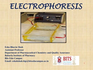
Electrophoresis
1. ELECTROPHORESIS
1
Esha Bhavin Shah
Assistant Professor
Department of Pharmaceutical Chemistry and Quality Assurance
Babaria Institute of Pharmacy
Bits Edu Campus
Email: eshahshah.bip@bitseducampus.ac.in
2. INTRODUCTION
In practical terms a positive electrode(anode)
and negative electrode(cathode) are placed in a
solution containing ions.
Then voltage is applied across the electrodes, so
solute ions of different charge for example,
anions (negative) and cations (positive) will
move through the solution towards the electrode
of opposite charge.
4. THEORY
Electrohoresis is the movement of ions under the
influence of electrical field. So, separation in
electrophoresis relies on differences in the speed of
migration (migration velocity) of ions or solutes.
• Migration Velocity (ν) =μe E
Where, μe=elecrophoretic mobility E=electrical field
strength
Electrophoretic mobility is a factor that determines
how fast ion or solute moves in a given medium.
• μe = q/ 6Лήr
• where, q=charge of ion ή=viscosity of solution
r=radius of ion
5. The instrumentation of capillary electrophoresis is very similar to
that of HPLC.
Power supply of E is equivalent to HPLC pump and capillary
equivalent to column.
COMPARISON OF ELECTROPHORETIC AND
CHROMATOGRAPHIC TERMS
5
7. Principle of Electrophoresis
• Electromigration :
– Ions migrating in electric field
Cations cathode (-ve)
Anions anode (+ve)
7
-+
8. Structure and Properties of Protein
• PROTEINS - polymers of amino acids
• STRUCTURE of Amino Acids
– aa's have a carboxyl group (-COOH) & amino group (-NH2) and
are often ionized at physiological pH
• Proteins are amphoteric compounds and are therefore either positively
or negatively charged.
8
9. Migration of proteins in electric field
negatively charged proteins move towards the
positive pole
directly proportional to the overall charge of
proteins
inversely proportional to protein size (molecular
weight)
10. Electropherogram
An Electropherogram is the
result of an electrophoresis
which gives the movement of
charged particles over time in
a gel, paper or another
medium.
10
18. Buffer solution added to the tank
This ensures that the electric current goes through the whole tank and that maintains
that ions can move in the solution
20. Electrical current applied to the chamber
Safety cover is put over the top and the current is switched on.
The dye will migrate through the gel toward the positive electrode, as will the DNA
Depending on how much voltage is applied and how warm the gel is and size and shape
of molecules will depend on how fast the ions move through the gel
Smaller fragments will move easier so they will be closer to the positive electrode
Once the dye has moved through the gel to the buffer, the electrical current is switched
off and gel is removed from the tray
21. VISUALISATION
• After the electrophoresis is complete, the molecules in the
gel can be stained to make them visible.
• Ethidium bromide, silver, or coomassie blue dye may
be used for this process.
• If the analyte molecules fluoresce under ultraviolet light,
a photograph can be taken of the gel under ultraviolet
lighting conditions.
• If the molecules to be separated contain radioactivity
added for visibility, an autoradiogram can be recorded of
the gel.
22. 22
There are molecular weight size
markers available that contain a
mixture of molecules of known
sizes. If such a marker was run on
one lane in the gel parallel to the
unknown samples, the bands
observed can be compared to
those of the unknown in order to
determine their size. The distance
a band travels is approximately
inversely proportional to the
logarithm of the size of the
molecule.
23. Quantification of separated protein band by Densitometer
Densitometer is a device that measures
the degree of darkness in photographic
or semi-transparent material.
24. Types of Electrophoresis:
Zone Electrophoresis:
a) Paper electrophoresis
b) Gel electrophoresis
c) Cellulose acetate electrophoresis
d) Thin layer electrophoresis
Moving boundary electrophoresis:
a) Capillary electrophoresis
b) Isoelectric focussing
c) Immuno electrophoresis
25. Zone electrophoresis:
The term Zone electrophoresis is applied to those systems in which ionic
mobilities are studied on strips of paper, cellulose acetate or acrylamide.
26. Zone electrophoresis
It involves the migration of the charged particle on the supporting media, paper ,
cellulose acetate membrane, starch gel, polyacrylamide.
The components separated are distributed into discrete zone on the support media.
Supporting media is saturated with buffer solution, small volume of the sample is
applied as narrow band.
On application of potential difference at the ends of a strip ,components migrate at the
rate determined by its electrophoretic mobility.
Advantages:
Useful in biochemical investigations.
Small quantity of sample can be analyzed
Low cost and easy maintenance
Disadvantages:
Unsuitable for accurate mobility and isoelectric point determination.
Due to the presence of supporting medium, technical complications such as capillary
flow, electrosmosis, adsorption and molecular sieving are introduced.
27. Paper Electrophoresis
Paper Electrophoresis is one of the zone electrophoresis. This is very important method
in all laboratories.
Paper of good quality should contain at least 95% α-cellulose and should have only a very
slight adsorption capacity.
This technique is useful for the separation of small charged molecules such as amino
acids and small proteins. A strip of filter paper is moistened with buffer and the ends of the
strip are immersed into buffer reservoirs containing the electrodes. The samples are
spotted in the center of the paper, high voltage is applied, and the spots migrate according
to their charges. After electrophoresis, the separated components can be detected by a
variety of staining techniques, depending upon their chemical identity.
28. Applications:
Serum analysis for diagnostic purpose is routinely carried about by paper
electrophoresis.
Muscle proteins, egg white proteins, milk proteins & snake, insect venom analysis
done by this technique.
29. Capillary electrophoresis
By using capillaries as distinct from
flat bead systems, it was possible to:
minimize zone spreading,
improve heat dissipation,
shorten separation times and
increase efficiency.
Applications:
Separation of steroids and complex drug mixtures within 10 to 15 minutes.
analysis of proteins found in serum, urine, CSF and body fluids, immunosubstraction
electrophoresis, hemoglobin variants, lipoproteins, carbohydrate-deficient transferrin (CDT)
forensic and therapeutic drug screening, and molecular diagnostics.
31. Gel electrophoresis is a method for separation and analysis of macromolecules (DNA,
RNA and proteins) and their fragments, based on their size and charge.
Applications:
It is used in clinical chemistry to separate proteins by charge and/or size (IEF agarose,
essentially size independent).
In biochemistry and molecular biology to separate a mixed population of DNA and RNA
fragments by length, to estimate the size of DNA and RNA fragments or to separate proteins
by charge.
Estimation of the size of DNA molecules following restriction enzyme digestion, e.g. in
restriction mapping of cloned DNA.
Analysis of PCR products, e.g. in molecular genetic diagnosis or genetic fingerprinting
Separation of restricted genomic DNA prior to Southern transfer, or of RNA prior to
Northern transfer.
Gel electrophoresis is used in forensics, molecular biology, genetics, microbiology and
biochemistry.
The results can be analyzed quantitatively by visualizing the gel with UV light and a gel
imaging device. The image is recorded with a computer operated camera, and the intensity
of the band or spot of interest is measured and compared against standard or markers
loaded on the same gel. The measurement and analysis are mostly done with specialized
software.
Depending on the type of analysis being performed, other techniques are often
implemented in conjunction with the results of gel electrophoresis, providing a wide range of
field-specific applications.
33. Principle
• Isoelectric focusing is an electrophoretic
method in which proteins are separated on the
basis of their pIs. (isoelectric pH) (1-12).
• It makes use of the property of the protein that
their net charges are determined by the pH of
their local environment.
• Proteins carry positive, negative or zero net
electrical charge, depending on the pH of their
surroundings.
34. • The net charge of any particular protein is the sum
of all its positive and negative charges.
• They are determined by the ionizable acidic and
basic side chains of the constituent amino acids
and prosthetic groups of the protein.
• If no. of acidic groups >> no. of basic groups, the
pI of that protein will be at a low pH value and
the protein is classified as acidic.
• If no. of basic groups >> no. of acidic groups, the
pI of that protein will be at a high pH value and
the protein is classified as basic.
• Proteins show variations in isoelectric points, but
pI values usually fall in between pH 3-12, mostly
between pH 4-7.
35. • Proteins are positively charged in solutions at pH
values below their pI and negatively charged
above pI.
• So,
pH<<pI ----> proteins carry net positive charge-
migrate towards cathode
pH>>pI ----> Proteins carry net negative charge-
migrate towards anode
• At pI a protein will not move in an electric
field.
36.
37. Mechanism
• When protein is placed in a medium with linear
pH gradient and subjected to electric field, it will
initially move towards the electrode with opposite
charge.
• During migration through pH gradient, the protein
will either pick up or lose protons.
• So, this results in change in its net charge and
mobility.
• Eventually, it will arrive at a point in the pH
gradient equaling to its pI.
• There being uncharged, it will stop migrating
results in sharp bands.
39. • Focusing is the steady state mechanism with
regard to pH.
• Proteins approach their respective pI values at
differing rates but remain relatively fixed at those
pH values for extended periods.
• This type of motion is in contrast to conventional
electrophoresis in which proteins continue to
move through the medium until electric field is
removed.
• In IEF, proteins migrate to their steady state
positions from any where in the system.
• Thus, sample application point is arbitary.
40. Establishing pH gradients
• Establishment of stable, linear pH gradients is
accomplished in two ways using
1. Carrier ampholytes
2. Acrylamido buffers
41. 1. Carrier ampholytes
• CA (amphoteric electrolytes) are mixtures of molecules
containing multiple aliphatic amino and carboxylic
groups.
• They are small (300-1000 Da) multi charged organic
buffer molecules with closely spaced pI values and high
conductivity.
• CA are included directly in IEF gels.
• In electric fields, CA partition in to smooth pH gradient
that linearly increase from anode to cathode.
• This is the most common and simplest way to obtain
pH gradients.
42. 2. Acrylamido buffers
• AB are derivatives of acrylamide containing both
reactive double bonds and buffering groups.
• Their general structure is CH2=CH-CO-NH-R, where
R contains either a carboxyl or 30 amine group.
• They are incorporated into PAGE at time of casting.
• Because the buffering compounds are fixed in place in
the separation medium, the gels are called
“immobilized pH gradients” or IPGs.
• IPGs have more stability but are cumbersome and
expensive.
43. • IEF is a high resolution technique that can resolve
proteins differing in pI value of less than 0.05pH
unit.
• Antibodies, antigens and enzymes retain their
activities during IEF.
• The pH range chosen should be centered on pI of
the proteins of interest.
• With CA, 2%w/v is best
• <1% -- unstable pH gradients
• >3% -- ampholytes difficult to remove at time
of staining
• With AB, conc of 3 meq is appropriate.
44. Gels for IEF
• The most common composition of gels for IEF
are
• Poly acrylamide gels with 5%T and 3%C
• Ammonium persulfate
• Urea
• 0.05% TEMED
• Now a days, pre cast IEF mini gels and IPG
sheets are also available.
45. Power conditions and resolution
• The pH gradient and applied electric field
determine the resolution of IEF run.
• pI = sqrt of pH gradient/ sqrt of voltage gradient
• Narrow pH range and high voltage = better
resolution.
• High voltage leads to shorter run time but has dis
adv of generating heat. (joule effect)
• So electric fields of order 100V/cm are preffered.
46. Protein solubilization at pI
• Some proteins tend to precipitate out at pI
values.
• To prevent this, most commonly Urea is used,
especially for those proteins that tend to
aggregate at their pI values.
• Urea works by disrupting hydrogen bonds. (7-
8 M).
• Membrane proteins require detergent
solubilizing agents like CHAPS and CHAPSO,
triton X-100.
47. Detection
• Mostly dyes like amidoblack and coomassive
blue and silver staining are used.
• Autoradiography, liquid scintillation counting
and Flourography can also be used for
radioactive proteins.
48. Capillary Isoelectric Focusing (CIEF)
It separates analytes according to differences in their isoelectric points or pI values.
Capillary filled with a solution of carrier ampholytes and sample.
Anode end of the capillary is placed into an acidic solution and cathode end into a
basic solution.
When electric field is applied, due to presence of ampholyte cause the pH gradient
into capillary.
Charged sample components then migrate through the capillay untill they reach a
region of pH equal to their pI, where they become neutral and therefore, cease to
migrate.
E
48
49. Applications of Electrophoresis
1. Forensics
DNA fingerprint of a criminal.
2. Molecular Biology To separate and organize DNA and RNA by
size
3. Genetics Provide clearer picture of DNA, it also helps prepare
DNA for cloning and genetic engineering.
4. Microbiology Information out about the organisms. Virology : to
help diagnose different strains of viruses.
5. Biochemistry Mapping of cellular components, particularly
proteins and nucleic acids.
49
50. 5. Protein and peptide determination:
(1) To check Purity
(2) Physical properties like MW, pI
(3) Binding studies
(4) Identification (CE-MS)
(5) Quantitation
(6) Immunoassays
50
6. Determination of additives in food and drug.
For example, Separation of Tartazine, Sunset yellow, amaranth,
indigo carmine
7. Separation and Quantification of Vitamins in fruits and
vegetable.
For example, Separation and quantification of Ascorbic acid in
vegetable and fruits
8. Used for separation of enatiomer from racemic mixture.