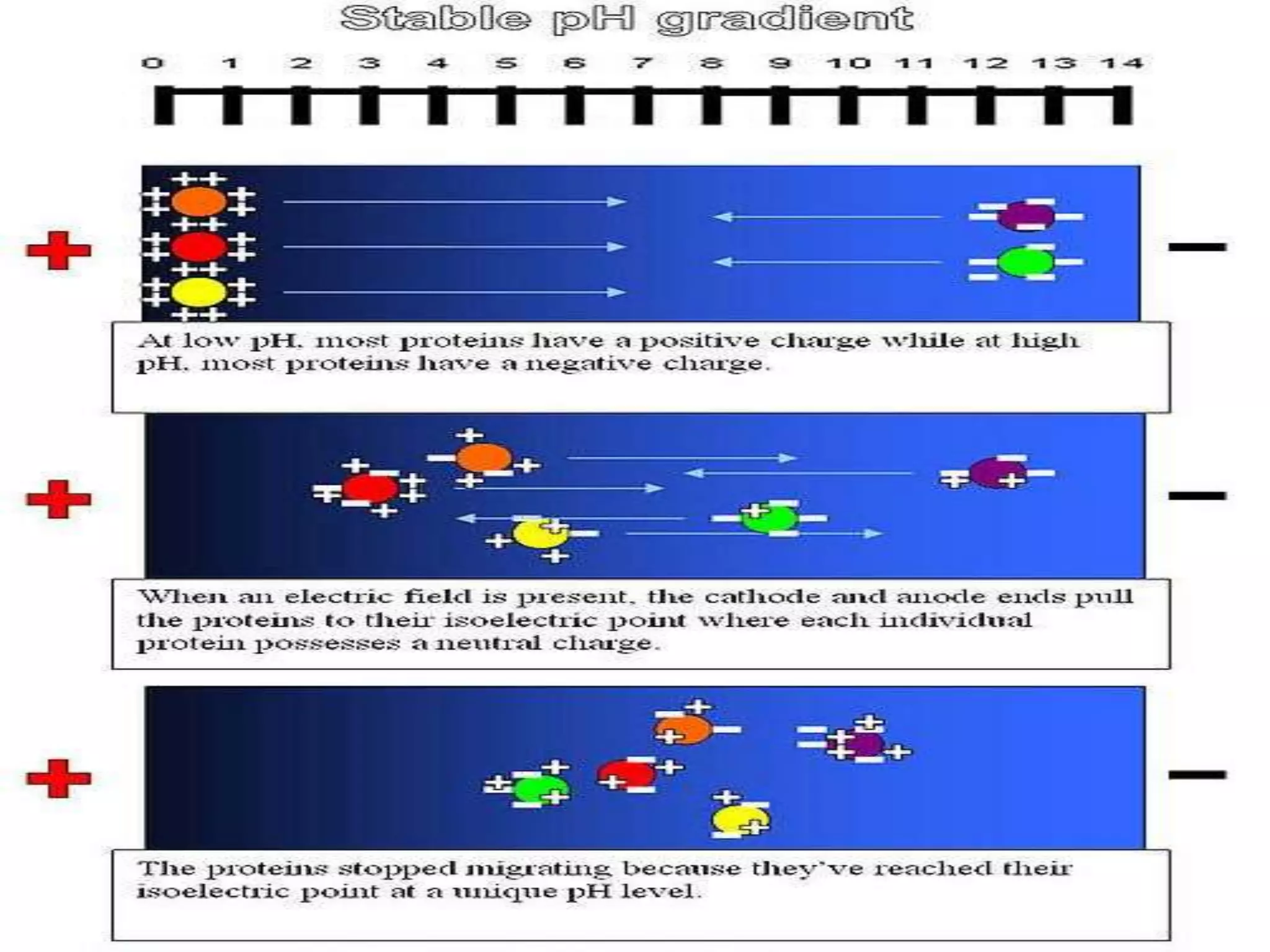
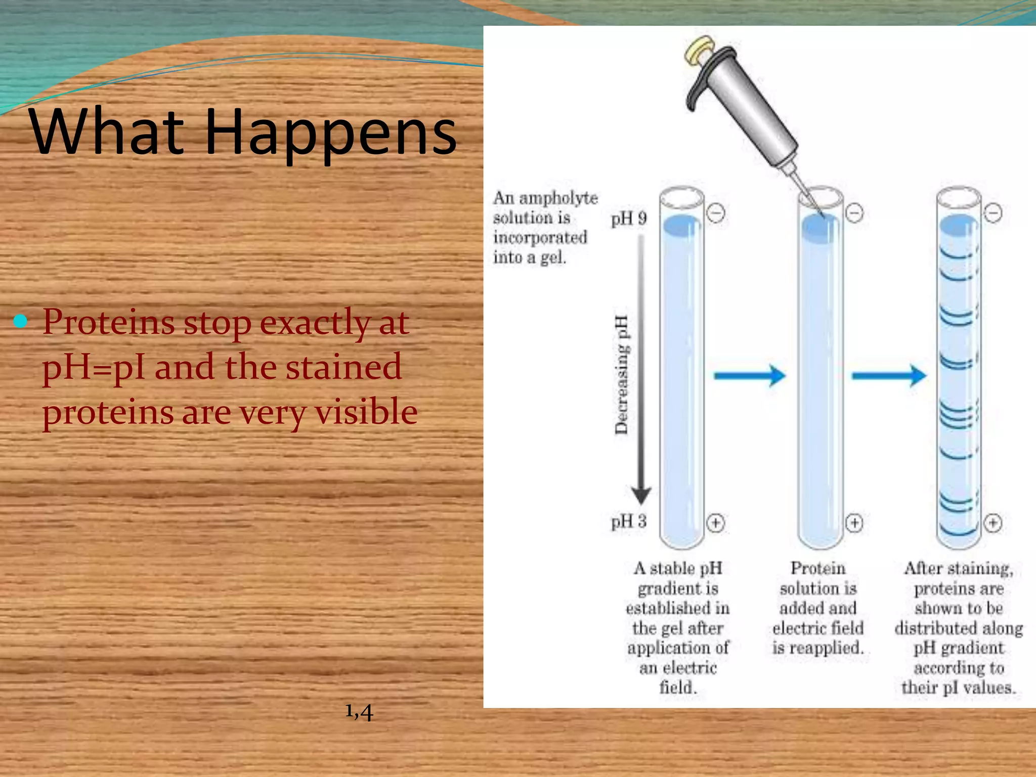
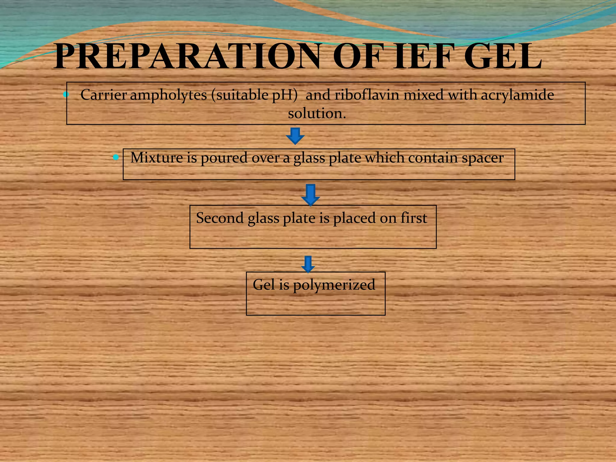
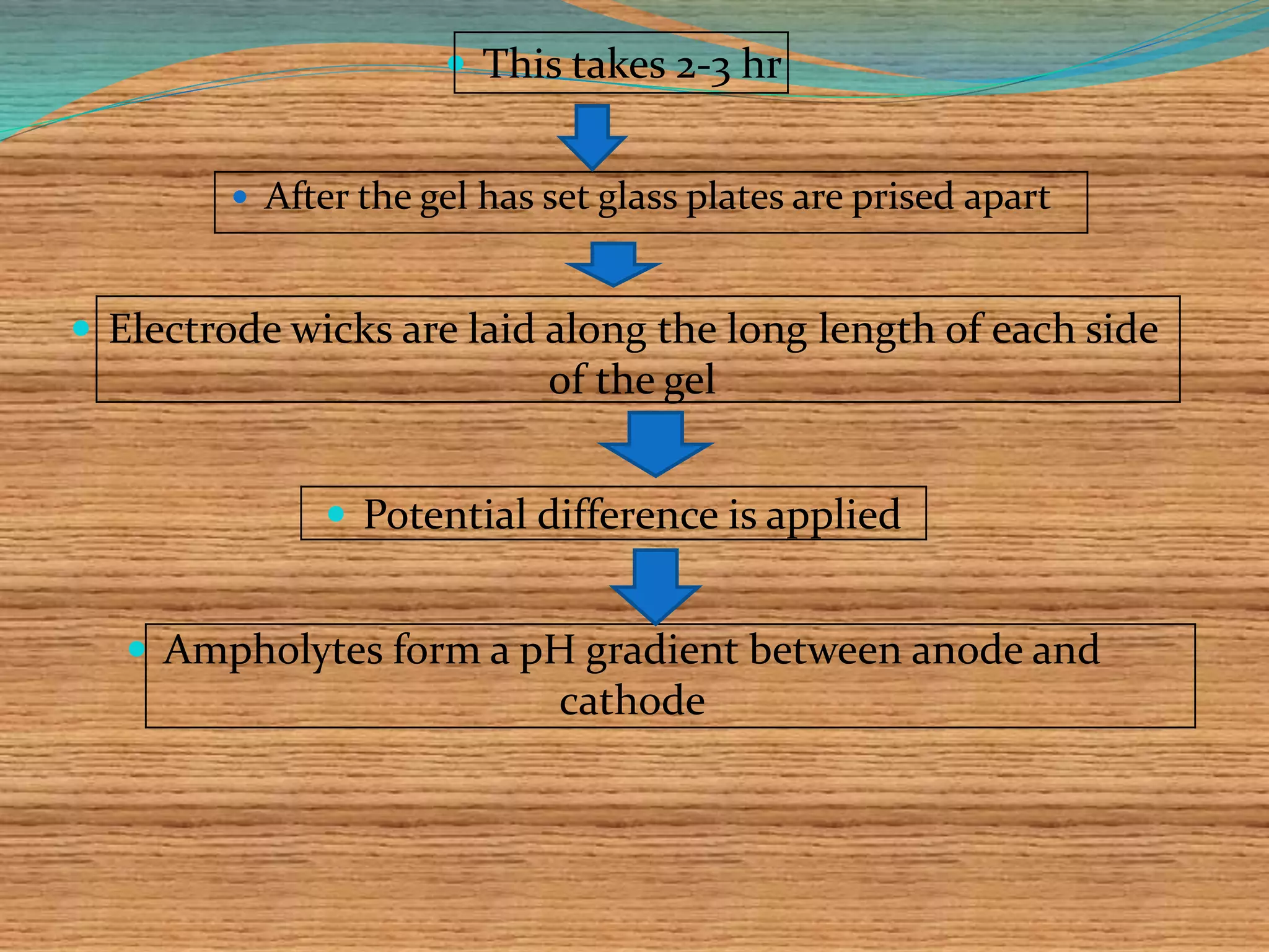
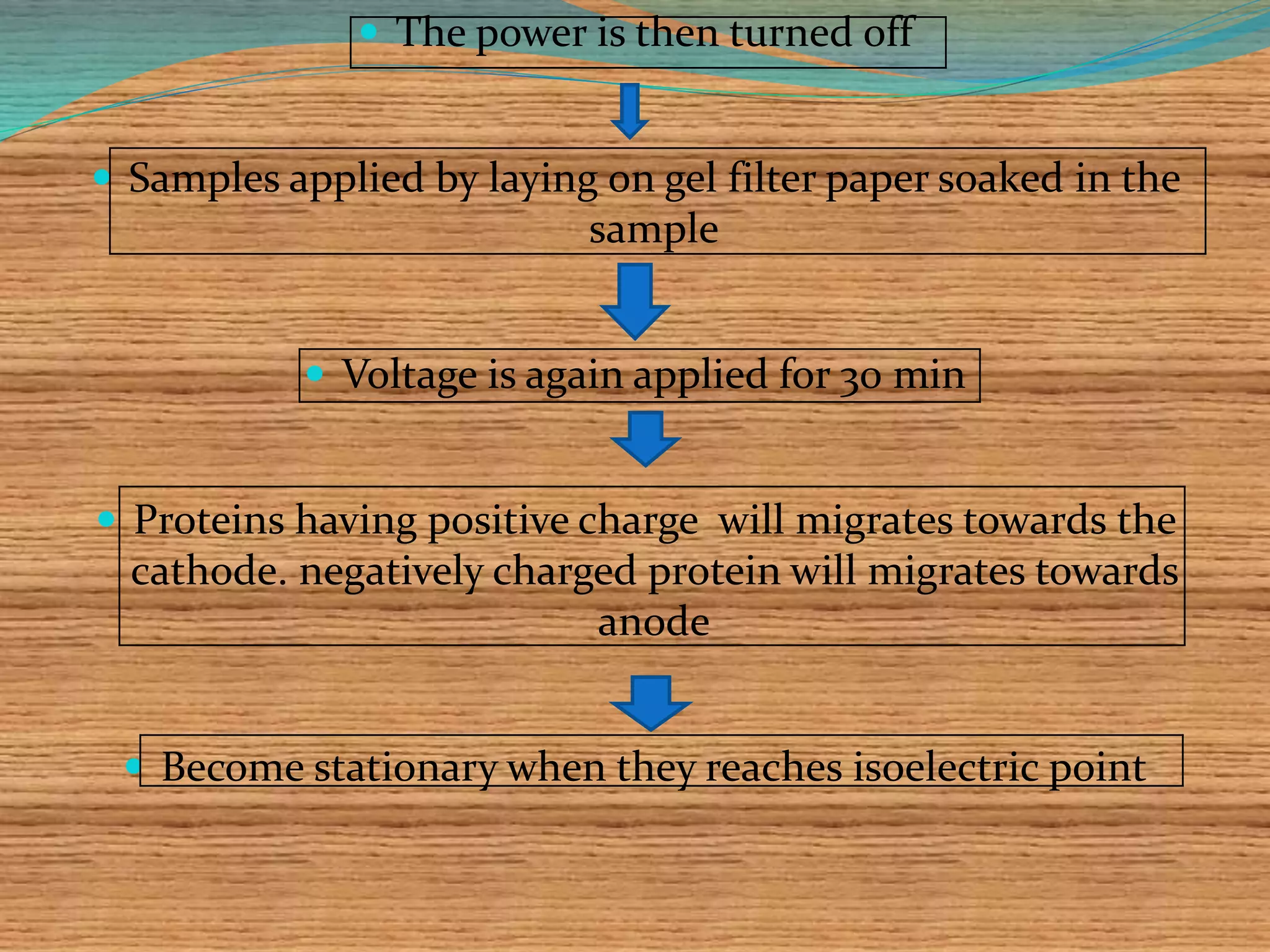
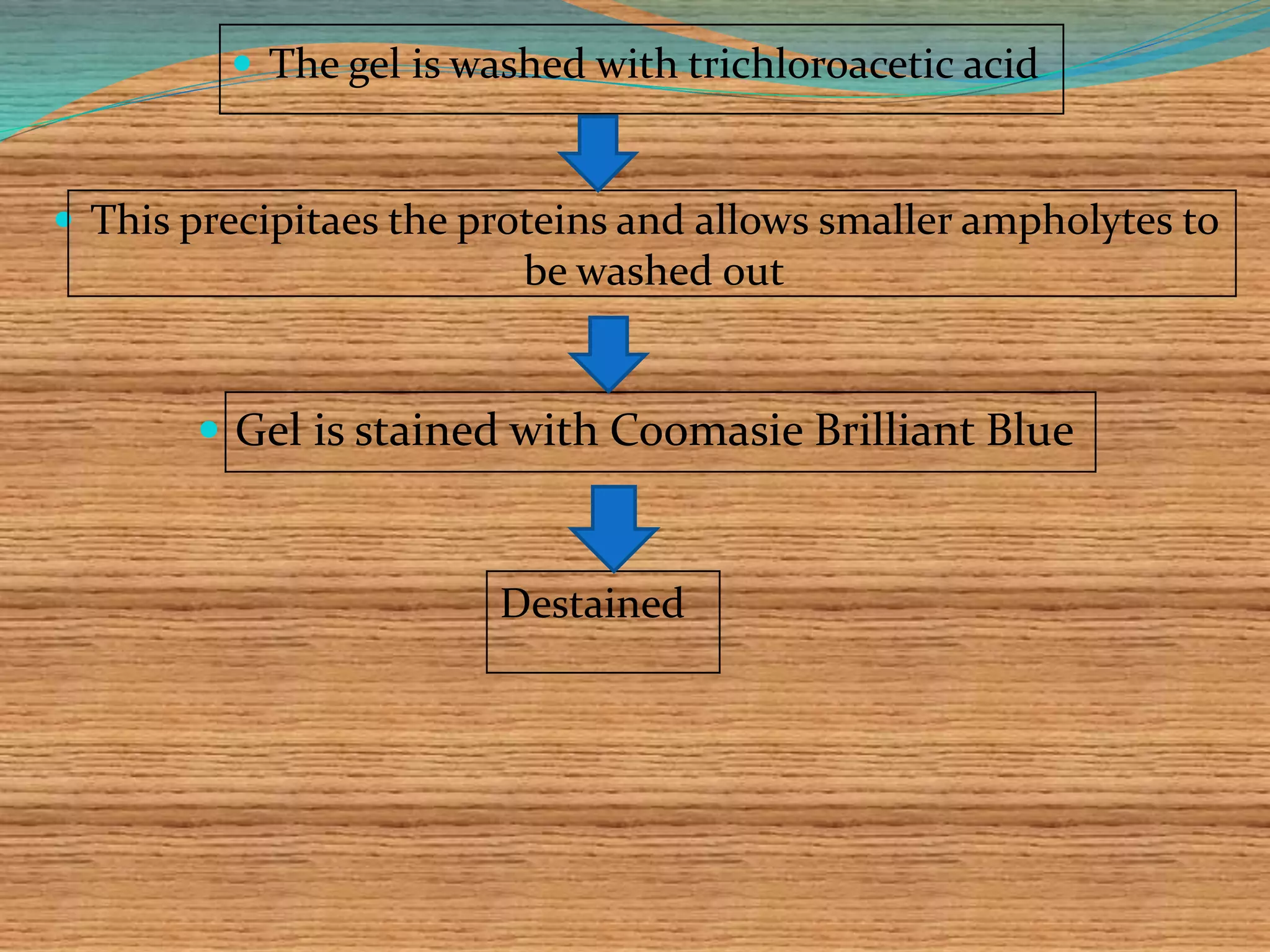
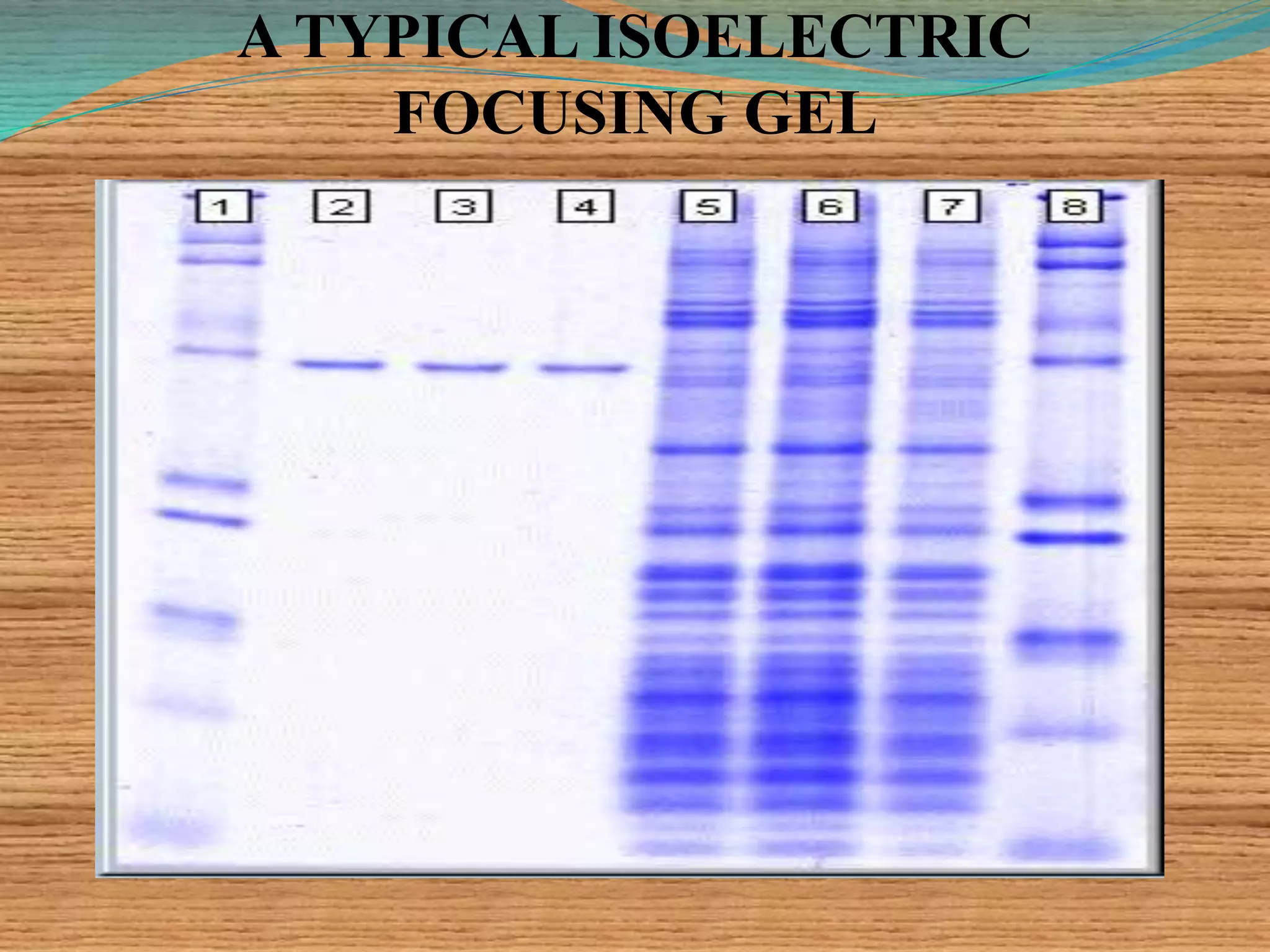
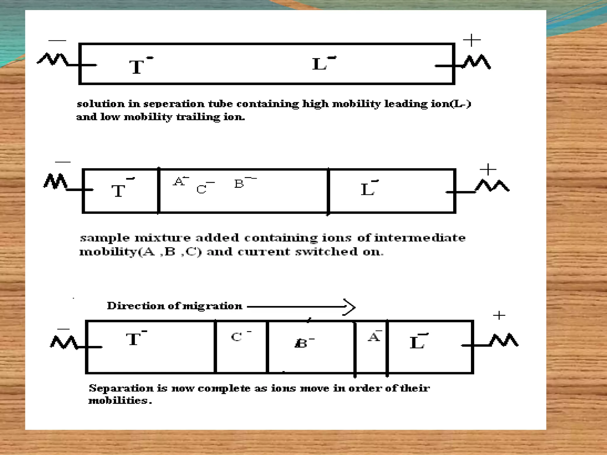
PRESENTED BY,
VINEETHA K
1ST M.PHARM
DEPT. OF PHARMACEUTICS
NGSMIPS.
CONTENTS
 ISOELECTIC FOCUSSING
 PRINCIPLE
 THEORETICAL ASPECTS
 PREPARATION OF IEF GEL
 DETERMINATION OF ISOELECTRIC POINT OF
PROTEIN
 APPLICATION
 ISOTACHOPHORESIS
 PRINCIPLE
 APPLICATION.
ISOELECTRIC FOCUSSING
 Electrophoretic method that separates proteins
according to the iso-electric points.
 Is ideal for separation of amphoteric substances.
 Separation is achieved by applying a potential
difference across a gel that contain a pH gradient.
 Isoelectric focusing requires solid support such as
agarose gel and polyacrylamide.
Isoelectric focusing gels contain synthetic buffers
called ampholytes that smooth the pH gradients.
Ampholytes are complex mixtures of synthetic
polyamino-polycarboxylic acids
Commercially available ampholytes are-
BIO-LYTE
PHARMALYTE
 It gives good separation with a high resolution
compared to any other method
 Resolution depends on
I. The pH gradient,
II. The thickness of the gel
III. Time of electrophoresis,
IV. The applied voltage,
V. Diffusion of the protein into the gel
pI:
 Isoelectric focusing uses the theory of
protein pI.
 pI is the pH at which a given protein has a
neutral overall charge.
 The pI is dependant on which type of
residues are present and how many bases
make proteins positive and acids
negative.
 pI is very specific for each protein.
PRINCIPLE
 Isoionic point is the point at which the net charge of protein
molecule is zero.
 In isoelectric focusing a stable pH gradient established.
 pH increases gradually from anode to cathode.
 A protein introduced at a point where the pH is lower than
the isoionic point will posses a net positive charge & will
migrate in the direction of cathode.
 Due to the presence of protein gradient, the protein will
migrate to an environment of higher pH values .
 Iso electric point of protein varies with the
environment.
 Isoelectric point is taken to be the pH where all the an
ions on the protein are exactly balanced by equal
number of cations;the net charge is zero & the protein
does not migrate in the electric field.
 The value of pH provides a rough idea about the
amino acid composition of a given protein.
•Finally protein will encounter a pH where its net charge
is zero & will stop migrating.
•This is the isoelectric point of protein.
THEORETICAL ASPECTS.
 The pH gradient forces a protein to remain in its isoelectric
point position, thus concentrating it ; this concentrating effect
is called "focusing".
 Increasing the applied voltage or reducing the sample load
result in improved separation of bands. The applied voltage is
limited by the heat generated, which must be dissipated.
 The use of thin gels and an efficient cooling plate controlled by
a thermostatic circulator prevents the burning of the gel whilst
allowing sharp focusing.
 The separation is estimated by determining the minimum pI
difference (ΔpI), which is necessary to separate 2 neighboring
bands:
What Happens
 Proteins stop exactly at
pH=pI and the stained
proteins are very visible
1,4
PREPARATION OF IEF GEL
 Carrier ampholytes (suitable pH) and riboflavin mixed with acrylamide
solution.
 Mixture is poured over a glass plate which contain spacer
Second glass plate is placed on first
Gel is polymerized
 This takes 2-3 hr
 After the gel has set glass plates are prised apart
 Electrode wicks are laid along the long length of each side
of the gel
 Potential difference is applied
 Ampholytes form a pH gradient between anode and
cathode
 The power is then turned off
 Samples applied by laying on gel filter paper soaked in the
sample
 Voltage is again applied for 30 min
 Proteins having positive charge will migrates towards the
cathode. negatively charged protein will migrates towards
anode
 Become stationary when they reaches isoelectric point
 The gel is washed with trichloroacetic acid
 This precipitaes the proteins and allows smaller ampholytes to
be washed out
 Gel is stained with Coomasie Brilliant Blue
Destained
A TYPICAL ISOELECTRIC
FOCUSING GEL
DETERMINATION OF ISOELECTRIC
POINT OF PROTEIN.
 To determine isoelectric point of protein (polypeptide),one has
to:
i. Measure the mobility of protein at several different pH values.
ii. Plot the mobility values so obtained against the pH values.
iii. Take reading of pH of that point where the point intersects
zero mobility line.
The pH at this intersection is then taken to be the isoelectric
point of the concerned protein.
Diagrammatic representation of the principle of pH determination.
The pH at zero mobility is the pI.
APPLICATION
 Useful for separating, purifying & characterizing
proteins.
 Identification of iso enzymes.
 Separation & identification of serum proteins.
 Used by food & agricultural industries, forensic &
human genetic laboratories.
 Used for research in enzymology, immunology &
membrane biochemistry.
ISO-TACHOPHORESIS
 This method is based on the principle of moving boundary
electrophoresis.
 Separation is achieved either in a horizontal or vertical way
based on the equipment employed.
 The name iso-tachophoresis is derived from Greek , refers
to the fact that the ions being separated all travel (phoresis)
at the same( iso)speed( tacho).
 Separation of ionic components of sample is achieved
through stacking them into discrete zones in order of their
mobilities, producing very high resolution.
 Migration of small ions
 Discontinuous electrolyte system
 leading electrolyte (L- ions) &
 trailing electrolyte (T- ions)
 Apply sample solution at interphase of L & T
 Apply electric field -> each type of ion arrange
between L and T ions -> discrete zones
 Separates small anions, cations, organic & amino
acids, peptides, nucleotides, nucleosides, proteins
 Separation of mixture of an ions involves:
 A leading anion (chloride) with higher mobility than the sample ion.
 A trailing an ion (glutamate) with lower mobility than the sample ion.
 A common cat ion for all the anions.
 When the current is switched on, the leading ions will move towards
appropriate electrode.
 The sample ions will follow in order of their mobilities .
 The trailing will follow the sample ions.
 Once equilibrium is achieved ions will all move at the same speed in
discrete bands in order of their mobilities.
 Where the mobilities of sample ions are similar, the
resolution can be enhanced by including with the
sample, synthetic ampholytes called spacer ions.
 These have mobilities intermediate to that of sample
ions and help to separate them by taking up positions
between the sample ions.
 The spacer ions are similar to ampholytes used in
isoelectric focusing.
APPLICATION
 Used for separating charged substances ranging from inorganic to
organic acids to proteins and nucleic acids.
 Used extensively in research laboratories.
 Find applications in industries in pollution control(detecting detergents
and inorganic ions in effluent water)
 In quality control in the food, brewing and pharmaceutical industries.
 Samples as small as few micrograms can be separated quantitatively,&
large quantities of sample can be separated preparatively .
REFERENCES:
Pictures:
2. http://www.science-tube.com/
3. http://www.zeitnews.org/
4.http://www.biochem.arizona.edu/classes/bioc462/462a/N
OTES/Protein_Properties/protein_purification.htm
3 .Introduction to chemical analysis, by “H.K.Kaur
4.S.M. Supriya .Instrumental methods of analysis.1st
edn.India:Popular prakashan;2010.343-347
1. Voet, D. Voet, J. G. Pratt. C. W. Fundamentals of
Biochemistry: Life at the Molecular Level. 3rd edition. John
Wiley and Sons. (2008)
THANK YOU

Theory and application of Isotachophoresis and Isoelectric focussing

  • 1.
    PRESENTED BY, VINEETHA K 1STM.PHARM DEPT. OF PHARMACEUTICS NGSMIPS.
  • 2.
    CONTENTS  ISOELECTIC FOCUSSING PRINCIPLE  THEORETICAL ASPECTS  PREPARATION OF IEF GEL  DETERMINATION OF ISOELECTRIC POINT OF PROTEIN  APPLICATION  ISOTACHOPHORESIS  PRINCIPLE  APPLICATION.
  • 3.
    ISOELECTRIC FOCUSSING  Electrophoreticmethod that separates proteins according to the iso-electric points.  Is ideal for separation of amphoteric substances.  Separation is achieved by applying a potential difference across a gel that contain a pH gradient.  Isoelectric focusing requires solid support such as agarose gel and polyacrylamide.
  • 4.
    Isoelectric focusing gelscontain synthetic buffers called ampholytes that smooth the pH gradients. Ampholytes are complex mixtures of synthetic polyamino-polycarboxylic acids Commercially available ampholytes are- BIO-LYTE PHARMALYTE
  • 5.
     It givesgood separation with a high resolution compared to any other method  Resolution depends on I. The pH gradient, II. The thickness of the gel III. Time of electrophoresis, IV. The applied voltage, V. Diffusion of the protein into the gel
  • 6.
    pI:  Isoelectric focusinguses the theory of protein pI.  pI is the pH at which a given protein has a neutral overall charge.  The pI is dependant on which type of residues are present and how many bases make proteins positive and acids negative.  pI is very specific for each protein.
  • 7.
    PRINCIPLE  Isoionic pointis the point at which the net charge of protein molecule is zero.  In isoelectric focusing a stable pH gradient established.  pH increases gradually from anode to cathode.  A protein introduced at a point where the pH is lower than the isoionic point will posses a net positive charge & will migrate in the direction of cathode.  Due to the presence of protein gradient, the protein will migrate to an environment of higher pH values .
  • 8.
     Iso electricpoint of protein varies with the environment.  Isoelectric point is taken to be the pH where all the an ions on the protein are exactly balanced by equal number of cations;the net charge is zero & the protein does not migrate in the electric field.  The value of pH provides a rough idea about the amino acid composition of a given protein. •Finally protein will encounter a pH where its net charge is zero & will stop migrating. •This is the isoelectric point of protein.
  • 10.
    THEORETICAL ASPECTS.  ThepH gradient forces a protein to remain in its isoelectric point position, thus concentrating it ; this concentrating effect is called "focusing".  Increasing the applied voltage or reducing the sample load result in improved separation of bands. The applied voltage is limited by the heat generated, which must be dissipated.  The use of thin gels and an efficient cooling plate controlled by a thermostatic circulator prevents the burning of the gel whilst allowing sharp focusing.  The separation is estimated by determining the minimum pI difference (ΔpI), which is necessary to separate 2 neighboring bands:
  • 12.
    What Happens  Proteinsstop exactly at pH=pI and the stained proteins are very visible 1,4
  • 13.
    PREPARATION OF IEFGEL  Carrier ampholytes (suitable pH) and riboflavin mixed with acrylamide solution.  Mixture is poured over a glass plate which contain spacer Second glass plate is placed on first Gel is polymerized
  • 14.
     This takes2-3 hr  After the gel has set glass plates are prised apart  Electrode wicks are laid along the long length of each side of the gel  Potential difference is applied  Ampholytes form a pH gradient between anode and cathode
  • 15.
     The poweris then turned off  Samples applied by laying on gel filter paper soaked in the sample  Voltage is again applied for 30 min  Proteins having positive charge will migrates towards the cathode. negatively charged protein will migrates towards anode  Become stationary when they reaches isoelectric point
  • 16.
     The gelis washed with trichloroacetic acid  This precipitaes the proteins and allows smaller ampholytes to be washed out  Gel is stained with Coomasie Brilliant Blue Destained
  • 17.
  • 18.
    DETERMINATION OF ISOELECTRIC POINTOF PROTEIN.  To determine isoelectric point of protein (polypeptide),one has to: i. Measure the mobility of protein at several different pH values. ii. Plot the mobility values so obtained against the pH values. iii. Take reading of pH of that point where the point intersects zero mobility line. The pH at this intersection is then taken to be the isoelectric point of the concerned protein.
  • 19.
    Diagrammatic representation ofthe principle of pH determination. The pH at zero mobility is the pI.
  • 20.
    APPLICATION  Useful forseparating, purifying & characterizing proteins.  Identification of iso enzymes.  Separation & identification of serum proteins.  Used by food & agricultural industries, forensic & human genetic laboratories.  Used for research in enzymology, immunology & membrane biochemistry.
  • 21.
    ISO-TACHOPHORESIS  This methodis based on the principle of moving boundary electrophoresis.  Separation is achieved either in a horizontal or vertical way based on the equipment employed.  The name iso-tachophoresis is derived from Greek , refers to the fact that the ions being separated all travel (phoresis) at the same( iso)speed( tacho).  Separation of ionic components of sample is achieved through stacking them into discrete zones in order of their mobilities, producing very high resolution.
  • 22.
     Migration ofsmall ions  Discontinuous electrolyte system  leading electrolyte (L- ions) &  trailing electrolyte (T- ions)  Apply sample solution at interphase of L & T  Apply electric field -> each type of ion arrange between L and T ions -> discrete zones  Separates small anions, cations, organic & amino acids, peptides, nucleotides, nucleosides, proteins
  • 23.
     Separation ofmixture of an ions involves:  A leading anion (chloride) with higher mobility than the sample ion.  A trailing an ion (glutamate) with lower mobility than the sample ion.  A common cat ion for all the anions.  When the current is switched on, the leading ions will move towards appropriate electrode.  The sample ions will follow in order of their mobilities .  The trailing will follow the sample ions.  Once equilibrium is achieved ions will all move at the same speed in discrete bands in order of their mobilities.
  • 25.
     Where themobilities of sample ions are similar, the resolution can be enhanced by including with the sample, synthetic ampholytes called spacer ions.  These have mobilities intermediate to that of sample ions and help to separate them by taking up positions between the sample ions.  The spacer ions are similar to ampholytes used in isoelectric focusing.
  • 26.
    APPLICATION  Used forseparating charged substances ranging from inorganic to organic acids to proteins and nucleic acids.  Used extensively in research laboratories.  Find applications in industries in pollution control(detecting detergents and inorganic ions in effluent water)  In quality control in the food, brewing and pharmaceutical industries.  Samples as small as few micrograms can be separated quantitatively,& large quantities of sample can be separated preparatively .
  • 27.
    REFERENCES: Pictures: 2. http://www.science-tube.com/ 3. http://www.zeitnews.org/ 4.http://www.biochem.arizona.edu/classes/bioc462/462a/N OTES/Protein_Properties/protein_purification.htm 3.Introduction to chemical analysis, by “H.K.Kaur 4.S.M. Supriya .Instrumental methods of analysis.1st edn.India:Popular prakashan;2010.343-347 1. Voet, D. Voet, J. G. Pratt. C. W. Fundamentals of Biochemistry: Life at the Molecular Level. 3rd edition. John Wiley and Sons. (2008)
  • 28.