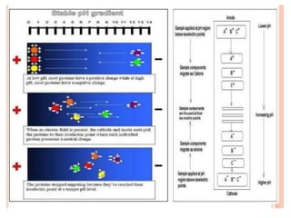
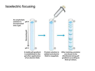
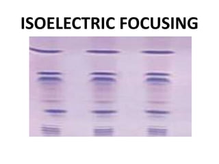
ISOELECTRIC FOCUSING
CONTENT
• INTRODUCTION
• ISOELECTRIC POINT(pI)
• PRINCIPLE
• CAPILLARY
ISOELECTRIC FOCUSING
• AMPHOLYTES
• WORKING PROCEDURE
• SET UP GEL
• SAMPLE PREPARATION
AND LOADING
• 2D GEL
ELECTROPHORESIS
• ADVANTAGES
• DISADVANTAGES
• APPLICATIONS
ISOELECTRIC FOCUSING
INTRODUCTION
• Proteins are separated in a pH gradient according
to their isoelectric points.
• Basic principle involved is electrophoresis.
• Proteins are subjected to electric field in a pH
gradient.
• Requires a solid surface normally Polyacrilamide.
ISOELECTRIC POINT (pI)
• The pH at which net charge on protein
becomes zero.
1. Below pI – Positive charge.
2. Above pI – Negative charge.
• Proteins move toward the electrode with the
opposite charge.
• During motion , proteins will either pick or
loose protons.
• Different from conventional electrophoresis
migrate to steady state.
PRINCIPLE
• All proteins have an isoelectric point pH.
• A procedure to determine the isoelectric point
of proteins thus , a mixture of proteins can be
electrophorised through a solution having a state
pH gradient in form the anode to the cathode and
a each protein will migrate to the position in the
pH gradient according to its isoelectric point.This
is called ISOELECTRIC FOCUSING.
• Protein migrate into the point where its net
charge is zero-isoelectric pH.
CAPILLARY ISOELECTRIC
FOCUSING
• pH gradient.
• Sample focusing and detection.
• Not useful for chiral compounds.
• Movement of gradient towards the detector.
AMPHOLYTES
• Establishment of stable pH gradient is important.
• Achieved by means of commercially available
synthetic carrier amphoteric electrolytes.
• 600-900 Da.
• Closely spaced pI and high conductivity.
• The curve is determined by pH interval covered
by the ampholyte and the distance between
electrodes.
WORKING PROCEDURE
• Following chemicals are required-
1. Acrylamide solution.
2. Water.
3. Ampholyte solution pH 3.5-10.
4. Ampholyte solution pH 4-6.
5. Urea.
• Spin gently to mix urea.
• Add 10% APS and TEMED at the end.
• Remove bubbles.
• Fill the cassette completely with solution.
• Allow to polymerize at room temperature.
7. Distilled water.
• Centrifuge the sample to remove any
aggregate.
• Apply the supernatant to the wells with a
disposable tip or Hamilton syringe.
• Run the get at 150v for 30 min and then at
200v for 2 hr.
ISOELECTRIC FOCUSING
SET UP GEL
• Remove the comb carefully after gel has
polymerized.
• Attach gel to the electrophoresis tank according
the instructions of manufacturer.
• Add catholyte (sodium hydroxide) to the upper
buffer chamber and anolyte (phosphoric acid) to
the lower buffer chamber.
SAMPLE PREPARATION AND
LOADING
• Mix protein sample with equal volume of 2x
loading buffer.
• Loading buffer includes the following reagents-
1. Urea.
2. Ampholyte solution pH 3.5-10.
3. Ampholyte solution pH 4-6.
4. Triton X-100.
5. 2- Mercaptoethanol.
6. 1% bromophenol blue.
2D GEL ELECTROPHORESIS
• Technique of IEF and SDS PAGE combined.
• Protein separated in two dimensions.
1. On the base of pI.
2. On the basis of molecular weight in normal SDS
PAGE.
• Procedure can be adapted by combining IEF and
PAGE.
• Series of spots formed in gel.
ADVANTAGES
• Proteins that by as little as 0.001 pH units can
be separated.
• As spreading of bands is minimized due to
application of the applied field and the pH
gradient , high resolution can be achieved.
• Isoelectric focusing (IEF) is a powerful analytical
tool for the separation of proteins.
• Performing IEF is easier because the placement
of sample application is not important
TRADITIONAL EQUIPMENT
FOR ISOELECTRIC FOCUSING
DISADVANTAGES
• Carrier ampholytes are generally used in high
concentration , a high voltage (upto 2000v) is
necessary. As a result the electrophoretic matrix
must be cooled which sometimes makes it
difficult.
• Limited stability of solutions.
• Lot-to-lot inconsistency.
• Inadequate purity for application as a standard.
APPLICATIONS
• For separating proteins and peptides.
• Used in limit test when the density of band is
compared with the density of band of std prep.
• For research in Taxonomy , Cytology and
Immunology etc.
• IEF gel is used as identity test when migration
pattern on gel is compared with std preparation.
• Isoelectric focusing (IEF) offers an effective
alternative to conventional electrophoresis for
genetic marker typing.

ISOELECTRIC FOCUSING PPT - SLIDE SHARE

  • 1.
  • 2.
    CONTENT • INTRODUCTION • ISOELECTRICPOINT(pI) • PRINCIPLE • CAPILLARY ISOELECTRIC FOCUSING • AMPHOLYTES • WORKING PROCEDURE • SET UP GEL • SAMPLE PREPARATION AND LOADING • 2D GEL ELECTROPHORESIS • ADVANTAGES • DISADVANTAGES • APPLICATIONS
  • 3.
  • 4.
    INTRODUCTION • Proteins areseparated in a pH gradient according to their isoelectric points. • Basic principle involved is electrophoresis. • Proteins are subjected to electric field in a pH gradient. • Requires a solid surface normally Polyacrilamide.
  • 5.
    ISOELECTRIC POINT (pI) •The pH at which net charge on protein becomes zero. 1. Below pI – Positive charge. 2. Above pI – Negative charge. • Proteins move toward the electrode with the opposite charge. • During motion , proteins will either pick or loose protons. • Different from conventional electrophoresis migrate to steady state.
  • 7.
    PRINCIPLE • All proteinshave an isoelectric point pH. • A procedure to determine the isoelectric point of proteins thus , a mixture of proteins can be electrophorised through a solution having a state pH gradient in form the anode to the cathode and a each protein will migrate to the position in the pH gradient according to its isoelectric point.This is called ISOELECTRIC FOCUSING. • Protein migrate into the point where its net charge is zero-isoelectric pH.
  • 9.
    CAPILLARY ISOELECTRIC FOCUSING • pHgradient. • Sample focusing and detection. • Not useful for chiral compounds. • Movement of gradient towards the detector.
  • 11.
    AMPHOLYTES • Establishment ofstable pH gradient is important. • Achieved by means of commercially available synthetic carrier amphoteric electrolytes. • 600-900 Da. • Closely spaced pI and high conductivity. • The curve is determined by pH interval covered by the ampholyte and the distance between electrodes.
  • 12.
    WORKING PROCEDURE • Followingchemicals are required- 1. Acrylamide solution. 2. Water. 3. Ampholyte solution pH 3.5-10. 4. Ampholyte solution pH 4-6. 5. Urea. • Spin gently to mix urea. • Add 10% APS and TEMED at the end. • Remove bubbles. • Fill the cassette completely with solution. • Allow to polymerize at room temperature.
  • 13.
    7. Distilled water. •Centrifuge the sample to remove any aggregate. • Apply the supernatant to the wells with a disposable tip or Hamilton syringe. • Run the get at 150v for 30 min and then at 200v for 2 hr.
  • 14.
  • 15.
    SET UP GEL •Remove the comb carefully after gel has polymerized. • Attach gel to the electrophoresis tank according the instructions of manufacturer. • Add catholyte (sodium hydroxide) to the upper buffer chamber and anolyte (phosphoric acid) to the lower buffer chamber.
  • 16.
    SAMPLE PREPARATION AND LOADING •Mix protein sample with equal volume of 2x loading buffer. • Loading buffer includes the following reagents- 1. Urea. 2. Ampholyte solution pH 3.5-10. 3. Ampholyte solution pH 4-6. 4. Triton X-100. 5. 2- Mercaptoethanol. 6. 1% bromophenol blue.
  • 17.
    2D GEL ELECTROPHORESIS •Technique of IEF and SDS PAGE combined. • Protein separated in two dimensions. 1. On the base of pI. 2. On the basis of molecular weight in normal SDS PAGE. • Procedure can be adapted by combining IEF and PAGE. • Series of spots formed in gel.
  • 19.
    ADVANTAGES • Proteins thatby as little as 0.001 pH units can be separated. • As spreading of bands is minimized due to application of the applied field and the pH gradient , high resolution can be achieved. • Isoelectric focusing (IEF) is a powerful analytical tool for the separation of proteins. • Performing IEF is easier because the placement of sample application is not important
  • 20.
  • 21.
    DISADVANTAGES • Carrier ampholytesare generally used in high concentration , a high voltage (upto 2000v) is necessary. As a result the electrophoretic matrix must be cooled which sometimes makes it difficult. • Limited stability of solutions. • Lot-to-lot inconsistency. • Inadequate purity for application as a standard.
  • 22.
    APPLICATIONS • For separatingproteins and peptides. • Used in limit test when the density of band is compared with the density of band of std prep. • For research in Taxonomy , Cytology and Immunology etc. • IEF gel is used as identity test when migration pattern on gel is compared with std preparation. • Isoelectric focusing (IEF) offers an effective alternative to conventional electrophoresis for genetic marker typing.