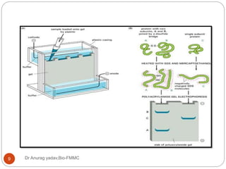
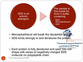
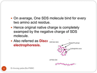
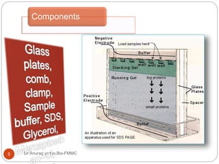
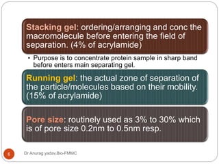
Presenter Dr Anurag Yadav
Post-graduate, biochemistry
Father Muller Medical college
Dr Anurag yadav,Bio-FMMC1
ELECTROPHORESIS
SDS-PAGE
Dr Anurag yadav,Bio-FMMC2
ď‚— Sodium dodecyl sulphate- polyacrylamide gel
electrophoresis.
ď‚— Most widely used method for analysing protein
mixture qualitatively.
 Useful for monitoring protein purification – as
separation of protein is based on the size of the
particle.
ď‚— Can also be used for determining the relative
molecular mass of a protein.
Dr Anurag yadav,Bio-FMMC3
ď‚— Mercaptoethanol will break the disulphide bridges.
ď‚— SDS binds strongly to and denatures the protein.
ď‚— Each protein is fully denatured and open into rod-
shape with series of negatively charged SDS
molecule on polypeptide chain.
SDS is an
anionic
detergent.
The sample is
first boiled for
5min in buffer
containing
• Beta-
Mercaptoethanol
• SDS
Dr Anurag yadav,Bio-FMMC4
ď‚— On average, One SDS molecule bind for every
two amino acid residue.
ď‚— Hence original native charge is completely
swamped by the negative charge of SDS
molecule.
ď‚— Also referred as Discontinuous gel
electrophoresis.
Components
Dr Anurag yadav,Bio-FMMC5
Dr Anurag yadav,Bio-FMMC6
Stacking gel: ordering/arranging and conc the
macromolecule before entering the field of
separation. (4% of acrylamide)
• Purpose is to concentrate protein sample in sharp band
before enters main separating gel.
Running gel: the actual zone of separation of
the particle/molecules based on their mobility.
(15% of acrylamide)
Pore size: routinely used as 3% to 30% which
is of pore size 0.2nm to 0.5nm resp.
Dr Anurag yadav,Bio-FMMC7
Movement of
particle
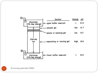
Dr Anurag yadav,Bio-FMMC8
[Cl] > [protein-SDS] >
[Glycinate]
Dr Anurag yadav,Bio-FMMC9
Dr Anurag yadav,Bio-FMMC10
ď‚— In separating gel, protein separate owing to
molecular sieving properties.
ď‚— Smaller proteins pass more easily, larger one
retarded by friction.
- Research tool
- Measuring molecular weight
- Peptide mapping
- Protein identification
- Determination of sample purity
- Identifying disulfide bonds
- Separation of proteins and establishing size
- Blotting
- Smaller fragments of DNA
- Separation of nucleic acids
- Major clinical use – ALP separation
APPLICATION:
ADVANTAGES:
- Clear, fairly easy to prepare
- Exhibit reasonable mechanical strength over
acrylamide conc
- Low endosmosis effect
DISADVANTAGES
- Gel preparation and casting- exacting n time-
consuming
- Complete reproducibility of gel preparation not
possible
STAINING:
Fluorescent stains - Ethidium bromide – Nucleic
acids
Silver stain for protein gel (sensitive 50 times dye
based)
Dye based – Coomassie blue – 50ng protein
band
Tracking dyes – BPB> xylene cyanol, Orange G
Dr Anurag yadav,Bio-FMMC14
References
Dr Anurag yadav,Bio-FMMC15
ď‚— Keith Wilson- Principles and techniques of
biochemistry and molecular biology.
ď‚— Upadhyay- biophysical chemistry.
ď‚— Tietz- Text book of clinical chemistry.
ď‚— Kaplan- clinical chemistry.
ď‚— YouTube and Google images.

SDS-PAGE electrophoresis by Dr. Anurag Yadav

  • 1.
    Presenter Dr AnuragYadav Post-graduate, biochemistry Father Muller Medical college Dr Anurag yadav,Bio-FMMC1 ELECTROPHORESIS
  • 2.
    SDS-PAGE Dr Anurag yadav,Bio-FMMC2 Sodium dodecyl sulphate- polyacrylamide gel electrophoresis.  Most widely used method for analysing protein mixture qualitatively.  Useful for monitoring protein purification – as separation of protein is based on the size of the particle.  Can also be used for determining the relative molecular mass of a protein.
  • 3.
    Dr Anurag yadav,Bio-FMMC3 Mercaptoethanol will break the disulphide bridges.  SDS binds strongly to and denatures the protein.  Each protein is fully denatured and open into rod- shape with series of negatively charged SDS molecule on polypeptide chain. SDS is an anionic detergent. The sample is first boiled for 5min in buffer containing • Beta- Mercaptoethanol • SDS
  • 4.
    Dr Anurag yadav,Bio-FMMC4 ď‚—On average, One SDS molecule bind for every two amino acid residue. ď‚— Hence original native charge is completely swamped by the negative charge of SDS molecule. ď‚— Also referred as Discontinuous gel electrophoresis.
  • 5.
  • 6.
    Dr Anurag yadav,Bio-FMMC6 Stackinggel: ordering/arranging and conc the macromolecule before entering the field of separation. (4% of acrylamide) • Purpose is to concentrate protein sample in sharp band before enters main separating gel. Running gel: the actual zone of separation of the particle/molecules based on their mobility. (15% of acrylamide) Pore size: routinely used as 3% to 30% which is of pore size 0.2nm to 0.5nm resp.
  • 7.
  • 8.
    Movement of particle Dr Anuragyadav,Bio-FMMC8 [Cl] > [protein-SDS] > [Glycinate]
  • 9.
  • 10.
    Dr Anurag yadav,Bio-FMMC10 ď‚—In separating gel, protein separate owing to molecular sieving properties. ď‚— Smaller proteins pass more easily, larger one retarded by friction.
  • 11.
    - Research tool -Measuring molecular weight - Peptide mapping - Protein identification - Determination of sample purity - Identifying disulfide bonds - Separation of proteins and establishing size - Blotting - Smaller fragments of DNA - Separation of nucleic acids - Major clinical use – ALP separation APPLICATION:
  • 12.
    ADVANTAGES: - Clear, fairlyeasy to prepare - Exhibit reasonable mechanical strength over acrylamide conc - Low endosmosis effect DISADVANTAGES - Gel preparation and casting- exacting n time- consuming - Complete reproducibility of gel preparation not possible
  • 13.
    STAINING: Fluorescent stains -Ethidium bromide – Nucleic acids Silver stain for protein gel (sensitive 50 times dye based) Dye based – Coomassie blue – 50ng protein band Tracking dyes – BPB> xylene cyanol, Orange G
  • 14.
  • 15.
    References Dr Anurag yadav,Bio-FMMC15 ď‚—Keith Wilson- Principles and techniques of biochemistry and molecular biology. ď‚— Upadhyay- biophysical chemistry. ď‚— Tietz- Text book of clinical chemistry. ď‚— Kaplan- clinical chemistry. ď‚— YouTube and Google images.