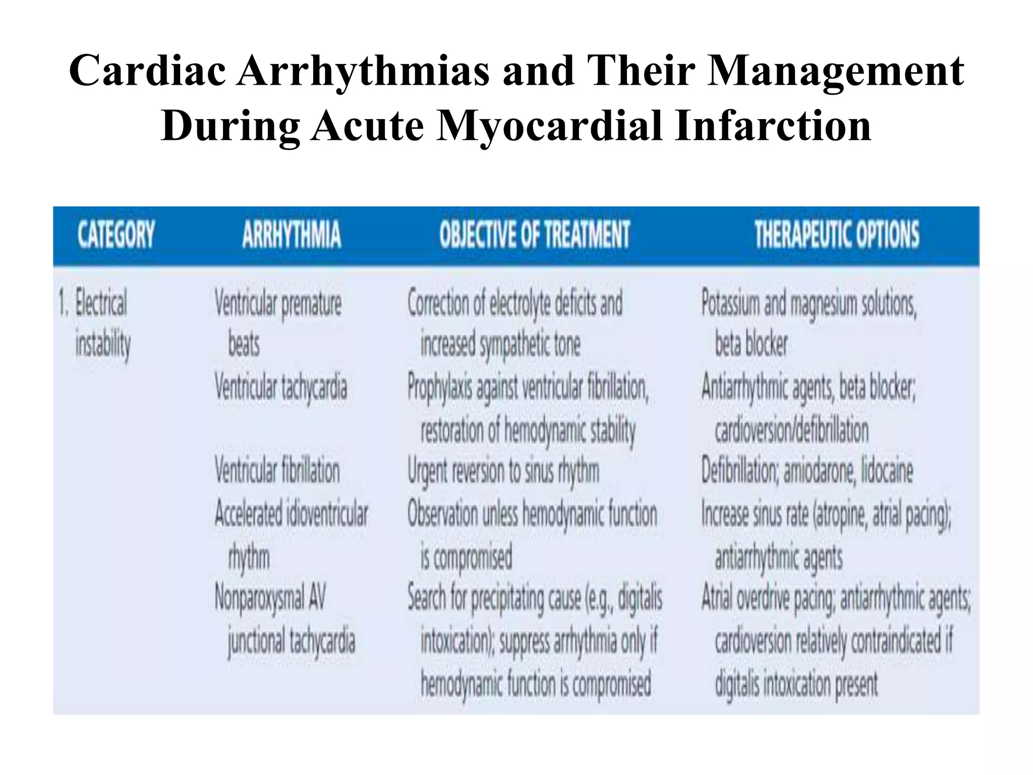
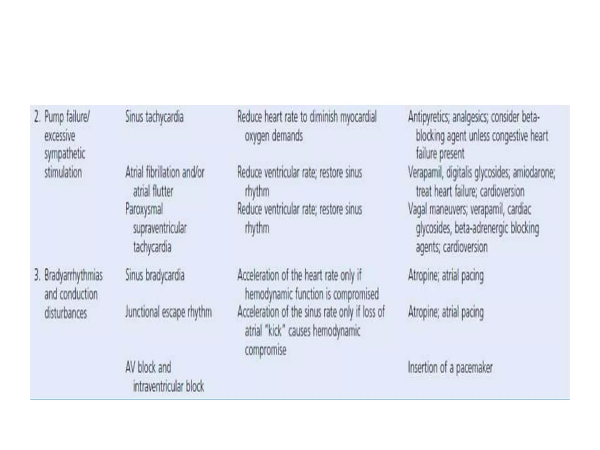
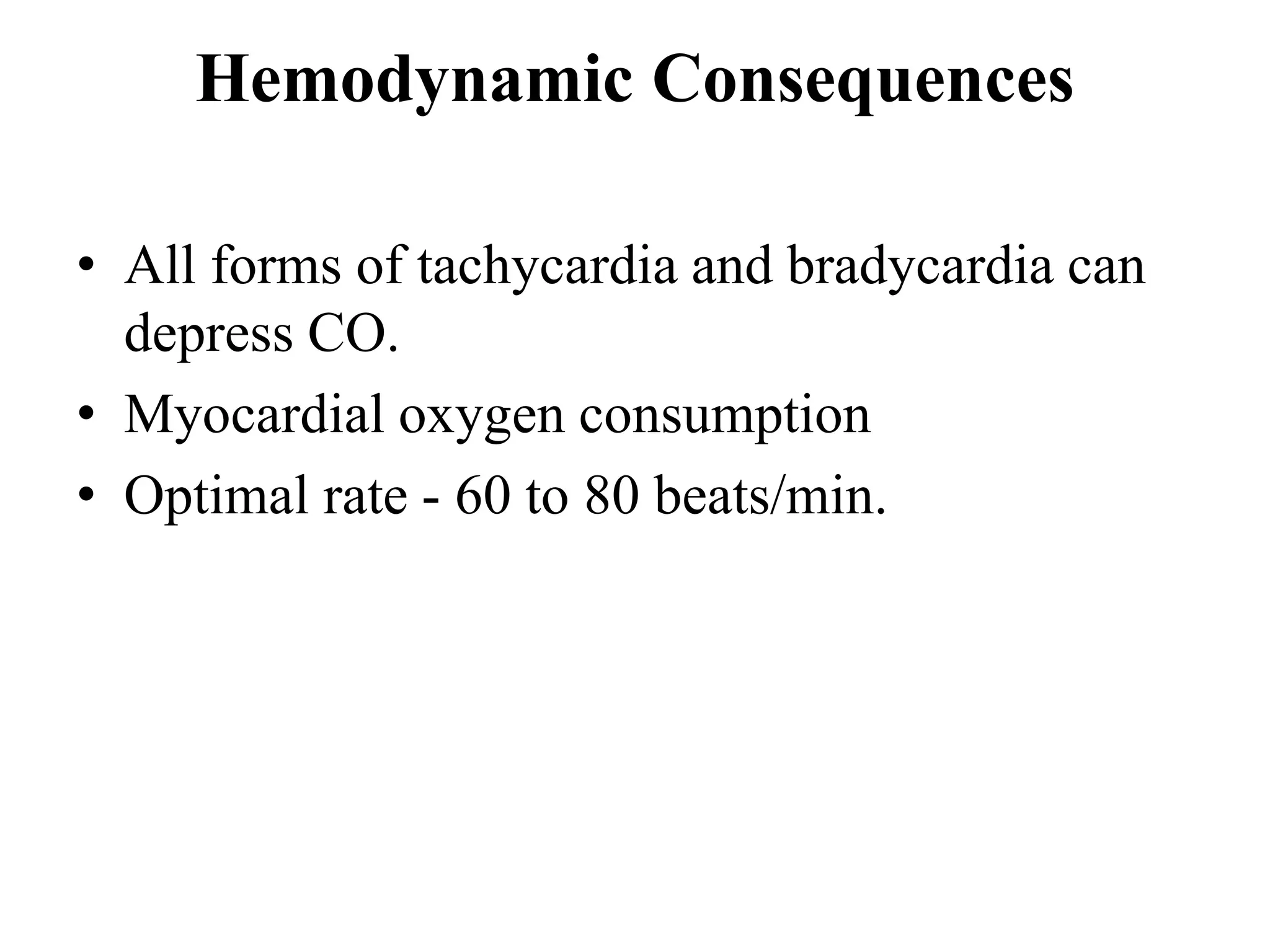
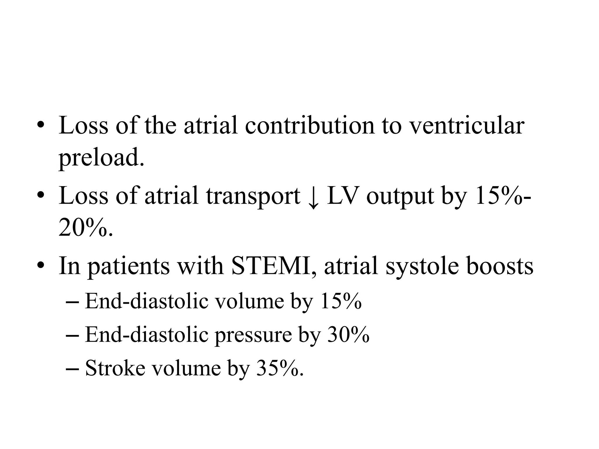
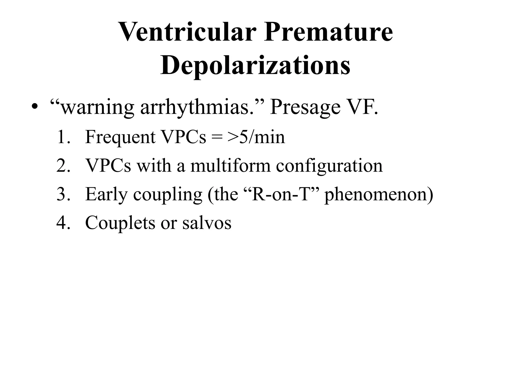
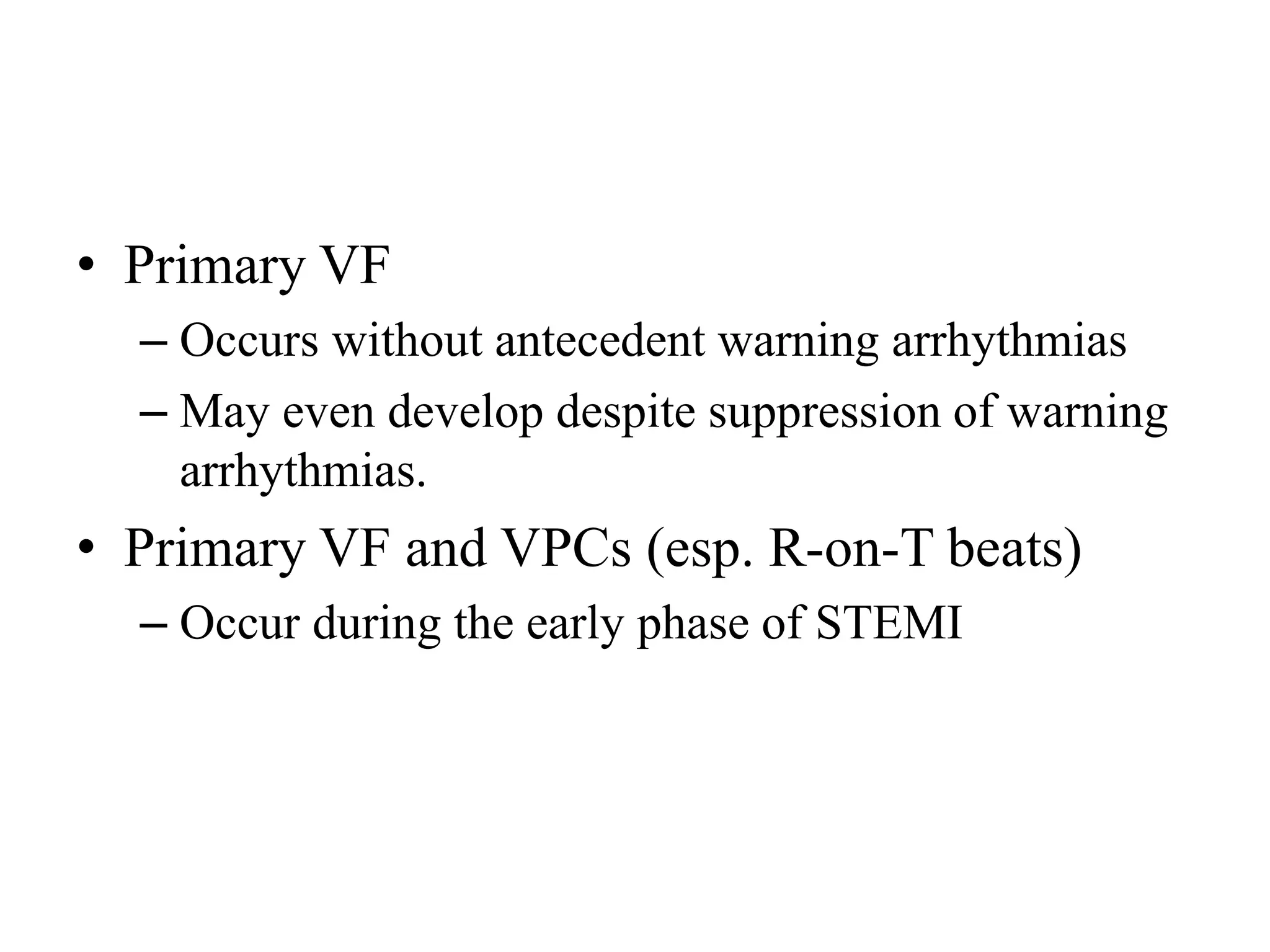
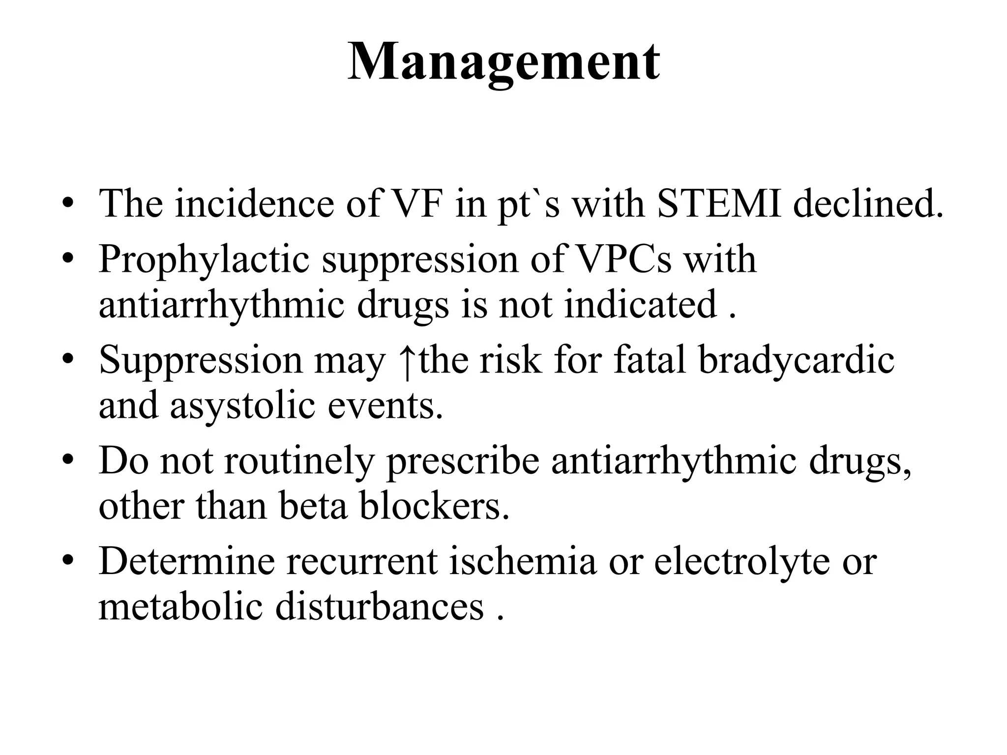
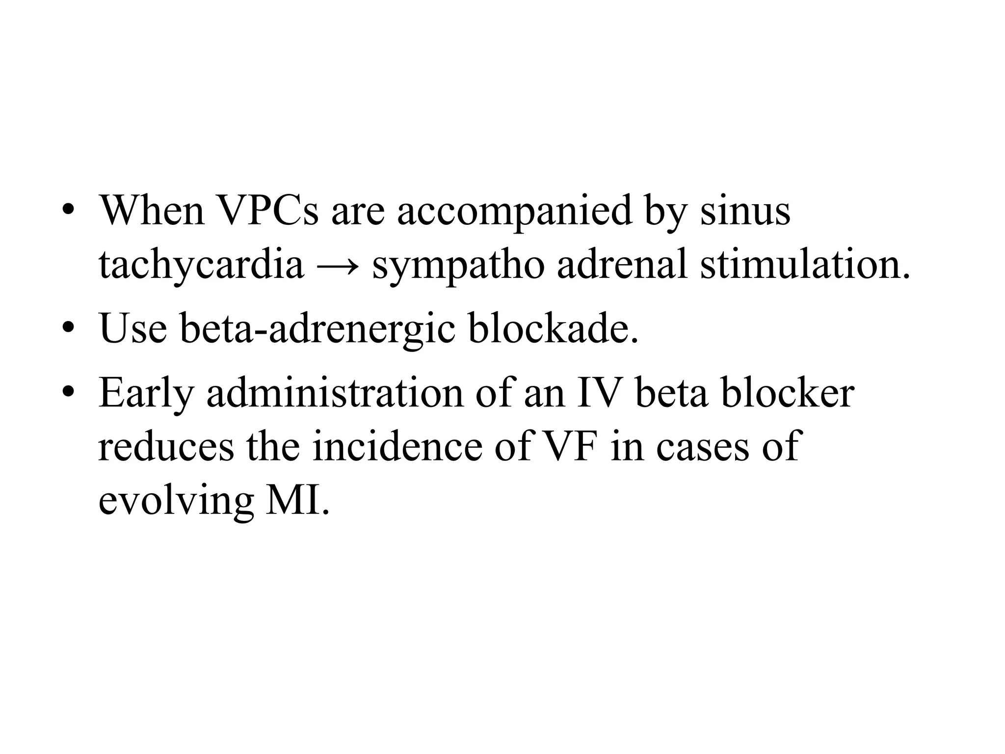
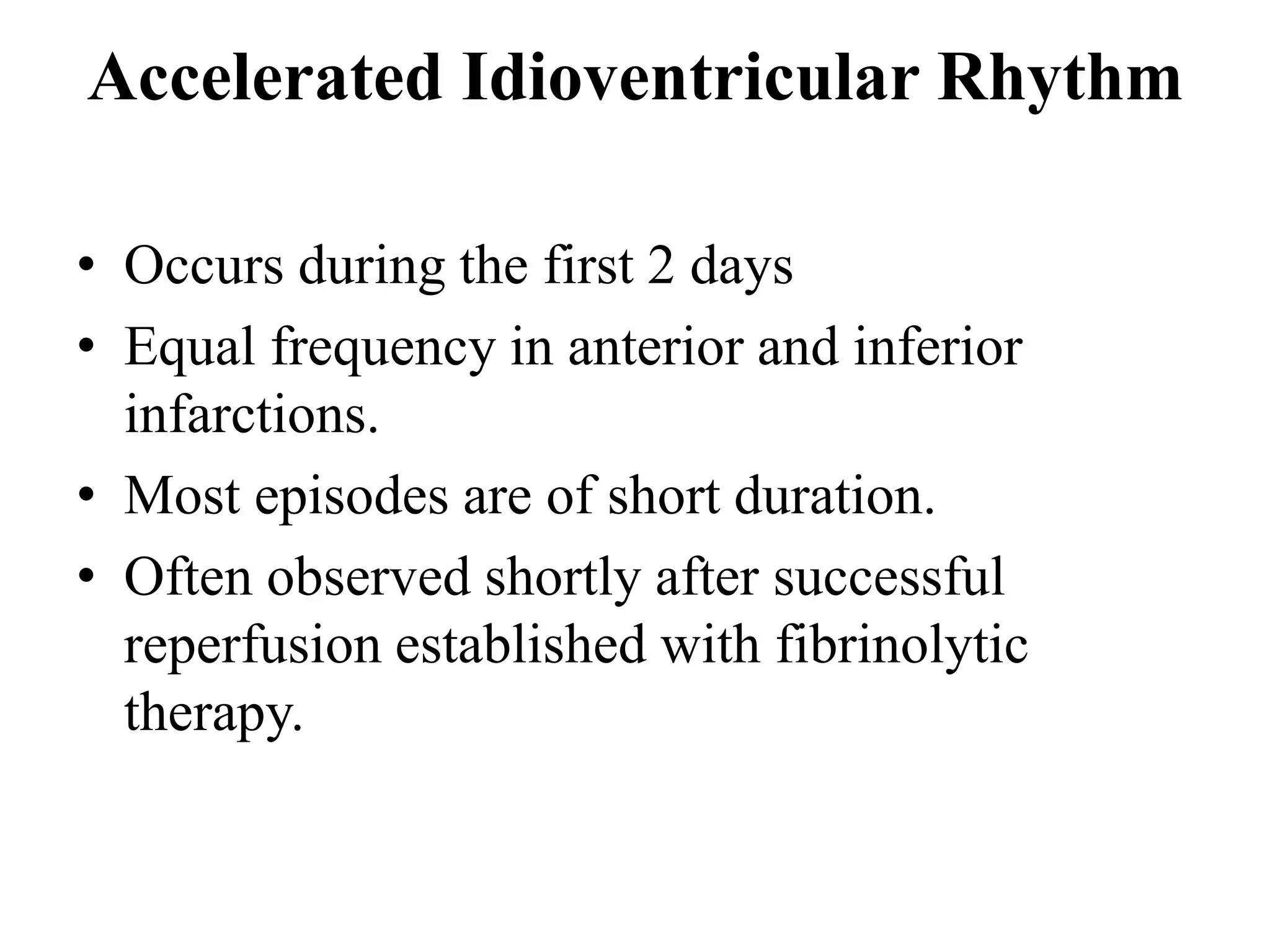
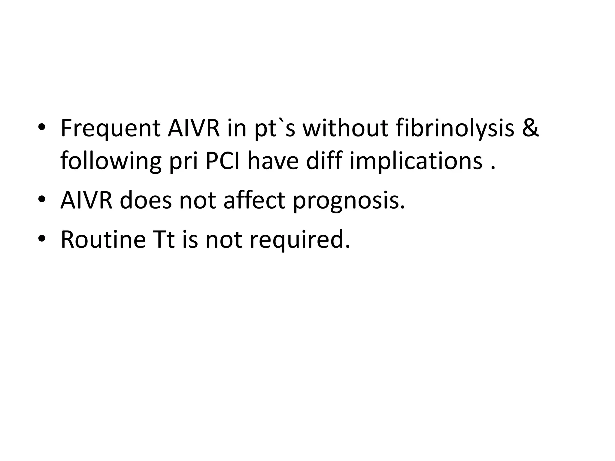
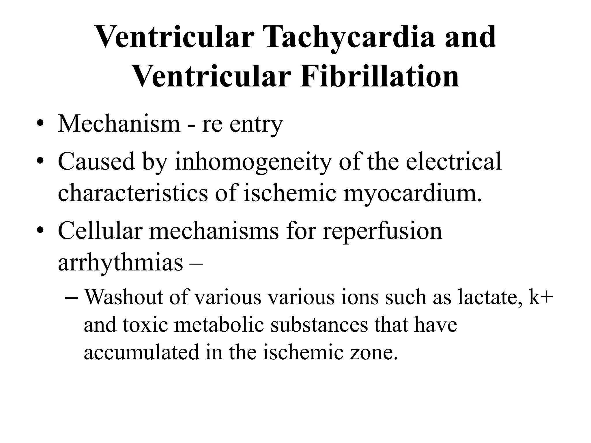
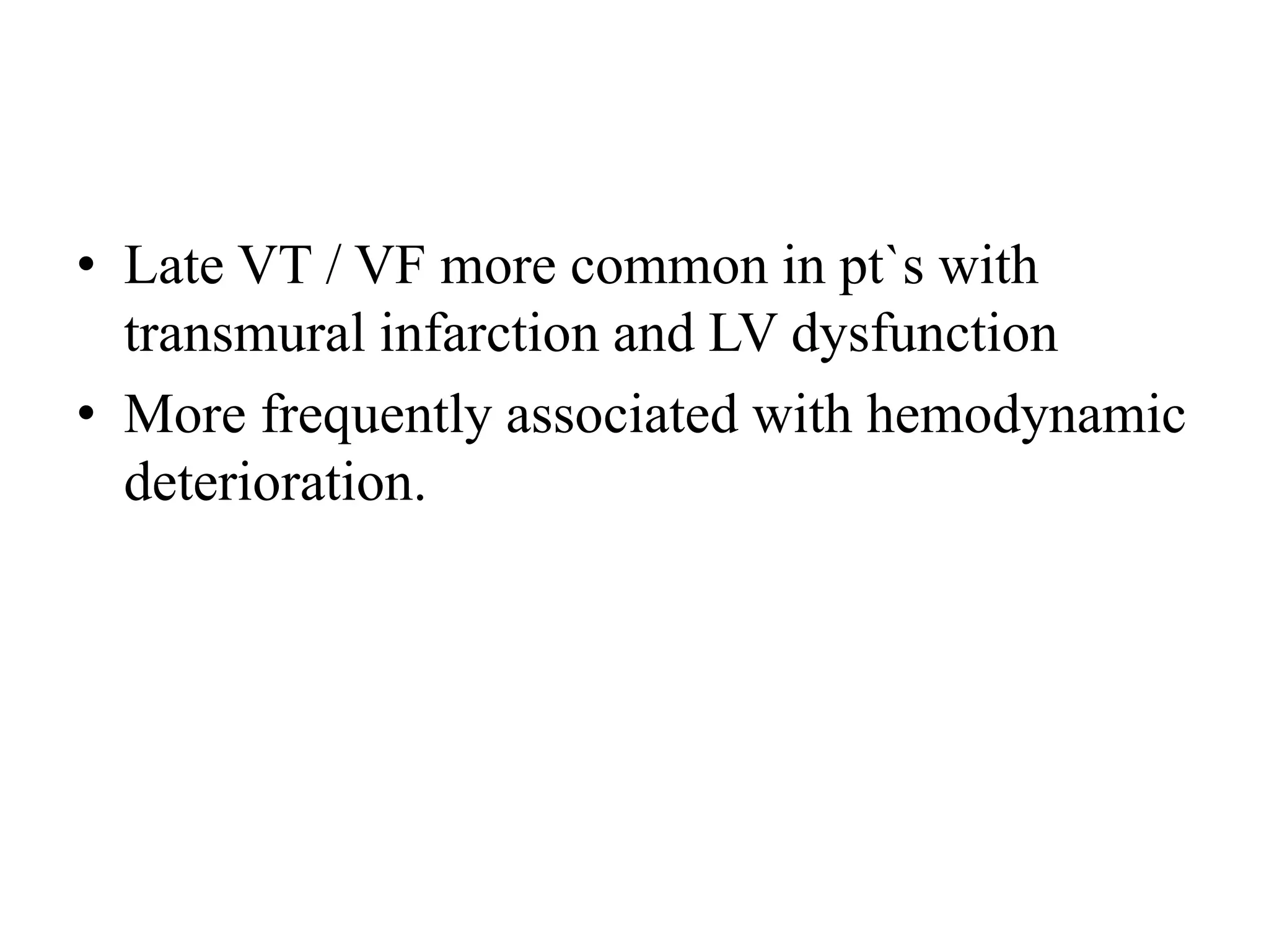
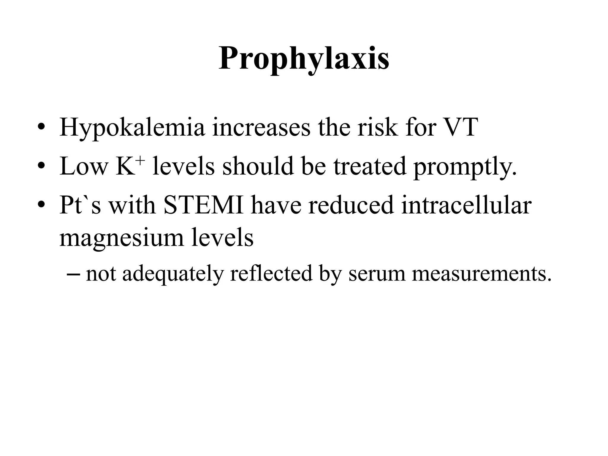
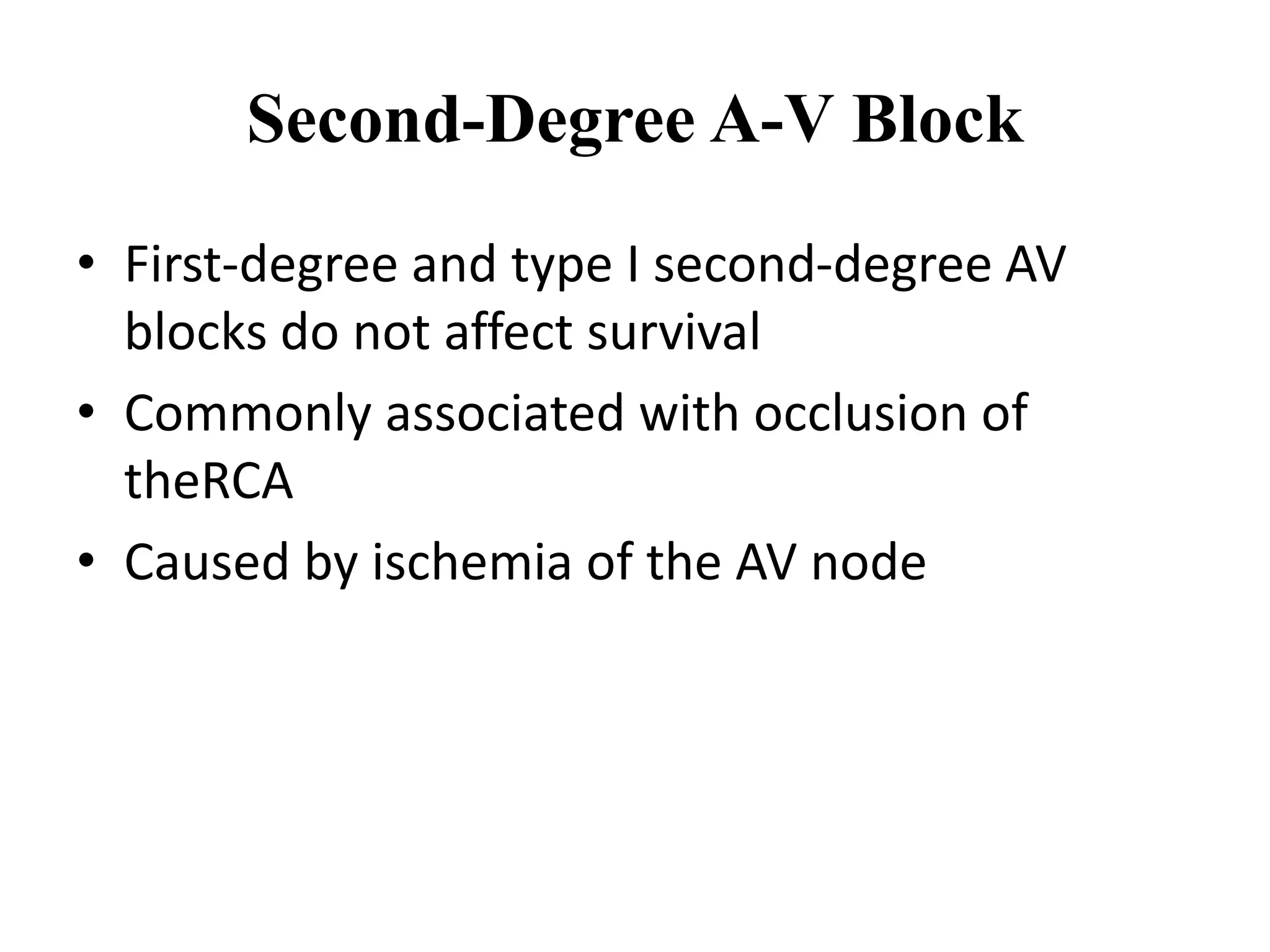
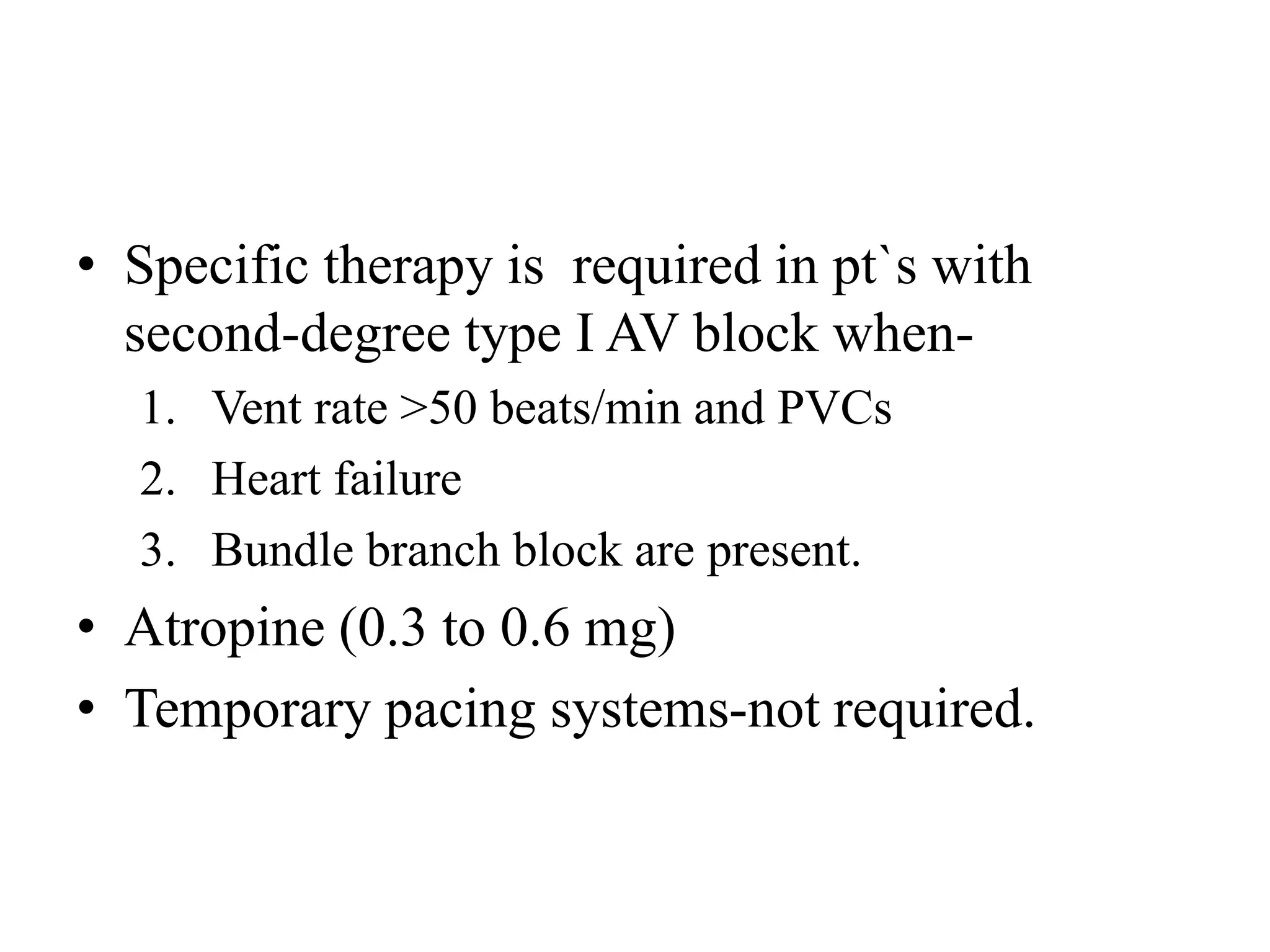
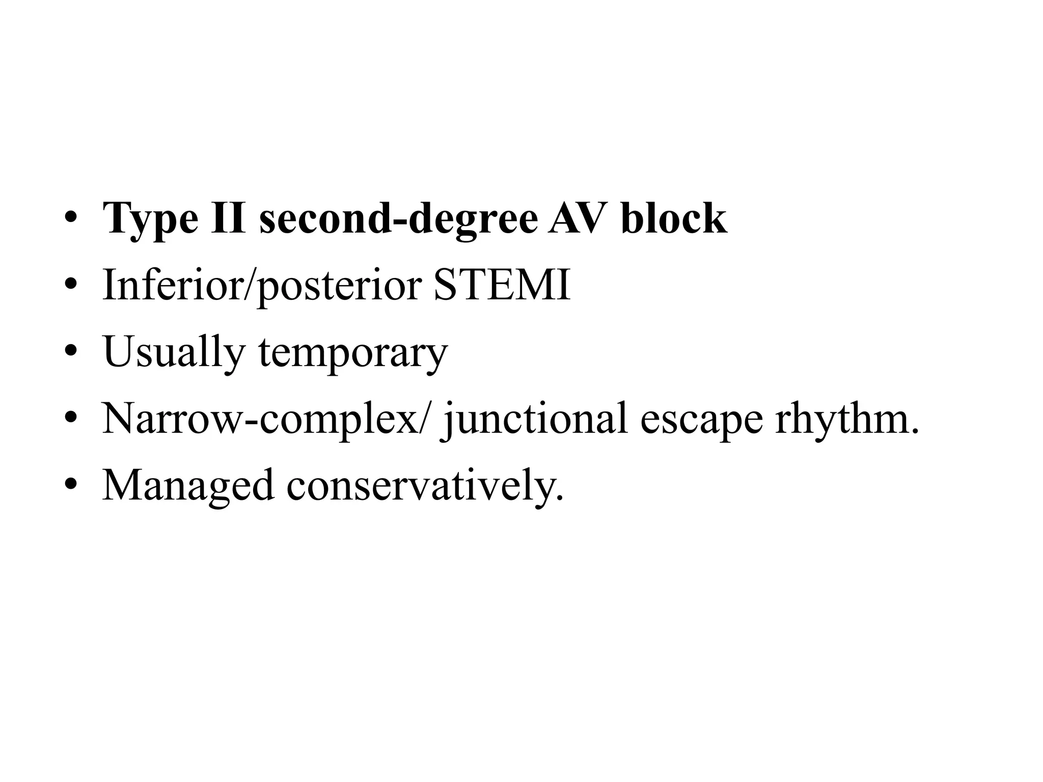
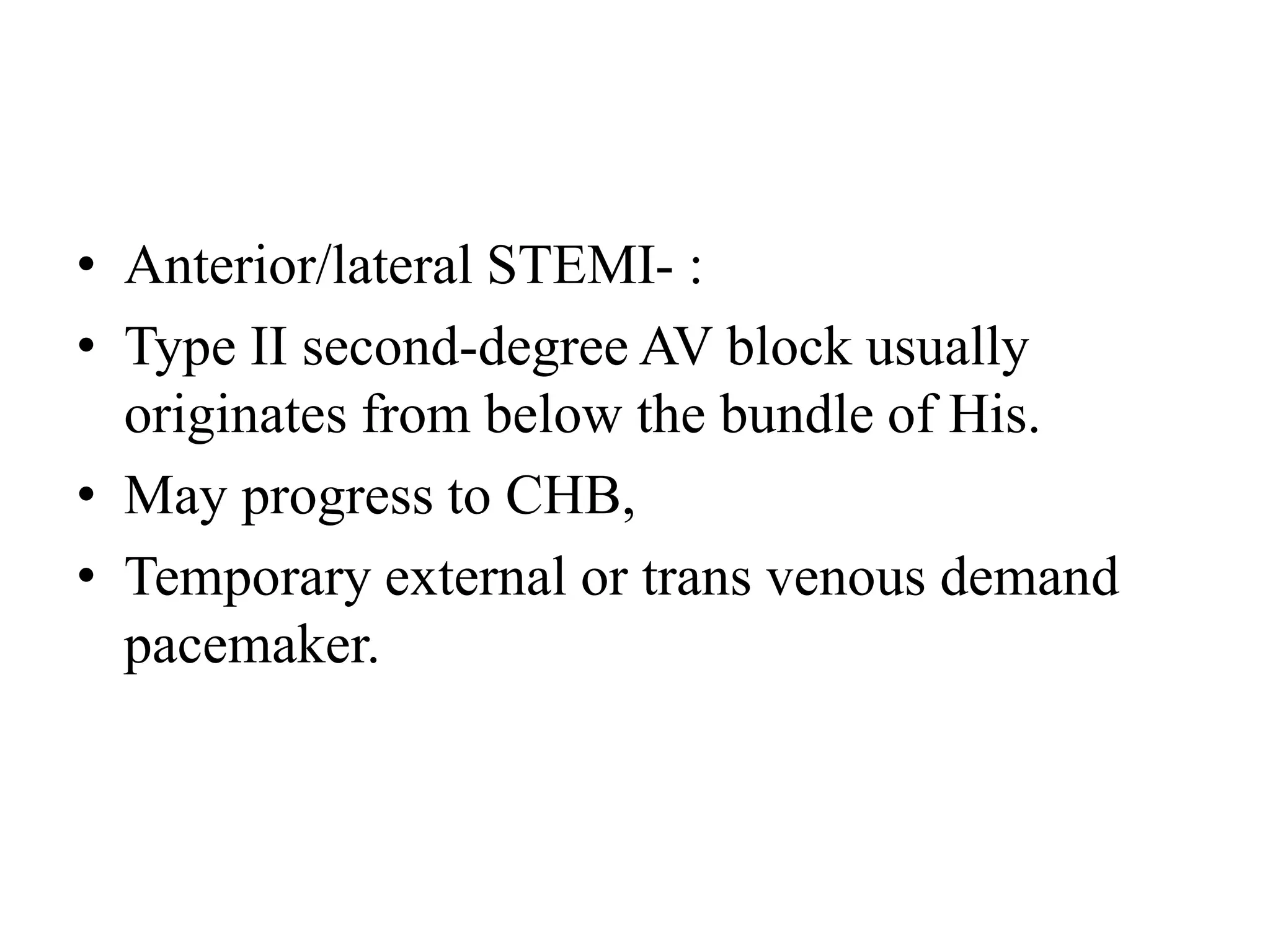
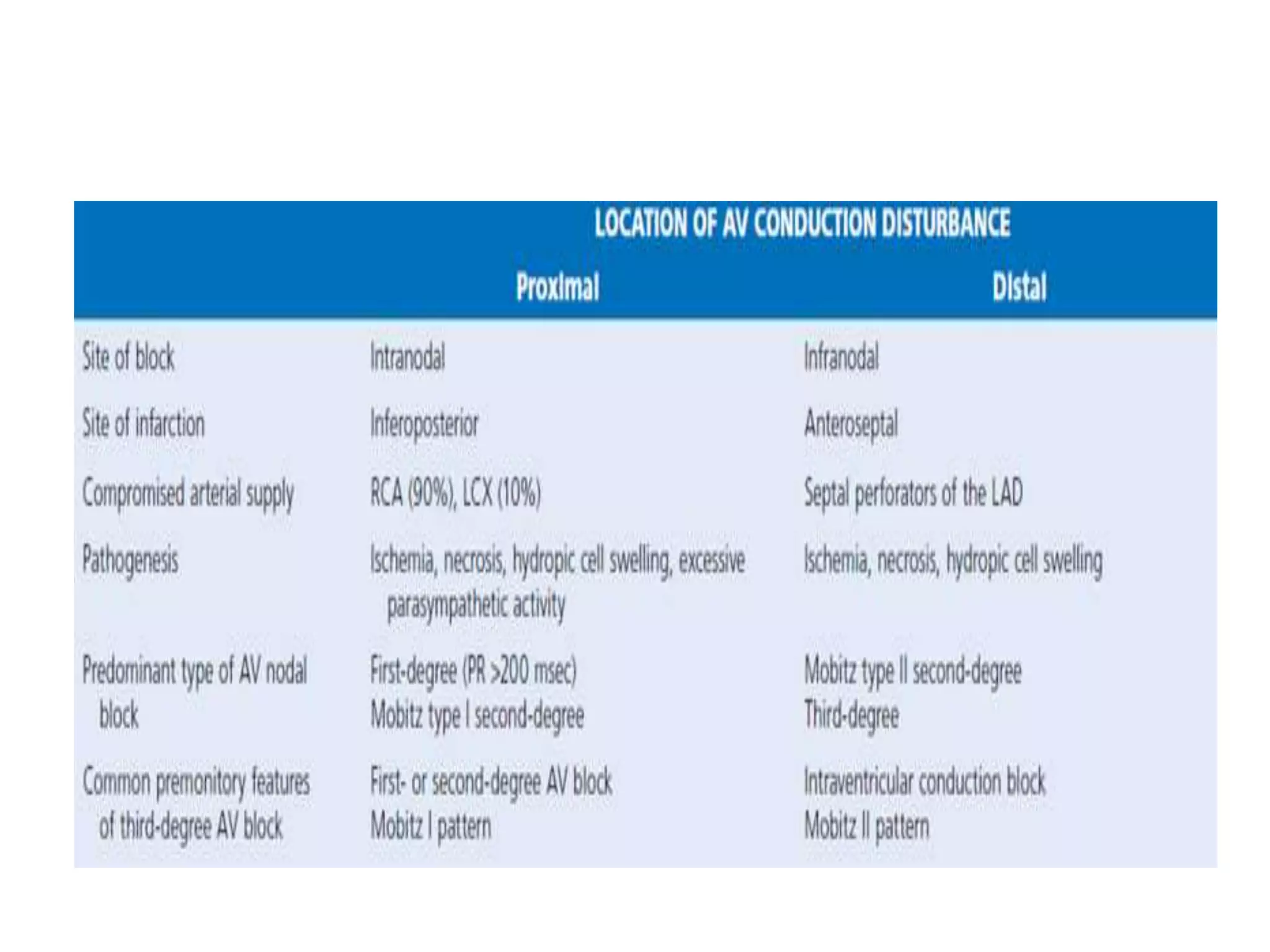
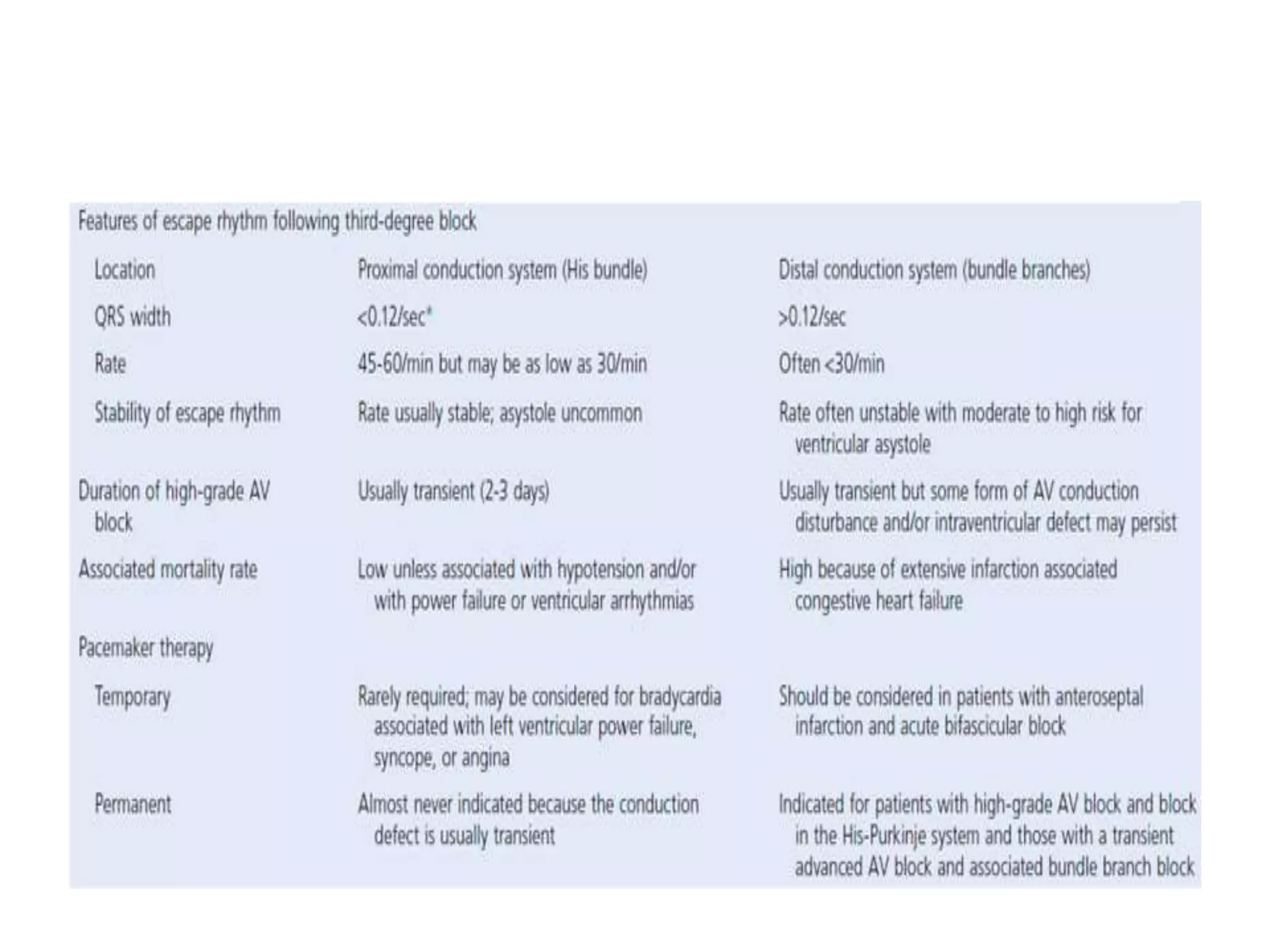
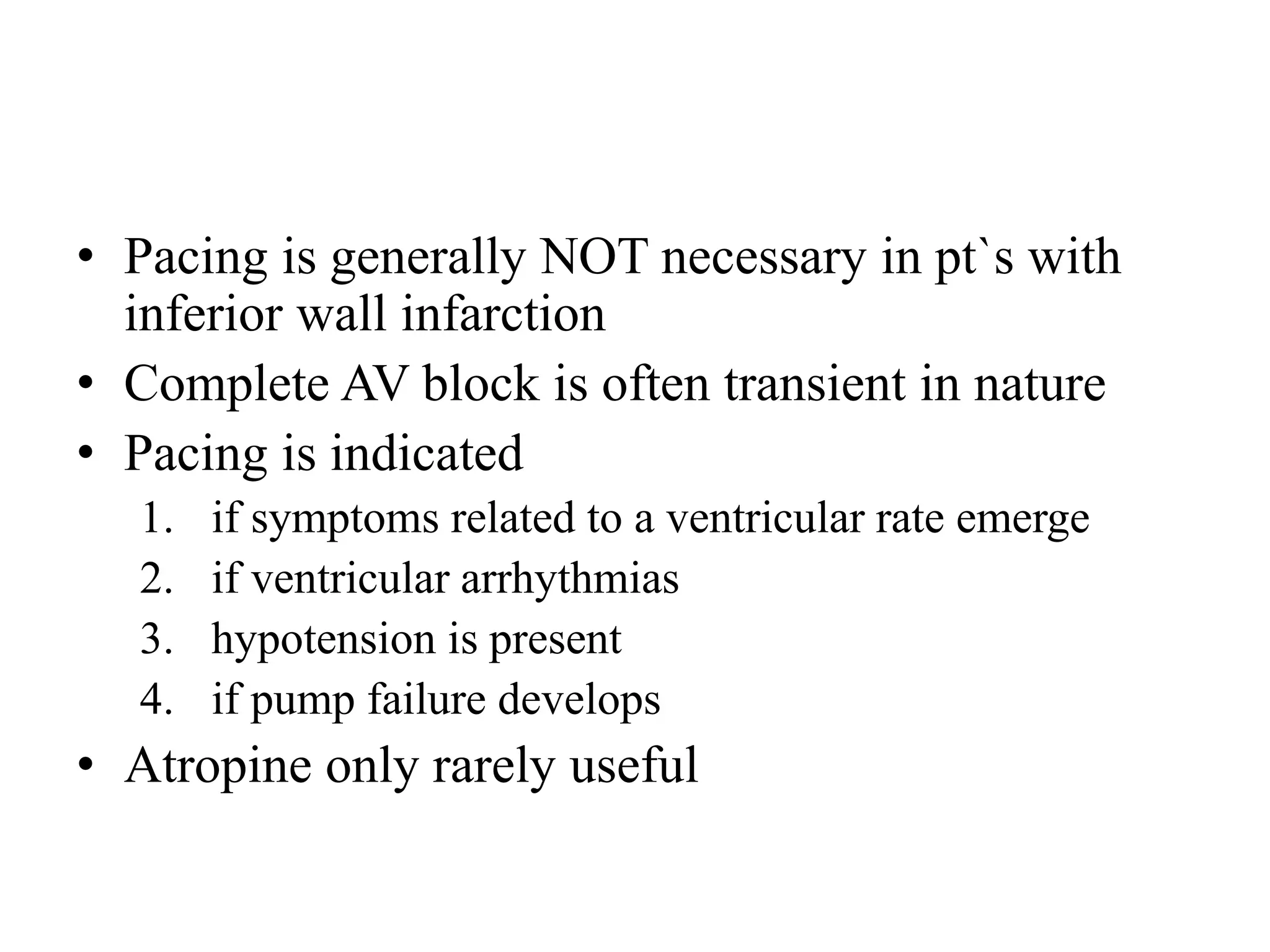
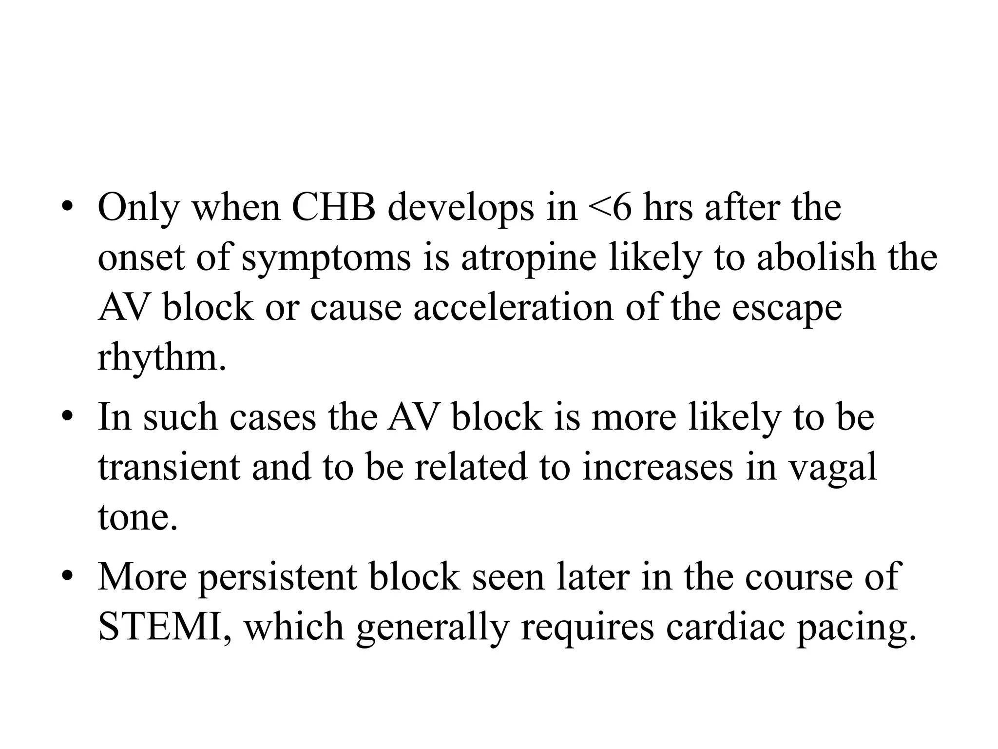
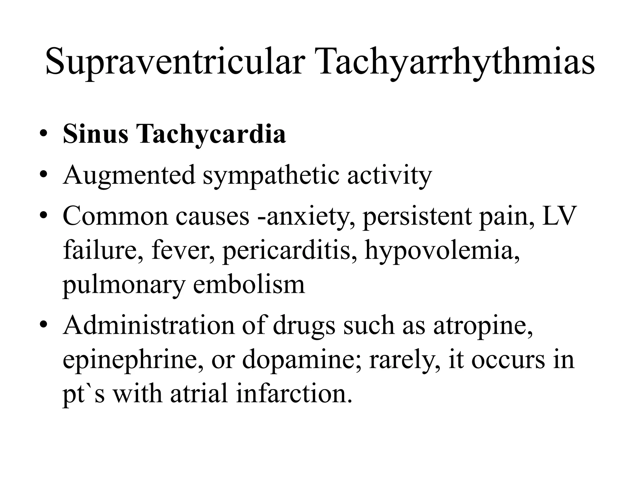
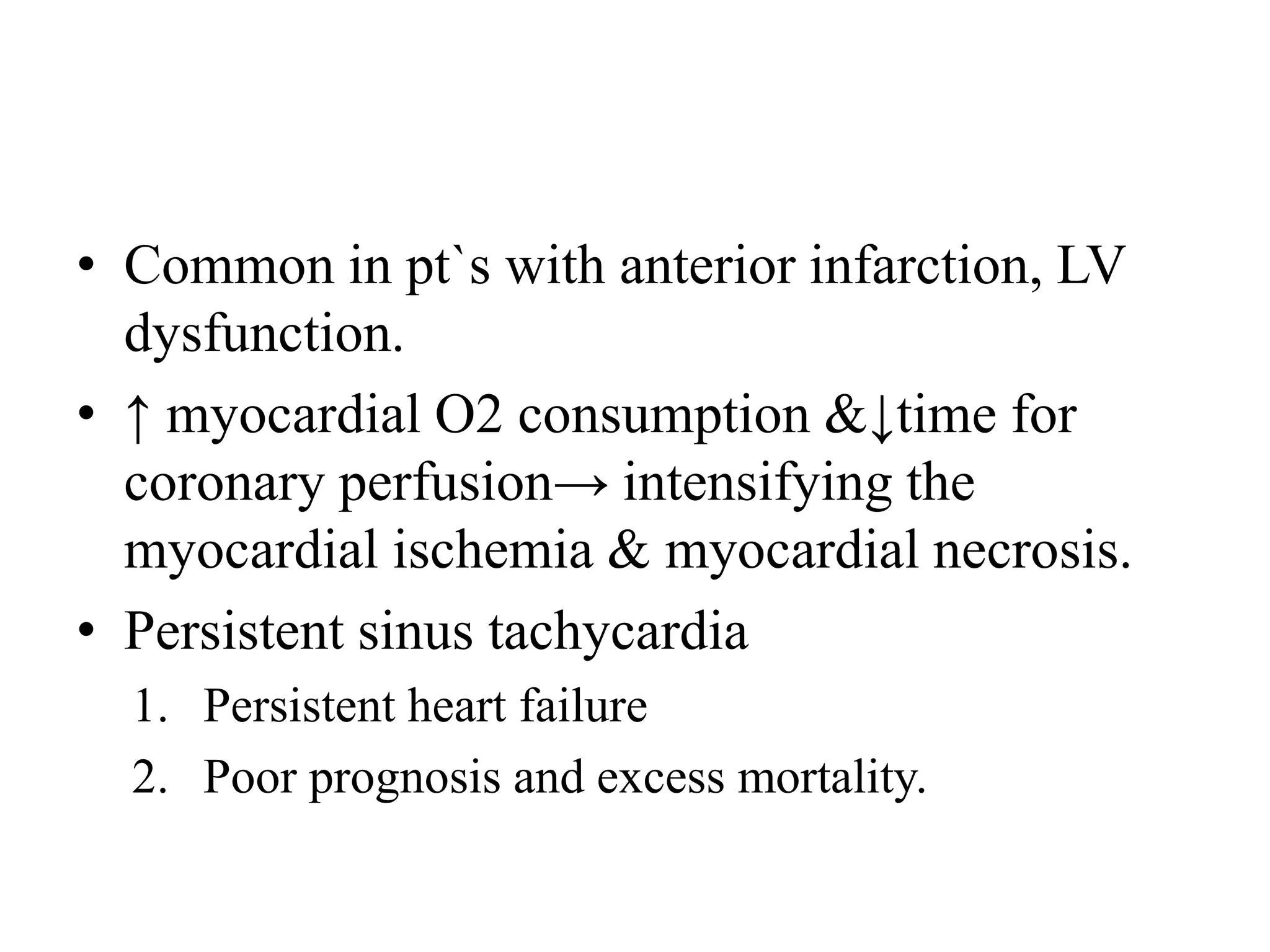
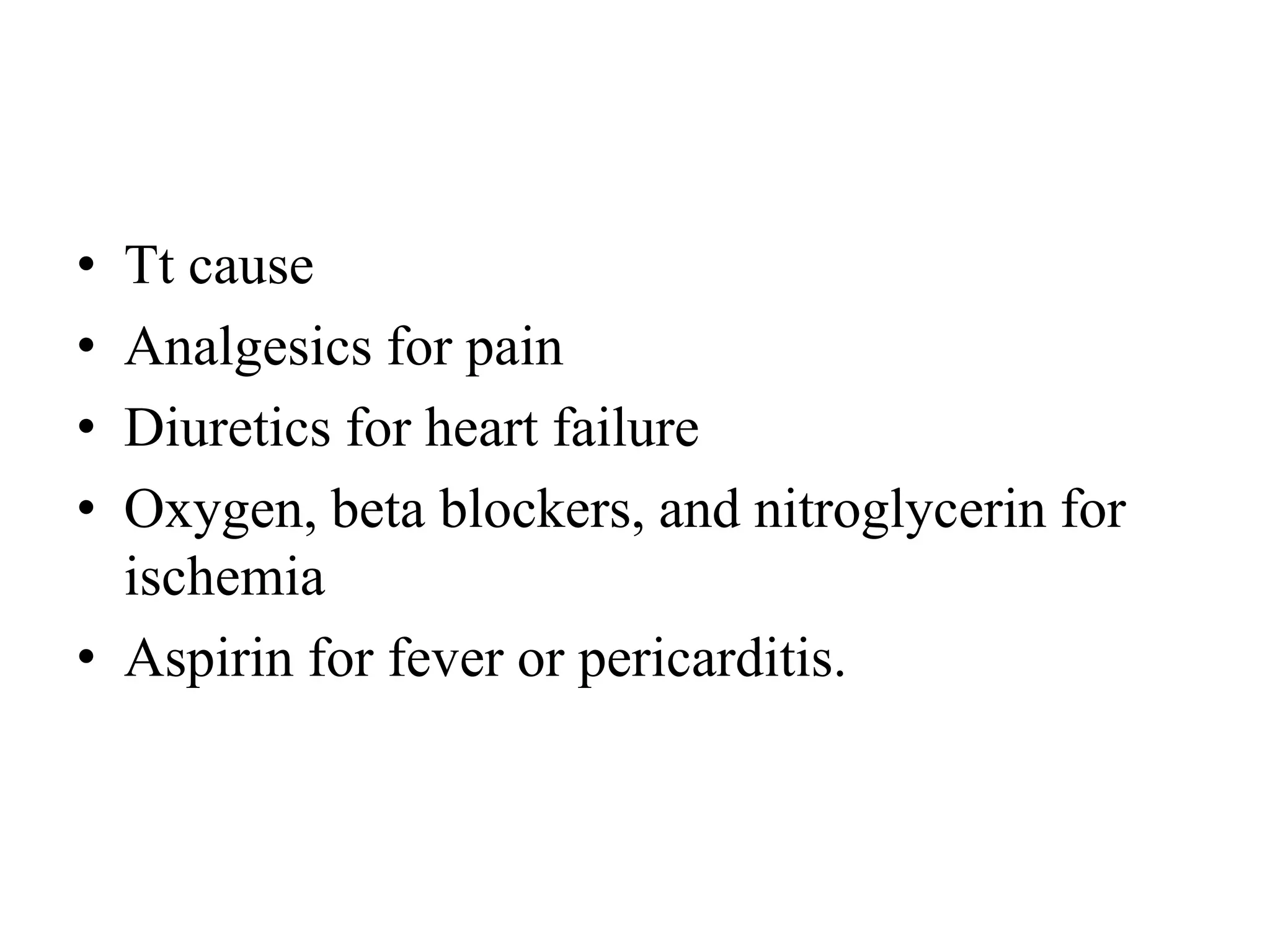
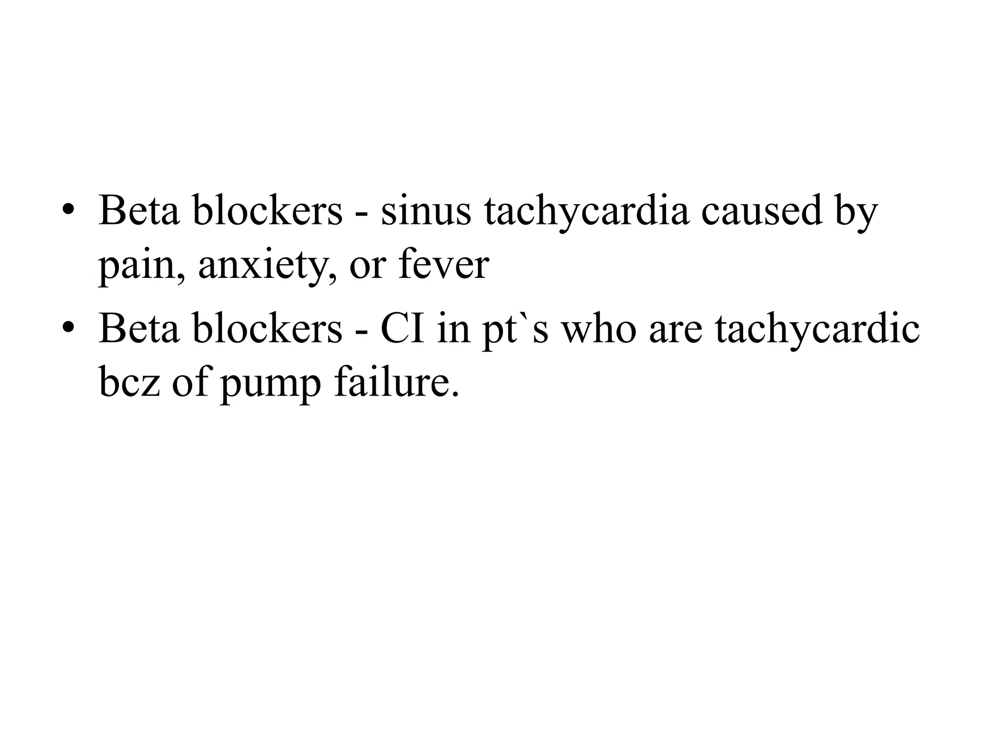
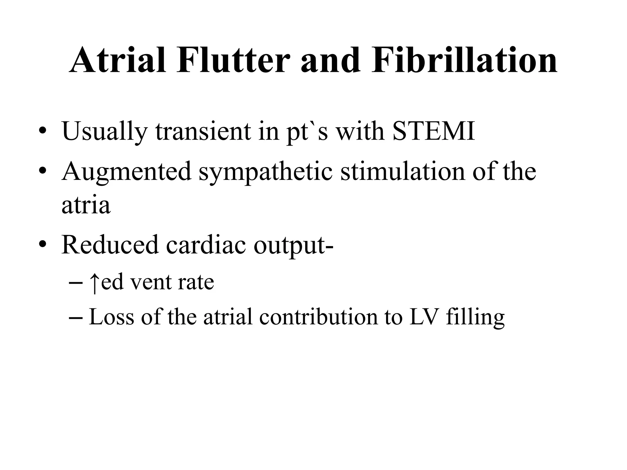
This document discusses cardiac arrhythmias and their management during acute myocardial infarction. It covers tachyarrhythmias like ventricular fibrillation and ventricular tachycardia as well as bradyarrhythmias including different types of atrioventricular block. It provides details on the mechanisms, risk factors, diagnosis and management of these arrhythmias with a focus on preventing complications through prompt treatment of underlying issues and use of drugs like beta blockers, amiodarone and pacemakers if needed.
Tachycardias are broadly categorized based upon the width of the QRS complex on the electrocardiogram (ECG). A narrow QRS complex (<120 milliseconds) reflects rapid activation of the ventricles via the normal His-Purkinje system, which in turn suggests that the arrhythmia originates above or within the His bundle (ie, a supraventricular tachycardia). The site of origin may be in the sinus node, the atria, the atrioventricular (AV) node, the His bundle, or some combination of these sites. A widened QRS (≥120 milliseconds) occurs when ventricular activation is abnormally slow. The most common reason that a QRS is widened is because the arrhythmia originates below the His bundle in the bundle branches, Purkinje fibers, or ventricular myocardium (eg, ventricular tachycardia). Alternatively, a supraventricular arrhythmia can produce a widened QRS if there are either pre-existing or rate-related abnormalities within the His-Purkinje system (eg, supraventricular tachycardia with aberrancy), or if conduction occurs over an accessory pathway. Thus, wide QRS complex tachycardias may be either supraventricular or ventricular in origin.
Its a medical presentation describing how to approach to various cardiac arrhythmias in systematic way. Illustrated with more ECG photographs from standard sources.
ECG-T wave inversion , Dr. Malala Rajapaksha ,Cardiology unit,General Hospit...malala720
This is a presentation on “What are the deferential Diagnosis a clinician think of when the clinician encounter T inversions in an ECG of a patient”. This will be help full in day today clinical practice and also in academic purposes.
Kindly leave your comment if you found this helpful ;)
Some of the slides, i hide it from my real presentations for my own reference. Download to see all of them.
IDENTIFICATION AND APPROACH TO BRADYARRHYTHMIAS .pptxDr Dravid m c
Explanation of SA Nodal and AV nodal block , ECG changes , identification clinical features and presentation of patients to emergency department, their approach and medical linea of treatment
New Drug Discovery and Development .....NEHA GUPTA
The "New Drug Discovery and Development" process involves the identification, design, testing, and manufacturing of novel pharmaceutical compounds with the aim of introducing new and improved treatments for various medical conditions. This comprehensive endeavor encompasses various stages, including target identification, preclinical studies, clinical trials, regulatory approval, and post-market surveillance. It involves multidisciplinary collaboration among scientists, researchers, clinicians, regulatory experts, and pharmaceutical companies to bring innovative therapies to market and address unmet medical needs.
- Video recording of this lecture in English language: https://youtu.be/lK81BzxMqdo
- Video recording of this lecture in Arabic language: https://youtu.be/Ve4P0COk9OI
- Link to download the book free: https://nephrotube.blogspot.com/p/nephrotube-nephrology-books.html
- Link to NephroTube website: www.NephroTube.com
- Link to NephroTube social media accounts: https://nephrotube.blogspot.com/p/join-nephrotube-on-social-media.html
Tom Selleck Health: A Comprehensive Look at the Iconic Actor’s Wellness Journeygreendigital
Tom Selleck, an enduring figure in Hollywood. has captivated audiences for decades with his rugged charm, iconic moustache. and memorable roles in television and film. From his breakout role as Thomas Magnum in Magnum P.I. to his current portrayal of Frank Reagan in Blue Bloods. Selleck's career has spanned over 50 years. But beyond his professional achievements. fans have often been curious about Tom Selleck Health. especially as he has aged in the public eye.
Follow us on: Pinterest
Introduction
Many have been interested in Tom Selleck health. not only because of his enduring presence on screen but also because of the challenges. and lifestyle choices he has faced and made over the years. This article delves into the various aspects of Tom Selleck health. exploring his fitness regimen, diet, mental health. and the challenges he has encountered as he ages. We'll look at how he maintains his well-being. the health issues he has faced, and his approach to ageing .
Early Life and Career
Childhood and Athletic Beginnings
Tom Selleck was born on January 29, 1945, in Detroit, Michigan, and grew up in Sherman Oaks, California. From an early age, he was involved in sports, particularly basketball. which played a significant role in his physical development. His athletic pursuits continued into college. where he attended the University of Southern California (USC) on a basketball scholarship. This early involvement in sports laid a strong foundation for his physical health and disciplined lifestyle.
Transition to Acting
Selleck's transition from an athlete to an actor came with its physical demands. His first significant role in "Magnum P.I." required him to perform various stunts and maintain a fit appearance. This role, which he played from 1980 to 1988. necessitated a rigorous fitness routine to meet the show's demands. setting the stage for his long-term commitment to health and wellness.
Fitness Regimen
Workout Routine
Tom Selleck health and fitness regimen has evolved. adapting to his changing roles and age. During his "Magnum, P.I." days. Selleck's workouts were intense and focused on building and maintaining muscle mass. His routine included weightlifting, cardiovascular exercises. and specific training for the stunts he performed on the show.
Selleck adjusted his fitness routine as he aged to suit his body's needs. Today, his workouts focus on maintaining flexibility, strength, and cardiovascular health. He incorporates low-impact exercises such as swimming, walking, and light weightlifting. This balanced approach helps him stay fit without putting undue strain on his joints and muscles.
Importance of Flexibility and Mobility
In recent years, Selleck has emphasized the importance of flexibility and mobility in his fitness regimen. Understanding the natural decline in muscle mass and joint flexibility with age. he includes stretching and yoga in his routine. These practices help prevent injuries, improve posture, and maintain mobilit
The Gram stain is a fundamental technique in microbiology used to classify bacteria based on their cell wall structure. It provides a quick and simple method to distinguish between Gram-positive and Gram-negative bacteria, which have different susceptibilities to antibiotics
Explore natural remedies for syphilis treatment in Singapore. Discover alternative therapies, herbal remedies, and lifestyle changes that may complement conventional treatments. Learn about holistic approaches to managing syphilis symptoms and supporting overall health.
Title: Sense of Smell
Presenter: Dr. Faiza, Assistant Professor of Physiology
Qualifications:
MBBS (Best Graduate, AIMC Lahore)
FCPS Physiology
ICMT, CHPE, DHPE (STMU)
MPH (GC University, Faisalabad)
MBA (Virtual University of Pakistan)
Learning Objectives:
Describe the primary categories of smells and the concept of odor blindness.
Explain the structure and location of the olfactory membrane and mucosa, including the types and roles of cells involved in olfaction.
Describe the pathway and mechanisms of olfactory signal transmission from the olfactory receptors to the brain.
Illustrate the biochemical cascade triggered by odorant binding to olfactory receptors, including the role of G-proteins and second messengers in generating an action potential.
Identify different types of olfactory disorders such as anosmia, hyposmia, hyperosmia, and dysosmia, including their potential causes.
Key Topics:
Olfactory Genes:
3% of the human genome accounts for olfactory genes.
400 genes for odorant receptors.
Olfactory Membrane:
Located in the superior part of the nasal cavity.
Medially: Folds downward along the superior septum.
Laterally: Folds over the superior turbinate and upper surface of the middle turbinate.
Total surface area: 5-10 square centimeters.
Olfactory Mucosa:
Olfactory Cells: Bipolar nerve cells derived from the CNS (100 million), with 4-25 olfactory cilia per cell.
Sustentacular Cells: Produce mucus and maintain ionic and molecular environment.
Basal Cells: Replace worn-out olfactory cells with an average lifespan of 1-2 months.
Bowman’s Gland: Secretes mucus.
Stimulation of Olfactory Cells:
Odorant dissolves in mucus and attaches to receptors on olfactory cilia.
Involves a cascade effect through G-proteins and second messengers, leading to depolarization and action potential generation in the olfactory nerve.
Quality of a Good Odorant:
Small (3-20 Carbon atoms), volatile, water-soluble, and lipid-soluble.
Facilitated by odorant-binding proteins in mucus.
Membrane Potential and Action Potential:
Resting membrane potential: -55mV.
Action potential frequency in the olfactory nerve increases with odorant strength.
Adaptation Towards the Sense of Smell:
Rapid adaptation within the first second, with further slow adaptation.
Psychological adaptation greater than receptor adaptation, involving feedback inhibition from the central nervous system.
Primary Sensations of Smell:
Camphoraceous, Musky, Floral, Pepperminty, Ethereal, Pungent, Putrid.
Odor Detection Threshold:
Examples: Hydrogen sulfide (0.0005 ppm), Methyl-mercaptan (0.002 ppm).
Some toxic substances are odorless at lethal concentrations.
Characteristics of Smell:
Odor blindness for single substances due to lack of appropriate receptor protein.
Behavioral and emotional influences of smell.
Transmission of Olfactory Signals:
From olfactory cells to glomeruli in the olfactory bulb, involving lateral inhibition.
Primitive, less old, and new olfactory systems with different path
These simplified slides by Dr. Sidra Arshad present an overview of the non-respiratory functions of the respiratory tract.
Learning objectives:
1. Enlist the non-respiratory functions of the respiratory tract
2. Briefly explain how these functions are carried out
3. Discuss the significance of dead space
4. Differentiate between minute ventilation and alveolar ventilation
5. Describe the cough and sneeze reflexes
Study Resources:
1. Chapter 39, Guyton and Hall Textbook of Medical Physiology, 14th edition
2. Chapter 34, Ganong’s Review of Medical Physiology, 26th edition
3. Chapter 17, Human Physiology by Lauralee Sherwood, 9th edition
4. Non-respiratory functions of the lungs https://academic.oup.com/bjaed/article/13/3/98/278874
Adv. biopharm. APPLICATION OF PHARMACOKINETICS : TARGETED DRUG DELIVERY SYSTEMSAkankshaAshtankar
MIP 201T & MPH 202T
ADVANCED BIOPHARMACEUTICS & PHARMACOKINETICS : UNIT 5
APPLICATION OF PHARMACOKINETICS : TARGETED DRUG DELIVERY SYSTEMS By - AKANKSHA ASHTANKAR
Recomendações da OMS sobre cuidados maternos e neonatais para uma experiência pós-natal positiva.
Em consonância com os ODS – Objetivos do Desenvolvimento Sustentável e a Estratégia Global para a Saúde das Mulheres, Crianças e Adolescentes, e aplicando uma abordagem baseada nos direitos humanos, os esforços de cuidados pós-natais devem expandir-se para além da cobertura e da simples sobrevivência, de modo a incluir cuidados de qualidade.
Estas diretrizes visam melhorar a qualidade dos cuidados pós-natais essenciais e de rotina prestados às mulheres e aos recém-nascidos, com o objetivo final de melhorar a saúde e o bem-estar materno e neonatal.
Uma “experiência pós-natal positiva” é um resultado importante para todas as mulheres que dão à luz e para os seus recém-nascidos, estabelecendo as bases para a melhoria da saúde e do bem-estar a curto e longo prazo. Uma experiência pós-natal positiva é definida como aquela em que as mulheres, pessoas que gestam, os recém-nascidos, os casais, os pais, os cuidadores e as famílias recebem informação consistente, garantia e apoio de profissionais de saúde motivados; e onde um sistema de saúde flexível e com recursos reconheça as necessidades das mulheres e dos bebês e respeite o seu contexto cultural.
Estas diretrizes consolidadas apresentam algumas recomendações novas e já bem fundamentadas sobre cuidados pós-natais de rotina para mulheres e neonatos que recebem cuidados no pós-parto em unidades de saúde ou na comunidade, independentemente dos recursos disponíveis.
É fornecido um conjunto abrangente de recomendações para cuidados durante o período puerperal, com ênfase nos cuidados essenciais que todas as mulheres e recém-nascidos devem receber, e com a devida atenção à qualidade dos cuidados; isto é, a entrega e a experiência do cuidado recebido. Estas diretrizes atualizam e ampliam as recomendações da OMS de 2014 sobre cuidados pós-natais da mãe e do recém-nascido e complementam as atuais diretrizes da OMS sobre a gestão de complicações pós-natais.
O estabelecimento da amamentação e o manejo das principais intercorrências é contemplada.
Recomendamos muito.
Vamos discutir essas recomendações no nosso curso de pós-graduação em Aleitamento no Instituto Ciclos.
Esta publicação só está disponível em inglês até o momento.
Prof. Marcus Renato de Carvalho
www.agostodourado.com
263778731218 Abortion Clinic /Pills In Harare ,sisternakatoto
263778731218 Abortion Clinic /Pills In Harare ,ABORTION WOMEN’S CLINIC +27730423979 IN women clinic we believe that every woman should be able to make choices in her pregnancy. Our job is to provide compassionate care, safety,affordable and confidential services. That’s why we have won the trust from all generations of women all over the world. we use non surgical method(Abortion pills) to terminate…Dr.LISA +27730423979women Clinic is committed to providing the highest quality of obstetrical and gynecological care to women of all ages. Our dedicated staff aim to treat each patient and her health concerns with compassion and respect.Our dedicated group ABORTION WOMEN’S CLINIC +27730423979 IN women clinic we believe that every woman should be able to make choices in her pregnancy. Our job is to provide compassionate care, safety,affordable and confidential services. That’s why we have won the trust from all generations of women all over the world. we use non surgical method(Abortion pills) to terminate…Dr.LISA +27730423979women Clinic is committed to providing the highest quality of obstetrical and gynecological care to women of all ages. Our dedicated staff aim to treat each patient and her health concerns with compassion and respect.Our dedicated group of receptionists, nurses, and physicians have worked together as a teamof receptionists, nurses, and physicians have worked together as a team wwww.lisywomensclinic.co.za/
5. Hemodynamic Consequences
• All forms of tachycardia and bradycardia can
depress CO.
• Myocardial oxygen consumption
• Optimal rate - 60 to 80 beats/min.
6. • Loss of the atrial contribution to ventricular
preload.
• Loss of atrial transport ↓ LV output by 15%-
20%.
• In patients with STEMI, atrial systole boosts
– End-diastolic volume by 15%
– End-diastolic pressure by 30%
– Stroke volume by 35%.
8. Ventricular Premature
Depolarizations
• “warning arrhythmias.” Presage VF.
1. Frequent VPCs = >5/min
2. VPCs with a multiform configuration
3. Early coupling (the “R-on-T” phenomenon)
4. Couplets or salvos
9. • Primary VF
– Occurs without antecedent warning arrhythmias
– May even develop despite suppression of warning
arrhythmias.
• Primary VF and VPCs (esp. R-on-T beats)
– Occur during the early phase of STEMI
10. Management
• The incidence of VF in pt`s with STEMI declined.
• Prophylactic suppression of VPCs with
antiarrhythmic drugs is not indicated .
• Suppression may ↑the risk for fatal bradycardic
and asystolic events.
• Do not routinely prescribe antiarrhythmic drugs,
other than beta blockers.
• Determine recurrent ischemia or electrolyte or
metabolic disturbances .
11. • When VPCs are accompanied by sinus
tachycardia → sympatho adrenal stimulation.
• Use beta-adrenergic blockade.
• Early administration of an IV beta blocker
reduces the incidence of VF in cases of
evolving MI.
12. Accelerated Idioventricular Rhythm
• Occurs during the first 2 days
• Equal frequency in anterior and inferior
infarctions.
• Most episodes are of short duration.
• Often observed shortly after successful
reperfusion established with fibrinolytic
therapy.
13. • Frequent AIVR in pt`s without fibrinolysis &
following pri PCI have diff implications .
• AIVR does not affect prognosis.
• Routine Tt is not required.
14. Ventricular Tachycardia and
Ventricular Fibrillation
• Mechanism - re entry
• Caused by inhomogeneity of the electrical
characteristics of ischemic myocardium.
• Cellular mechanisms for reperfusion
arrhythmias –
– Washout of various various ions such as lactate, k+
and toxic metabolic substances that have
accumulated in the ischemic zone.
15. • Late VT / VF more common in pt`s with
transmural infarction and LV dysfunction
• More frequently associated with hemodynamic
deterioration.
16. Prophylaxis
• Hypokalemia increases the risk for VT
• Low K+ levels should be treated promptly.
• Pt`s with STEMI have reduced intracellular
magnesium levels
– not adequately reflected by serum measurements.
17. • Magnesium should be repleted, achieve a
serum level of 2 mEq/liter.
• Early beta blocker use reduces VF and can be
instituted in pt`s without CI.
• Lidocaine prophylaxis to prevent primary VF
is no longer advised.
18. Management
• Unstable VT or VF -Electrical cardioversion
• I/V Amiodarone- prevention of refractory
recurrent episodes
• Sod bicarbonate- X, bcz of high osmotic load
• Hyperventilation is a more suitable means of
clearing the acidosis.
20. • Revascularization- Urgent, if caused by
ischemia.
• Antiarrhythmic drug- extended therapy
– Amiodarone
– Lidocaine
• Defibrillator
21. • Failure of electrical cardioversion
1. Rapidly recurrent VT or VF
2. Electromechanical dissociation
3. Electrical asystole.
22. Prognosis
• GUSTO-I study, among pt`s who under went
fibrinolysis ~10% experienced VT/VF.
• APEX-AMI study,-pt`s treated with pri PCI,
sustained VT/VF developed in 5.7%.
• Worse clinical outcomes in pts with VT/VF
than in those without VT/VF.
23. • Early versus late VT/VF, at 90 days doubles.
• VT/VF after(> 48 hrs) without reversible
cause, ICD for sec preventions before
discharge.
• VT/VF before reperfusion therapy-only beta
blockers.
25. Sinus Bradycardia
• Inf. & post. infarctions
• ↑ vagal tone produces sinus bradycardia
• May be beneficial
• Reduces myocardial O2 demand.
• Mortality rate similar
26. Management
• No hypotension or VPCs- observed
• IV atropine –In the first 4 to 6 hrs after
infarction
• If the sinus rate <40/min and associated with
hypotension
• Doses 0.3 -0.6 mg every 3 to 10 min.
• Total dose should not exceed 3 mg.
27. Atrioventricular and
Intraventricular Block
• Can produce conduction block at any level
• AV or intraventricular conduction system.
• Blocks-
– AV node
– Bundle of His
• Produce various grades of AV block
• RBBB
• LBBB –
– LAHB
– LPHB
28. First-Degree Atrioventricular Block
• Does not generally require specific Tt.
• Beta blockers & CCB- D/c could ↑ischemia
• Do not ↓the dosage unless the PR> 0.24 sec
• Stopped if a higher-degree block or
hemodynamic impairment.
29. • Atropine- If excessive vagotonia associated
with sinus bradycardia and hypotension
• Continued ECG monitoring
30. Second-Degree A-V Block
• First-degree and type I second-degree AV
blocks do not affect survival
• Commonly associated with occlusion of
theRCA
• Caused by ischemia of the AV node
31. • Specific therapy is required in pt`s with
second-degree type I AV block when-
1. Vent rate >50 beats/min and PVCs
2. Heart failure
3. Bundle branch block are present.
• Atropine (0.3 to 0.6 mg)
• Temporary pacing systems-not required.
32. • Type II second-degree AV block
• Inferior/posterior STEMI
• Usually temporary
• Narrow-complex/ junctional escape rhythm.
• Managed conservatively.
33. • Anterior/lateral STEMI- :
• Type II second-degree AV block usually
originates from below the bundle of His.
• May progress to CHB,
• Temporary external or trans venous demand
pacemaker.
34. Complete (Third-Degree)
Atrioventricular Block
• Inferior or anterior infarction
• More common in inferior than in anterior MI.
• Often progressing from a first-degree or type I
second-degree block –
– In pt`s with inf infarction
35. • The escape rhythm is typically stable without
asystole and often junctional.
• This form of complete AV block is often
transient.
• May respond to pharmacologic antagonism of
adenosine with methylxanthines
• Resolves in most patients within a few days
36.
37.
38. • Pt`s with inferior infarction often have
concomitant ischemia or infarction of the AV
node secondary to hypoperfusion of the AV node
artery, but the His-Purkinje system usually
escapes injury in such individuals.
• Pt`s with inf STEMI and AV block have larger
infarcts and more depressed RV and LV function
than do pts with an inf infarct and no AV block.
• junctional escape rhythms with narrow QRS
complexes occur commonly in this setting.
39. • Pacing is generally NOT necessary in pt`s with
inferior wall infarction
• Complete AV block is often transient in nature
• Pacing is indicated
1. if symptoms related to a ventricular rate emerge
2. if ventricular arrhythmias
3. hypotension is present
4. if pump failure develops
• Atropine only rarely useful
40. • Only when CHB develops in <6 hrs after the
onset of symptoms is atropine likely to abolish the
AV block or cause acceleration of the escape
rhythm.
• In such cases the AV block is more likely to be
transient and to be related to increases in vagal
tone.
• More persistent block seen later in the course of
STEMI, which generally requires cardiac pacing.
41. • In pt`s with ant infarction, third-degree AV block
can occur suddenly 12 to 24 hours after the onset
of infarction.
• It is usually preceded by an intraventricular block
and often a type II (not first-degree or type I) AV
block.
• Such pt`s typically have unstable escape rhythms
with wide QRS complexes and rates <40
beats/min
• ventricular asystole may occur quite suddenly.
42. • In pt`s with ant infarction, AV block generally
develops as a result of extensive septal
necrosis involving the bundle branches.
• The high rate of mortality in this group of pts
is the consequence of extensive myocardial
necrosis resulting in severe left ventricular
failure and frequently shock .
43. • Whether temporary transvenous pacing per se
improves survival in pt`s with anterior STEMI
remains controversial.
• Pacing protects against asystole and may
protect against transient hypotension, with its
attendant risks of extending the infarction and
precipitating malignant ventricular
tachyarrhythmias.
44. Intraventricular Block
• The right bundle branch and the left posterior
division have a dual blood supply from the
LAD and RCA
• Left anterior division is supplied by septal
perforators originating from the LAD.
• Not all conduction blocks in patients with
STEMI are complications of infarcts.
45. • STEMI and bundle branch blocks have higher
peak biomarker levels, lower EF, and ↑ed in-
hospital and long-term mortality rates.
• In the prefibrinolytic era, IVCD of the His-
Purkinje system occurred in 5% -10% of pt`s
with STEMI.
• In the reperfusion era intraventricular blocks
occur in ~2% - 5% of pt`s with MI.
46. Isolated Fascicular Blocks
• An isolated left anterior divisional block is
unlikely to progress to a complete AV block.
• Mortality is increased in these pt`s
• The post. fascicle is larger than the ant.
fascicle, and in general, a larger infarct is reqd
to block it.
• Complete AV block is not a frequent
complication of either form of isolated
divisional block.
47. Right Bundle Branch Block
• Can lead to AV block bcz it is often a new
lesion associated with antero septal infarction.
• Isolated RBBB is associated with an increased
risk for mortality in patients with anterior
STEMI
– Even if complete AV block does not occur,
– But if accompanied by CHF
48. Bifascicular Block, Including Left
Bundle Branch Block
• The combination of RBBB with either Lt. ant. or
post. divisional block is known as bidivisional or
bifascicular block.
• If a new block occurs in two of the three divisions
of the conduction system, the risk for
development of a complete AV block is quite
high.
• Mortality is also high because of the occurrence
of severe pump failure secondary to the extensive
myocardial necrosis required to produce such an
extensive intraventricular block.
49. • Preexisting BBB or divisional block is less
often associated with the development of
complete AV block in pt`s with STEMI.
• Bidivisional block in the presence of
prolongation of the PR interval may indicate
disease of the third subdivision rather than
disease of the AV node and is associated with a
greater risk for complete heart block than if
first-degree AV block is absent
50. • Complete BBB (either Lt or Rt), the combination
of RBBB and left anterior fascicular block, and any
of the various forms of trifascicular block are all
more often associated with anterior than with
inferoposterior infarction.
• All these forms are more frequent with large
infarcts.
• Older patients and have a higher incidence of
other accompanying arrhythmias.
51. Use of Pacemakers in Patients with
Acute Myocardial Infarction
• Temporary Pacing
• Just as is the case for complete AV block,
transvenous ventricular pacing has not resulted
in a statistically demonstrable improvement in
prognosis in patients with STEMI in whom
intraventricular conduction defects develop.
• Temporary pacing is advisable in some of
these patients, however, because of the high
risk for development of a complete AV block.
52. • Temporary pacing
• Pt`s with new bifascicular bundle branch block
(i.e., RBBB with Lt. ant. or post. divisional
block and alternating Rt. and Lt. BBB)
53. • An isolated new block in only one of the three
fascicles, even with PR prolongation and pre
existing bifascicular block and a normal PR
interval, poses somewhat less risk;
• These pt`s should be monitored closely, with
insertion of a temporary pacemaker deferred
unless a higher-degree AV block occurs.
54. • Asystole
• The presence of apparent ventricular asystole on
monitor displays of continuously recorded ECGs
may be misleading in that the rhythm may
actually be fine VF.
• The predominance of VF as the cause of cardiac
arrest in this setting suggests electrical counter
shock as initial therapy, even if definitive
electrocardiographic documentation of this
arrhythmia is not available.
55. • Permanent Pacing
• The advisability of permanent pacemaker
insertion is complicated because not all sudden
deaths in pt`s with STEMI and conduction defects
are caused by high-grade AV block.
• A high incidence of late VF occurs in CCU
survivors with anterior STEMI complicated by
either right or left bundle branch block.
• VF rather than asystole caused by failure of AV
conduction and infranodal pacemakers could be
responsible for late sudden death.
56. • Permanent Pacing
1. when CHB persists throughout the hospital phase in a pt
with STEMI,
2. when sinus node function is markedly impaired, or
3. when type II second-degree or third-degree block occurs
intermittently.
4. When high-grade AV block is associated with newly
acquired BBB or other criteria for conduction system
impairment, prophylactic long-term pacing may be
justified as well.
• Pt. who is candidate for an ICD or has severe heart
failure might improve with biventricular pacing.
57. Supraventricular Tachyarrhythmias
• Sinus Tachycardia
• Augmented sympathetic activity
• Common causes -anxiety, persistent pain, LV
failure, fever, pericarditis, hypovolemia,
pulmonary embolism
• Administration of drugs such as atropine,
epinephrine, or dopamine; rarely, it occurs in
pt`s with atrial infarction.
58. • Common in pt`s with anterior infarction, LV
dysfunction.
• ↑ myocardial O2 consumption &↓time for
coronary perfusion→ intensifying the
myocardial ischemia & myocardial necrosis.
• Persistent sinus tachycardia
1. Persistent heart failure
2. Poor prognosis and excess mortality.
59. • Tt cause
• Analgesics for pain
• Diuretics for heart failure
• Oxygen, beta blockers, and nitroglycerin for
ischemia
• Aspirin for fever or pericarditis.
60. • Beta blockers - sinus tachycardia caused by
pain, anxiety, or fever
• Beta blockers - CI in pt`s who are tachycardic
bcz of pump failure.
61. Atrial Flutter and Fibrillation
• Usually transient in pt`s with STEMI
• Augmented sympathetic stimulation of the
atria
• Reduced cardiac output-
– ↑ed vent rate
– Loss of the atrial contribution to LV filling
62. • AF during STEMI is associated with ↑ed
mortality and stroke
– esp. in pt`s with AWMI
63. Management
• Cardioversion-
1. Hypotension
2. Ongoing ischemia
3. Heart failure
• Beta blocker- In stabilized pt`s and in the
absence of CI
• Digitalis - when AF with ventricular
dysfunction.
• Amiodarone
64. • Oral anticoagulants -to reduce the risk for
stroke
• Even if sinus rhythm is present at the time of
discharge.
(Global Utilization of Streptokinase and Tissue Plasminogen Activator for Occluded Coronary Arteries), (Assessment of Pexelizumab in Acute Myocardial Infarction).
(See Chapters 36 and 37)
Vagotonia refers to a condition of excessive stimulation of the parasympathetic nerve fibers carried by the vagus. nerve increased activity in the vagus nerve tends to slow the heart, constrict bronchial passages in the airway, and increase digestive activity.