Downloaded 705 times
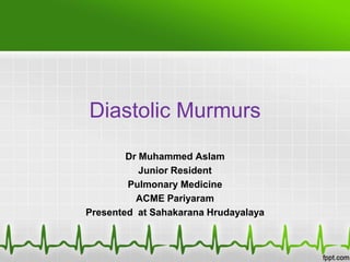


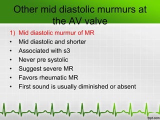


![Other Mid Diastolic Murmur
• Carey Coomb’s murmurs
– Acute rheumatic fever, mitral valve structures acutely inflamed with
some thickening and edema turbulence of flow during the rapid filling
phase + moderate MR [increased mitral inflow in diastole]
– Low pitched short MDM.
– Distinguished from MS MDM by the absence of opening snap before
the murmur
– good evidence of active carditis](https://image.slidesharecdn.com/diastolicmurmurs-131025004732-phpapp02/85/Diastolic-murmurs-32-320.jpg)


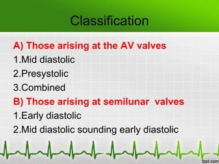
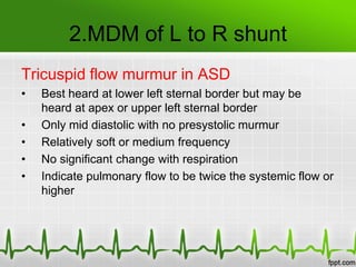
![Other diastolic murmurs
• Cabot– Locke Murmur- [Diastolic Flow murmur]
- in severe anemia
– The Cabot–Locke murmur is a diastolic murmur that sounds similar to
aortic insufficiency but does not have a decrescendo; it is heard best at
the left sternal border. [High flow thru coronary vessels, LMCA, LAD]
– The murmur resolves with treatment of anaemia.
• Dock’s murmur
– diastolic crescendo-decrescendo, with late accentuation, [consistent
with blood flow through the coronary] in a sharply localized area, 4 cm
left of the sternum in the 3LICS, detectable only when the patient was
sitting upright.
– Due to stenosis of LAD](https://image.slidesharecdn.com/diastolicmurmurs-131025004732-phpapp02/85/Diastolic-murmurs-45-320.jpg)
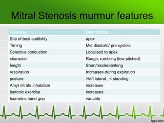
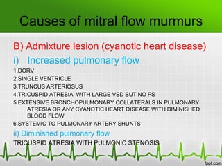
![Other diastolic murmurs
• Key–Hodgkin murmur
– EDM of AR; it has a raspy quality, [sound of a saw cutting through
wood]. Hodgkin correlated the murmur with retroversion of the aortic
valve leaflets in syphilitic disease.
• Rytand’s murmur
– Late diastolic murmur in complete heart block](https://image.slidesharecdn.com/diastolicmurmurs-131025004732-phpapp02/85/Diastolic-murmurs-46-320.jpg)


The document discusses diastolic murmurs, their classification, and mechanisms, emphasizing their clinical significance in cardiovascular conditions. It details the features, causes, and assessment of murmurs originating from atrioventricular and semilunar valves, including mitral and tricuspid valves. Furthermore, it elaborates on specific murmurs like the Austin Flint murmur and discusses their relation to physiological changes and various heart pathologies.


