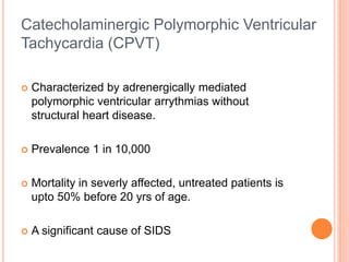
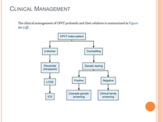
CATECHOLAMINERGIC POLYMORPHIC
VENTRICULAR TACHYCARDIA
Dr Abhishek Rathore
Sri Jayadeva Institute of Cardiovascular Science and
research, Bangalore
 Characterized by adrenergically mediated
polymorphic ventricular arrythmias without
structural heart disease.
 Prevalence 1 in 10,000
 Mortality in severly affected, untreated patients is
upto 50% before 20 yrs of age.
 A significant cause of SIDS
Catecholaminergic Polymorphic Ventricular
Tachycardia (CPVT)
DEFINITION (ESC 2015)
 A definitive diagnosis of CPVT requires the presence of a
structurally normal heart, normal ECG and exercise- or emotion-
induced bidirectional or polymorphic VT.
 CPVT is diagnosed in patients who are carriers of a pathogenic
mutation(s) in the genes RyR2 or CASQ2.
 The hallmark of CPVT is bidirectional VT,
characterized by beat-to-beat 180-degree
alternating QRS axis (observed in minority of
patients).
CLINICAL PRESENTATION
 Syncope.
 Aborted cardiac arrest.
 Sudden cardiac death.
 Some misdiagnosed as epilepsy (because CPVT-related syncope
may include convulsive movements and urinary or fecal
incontinence).
 Symptom onset in childhood ( between 7 to 9 yrs)
 Classical family history under similar conditions, but not always.
CARDIOLOGIC EXAMINATION
 Most patients have an unremarkable 12-lead resting ECG
including a normal QTc interval.
 However, sinus bradycardia and prominent U waves on
resting ECG can present.
The gold standard for diagnosis is
provocative testing.
 Preferably through exercise.
 Typically, a gradual increase in ventricular arrhythmia burden
and complexity is observed, starting with isolated VPBs at HR
of 110 to 130 per minute.
 VPBs are late-coupled with a coupling interval of
approximately 400 milliseconds.
 LBBB inferior axis and RBBB superior axis morphologies have
consistently been shown in CPVT & VPB morphologies are
reproducible in an individual patient.
 Further exercise increases the number of isolated VPBs &
eventually, polymorphic couplets or NSVT including
bidirectional VT may be induced.
 In rare cases, this may further escalate to polymorphic
sustained VT or VF.
 Ventricular arrhythmias rapidly recedes after exercise is
terminated.
 Holter monitoring, during which a patient should be
encouraged to perform exercise, although its sensitivity is low.
 For example, it may be useful in young children or in other
patients who are unable to perform an adequate exercise test.
 Adrenaline infusion (initiated at a dose of 0.05 µg/kg/min and
then titrated at 5-minute intervals to a maximum dose of
0.2 µg/kg/min) in CPVT has been advocated. But NOT
RECOMMENDED
 Adrenaline infusion–provoked VAs consistent with CPVT
when exercise testing or Holter monitoring is inconclusive.
 Sensitivity (28%) is less bec Max HR achieved was markedly
lower than during exercise testing but specificity (98%) is
more.
ROLE OF EP STUDY ??
 No role in CPVT diagnosis.
 Because ventricular arrhythmias cannot be triggered other
than by adrenergic stimulation.
ROLE OF CARDIAC IMAGING ??
 In an index patient cardiac imaging is mandatory to exclude
other causes of exercise-induced polymorphic
tachyarrhythmias.
 Cardiac imaging by definition, unremarkable in CPVT.
 However some exceptions; Mutations in RYR2 in patients
with fibrofatty myocardial replacement in the right ventricle,
mimicking ARVD.
SUPRAVENTRICULAR DISEASE
MANIFESTATIONS
 The most common supraventricular manifestation of CPVT is
sinus bradycardia and/or sinus node dysfunction.
 RYR2 mutation carriers had a lower average resting heart rate.
 Heart rates tended to be lowest in males and in RYR2 mutation
carriers.
 Other supraventricular dysrhythmias (16%): Intermittent
ectopic atrial rhythm.
GENETIC BASIS
CARDIAC RYANODINE RECEPTOR GENE
(CPVT 1)
 Disease locus: Ch 1q42-q43.
 AD
 RYR2 governs the release of calcium from SR, which initiates
cardiac muscle contraction.
 More than 130 missense mutations in RYR2 are identified.
 Mutations in RYR2 are identified in approximately 60% of
patients with a strong CPVT phenotype
CARDIAC CALSEQUESTRIN GENE (CPVT
2)
 AR
 CASQ2 - pivotal role in calcium homeostasis.
 Less than 5% of CPVT index cases.
OTHER CPVT TYPES
 Another autosomal recessive form, caused by mutations in
triadin (TRDN).
 Triadin is a transmembrane SR protein and a component of calcium
homeostasis.
 Skeletal muscle weakness is an associated feature of this CPVT form.
 Mutations in the gene encoding the potassium inwardly rectifying channel
Kir2.1 (KCNJ2), which generally are associated with Andersen-Tawil
syndrome, may cause a CPVT phenocopy, including the typical bidirectional
ventricular tachycardia .
 Others- Ank 2, CALM1 gene mutation
 However, at the present time it is not clear whether they are
phenocopies of CPVT
GENETIC TESTING IN CPVT
 Genetic testing has diagnostic but no therapeutic or
prognostic value.
 According to HRS/EHRA, comprehensive CPVT genetic
testing is indicated in probands in whom “a cardiologist has
established a clinical index of suspicion for CPVT based on
patient’s clinical history, FH & expressed ECG phenotype
during provocative stress testing with cycle, treadmill or
catecholamine infusion.”
Genetic testing should be performed in young children, possibly even at birth
because of young age of manifestation and its association with SIDS.
 CASQ2 and TRDN screening is performed in consanguineous
families.
 In mutation-negative cases with a strong CPVT phenotype
from nonconsanguineous families, the remaining exons
of RYR2 and CASQ2 are screened.
 KCNJ2 genetic testing is considered when an abundance of
supraventricular or ventricular ectopy is present on Holter
monitoring and/or a resting ECG correspond to Andersen-Tawil
syndrome.
 In relatives, genetic testing is critically important for
identifying asymptomatic mutation-carrying relatives.
 Relatives who are noncarriers of the familial CPVT-causing
mutation can be reassured and dismissed from further
cardiologic evaluation.
DIFFERENTIAL DIAGNOSIS
 Long QT syndrome.
 Andersen-Tawil syndrome.
 Concealed structural heart disease (arrhythmogenic
or hypertrophic cardiomyopathy , MVP &
myocardial ischemia may be alternate diagnoses).
CLINICAL MANAGEMENT
CPVT THERAPEUTIC INTERVENTIONS:
RECOMMENDATIONS (ESC 2015)
Class I
1. lifestyle changes:
Avoid competitive sports/ strenuous exercise/
stressful environments.
2. Beta-blockers (either spontaneous or stress
induced VAs).
3. ICD in addition to BB with or without flecainide in
those who experience cardiac arrest, recurrent
syncope or polymorphic/bidirectional VT despite OMT.
CPVT THERAPEUTIC INTERVENTIONS: RECOMMENDATIONS
CONT... (ESC 2015)
Class IIa
Flecainide in addition to BB who experience recurrent syncope or
polymorphic/bidirectional VT while on BB.
Flecainide in addition to BB in patients with a diagnosis of CPVT and
carriers of an ICD to reduce appropriate ICD shocks.
BB in carriers of a pathogenic CPVT mutation even after negative
exercise stress test.
Class IIb
LCSD in patients who experience recurrent syncope or
polymorphic/bidirectional VT/several appropriate ICD shocks while on
BB or BB plus Flecainide and in patients who are intolerant or with
contraindication to BB.
Class III
Invasive EPS with PVS is not recommended for stratification of SCD risk.
RISK STRATIFICATION
 At present, very little is known about risk stratification.
 In general, CASQ2 mutation–related CPVT display a more
severe phenotype as compared with RYR2mutation carriers.
 Young age at diagnosis & history of aborted cardiac arrest
were associated with future cardiac events.
 Two studies have reported promising results with verapamil in
CPVT patients, but its long-term efficacy has been
disappointing.
 When ventricular arrhythmias cannot be controlled by drug
therapy, left cardiac sympathetic denervation (LCSD), is
indicated.
 The lower half of the left stellate ganglion and thoracic ganglia
T2 to T4 are removed.
THANK YOU

Catecholaminergic Polymorphic VT

  • 1.
    CATECHOLAMINERGIC POLYMORPHIC VENTRICULAR TACHYCARDIA DrAbhishek Rathore Sri Jayadeva Institute of Cardiovascular Science and research, Bangalore
  • 2.
     Characterized byadrenergically mediated polymorphic ventricular arrythmias without structural heart disease.  Prevalence 1 in 10,000  Mortality in severly affected, untreated patients is upto 50% before 20 yrs of age.  A significant cause of SIDS Catecholaminergic Polymorphic Ventricular Tachycardia (CPVT)
  • 3.
    DEFINITION (ESC 2015) A definitive diagnosis of CPVT requires the presence of a structurally normal heart, normal ECG and exercise- or emotion- induced bidirectional or polymorphic VT.  CPVT is diagnosed in patients who are carriers of a pathogenic mutation(s) in the genes RyR2 or CASQ2.
  • 4.
     The hallmarkof CPVT is bidirectional VT, characterized by beat-to-beat 180-degree alternating QRS axis (observed in minority of patients).
  • 5.
    CLINICAL PRESENTATION  Syncope. Aborted cardiac arrest.  Sudden cardiac death.  Some misdiagnosed as epilepsy (because CPVT-related syncope may include convulsive movements and urinary or fecal incontinence).  Symptom onset in childhood ( between 7 to 9 yrs)  Classical family history under similar conditions, but not always.
  • 6.
    CARDIOLOGIC EXAMINATION  Mostpatients have an unremarkable 12-lead resting ECG including a normal QTc interval.  However, sinus bradycardia and prominent U waves on resting ECG can present.
  • 7.
    The gold standardfor diagnosis is provocative testing.  Preferably through exercise.  Typically, a gradual increase in ventricular arrhythmia burden and complexity is observed, starting with isolated VPBs at HR of 110 to 130 per minute.  VPBs are late-coupled with a coupling interval of approximately 400 milliseconds.  LBBB inferior axis and RBBB superior axis morphologies have consistently been shown in CPVT & VPB morphologies are reproducible in an individual patient.
  • 9.
     Further exerciseincreases the number of isolated VPBs & eventually, polymorphic couplets or NSVT including bidirectional VT may be induced.  In rare cases, this may further escalate to polymorphic sustained VT or VF.  Ventricular arrhythmias rapidly recedes after exercise is terminated.
  • 10.
     Holter monitoring,during which a patient should be encouraged to perform exercise, although its sensitivity is low.  For example, it may be useful in young children or in other patients who are unable to perform an adequate exercise test.
  • 11.
     Adrenaline infusion(initiated at a dose of 0.05 µg/kg/min and then titrated at 5-minute intervals to a maximum dose of 0.2 µg/kg/min) in CPVT has been advocated. But NOT RECOMMENDED  Adrenaline infusion–provoked VAs consistent with CPVT when exercise testing or Holter monitoring is inconclusive.  Sensitivity (28%) is less bec Max HR achieved was markedly lower than during exercise testing but specificity (98%) is more.
  • 12.
    ROLE OF EPSTUDY ??  No role in CPVT diagnosis.  Because ventricular arrhythmias cannot be triggered other than by adrenergic stimulation.
  • 13.
    ROLE OF CARDIACIMAGING ??  In an index patient cardiac imaging is mandatory to exclude other causes of exercise-induced polymorphic tachyarrhythmias.  Cardiac imaging by definition, unremarkable in CPVT.  However some exceptions; Mutations in RYR2 in patients with fibrofatty myocardial replacement in the right ventricle, mimicking ARVD.
  • 14.
    SUPRAVENTRICULAR DISEASE MANIFESTATIONS  Themost common supraventricular manifestation of CPVT is sinus bradycardia and/or sinus node dysfunction.  RYR2 mutation carriers had a lower average resting heart rate.  Heart rates tended to be lowest in males and in RYR2 mutation carriers.  Other supraventricular dysrhythmias (16%): Intermittent ectopic atrial rhythm.
  • 15.
  • 16.
    CARDIAC RYANODINE RECEPTORGENE (CPVT 1)  Disease locus: Ch 1q42-q43.  AD  RYR2 governs the release of calcium from SR, which initiates cardiac muscle contraction.  More than 130 missense mutations in RYR2 are identified.  Mutations in RYR2 are identified in approximately 60% of patients with a strong CPVT phenotype
  • 17.
    CARDIAC CALSEQUESTRIN GENE(CPVT 2)  AR  CASQ2 - pivotal role in calcium homeostasis.  Less than 5% of CPVT index cases.
  • 18.
    OTHER CPVT TYPES Another autosomal recessive form, caused by mutations in triadin (TRDN).  Triadin is a transmembrane SR protein and a component of calcium homeostasis.  Skeletal muscle weakness is an associated feature of this CPVT form.  Mutations in the gene encoding the potassium inwardly rectifying channel Kir2.1 (KCNJ2), which generally are associated with Andersen-Tawil syndrome, may cause a CPVT phenocopy, including the typical bidirectional ventricular tachycardia .  Others- Ank 2, CALM1 gene mutation  However, at the present time it is not clear whether they are phenocopies of CPVT
  • 19.
    GENETIC TESTING INCPVT  Genetic testing has diagnostic but no therapeutic or prognostic value.  According to HRS/EHRA, comprehensive CPVT genetic testing is indicated in probands in whom “a cardiologist has established a clinical index of suspicion for CPVT based on patient’s clinical history, FH & expressed ECG phenotype during provocative stress testing with cycle, treadmill or catecholamine infusion.” Genetic testing should be performed in young children, possibly even at birth because of young age of manifestation and its association with SIDS.
  • 20.
     CASQ2 andTRDN screening is performed in consanguineous families.  In mutation-negative cases with a strong CPVT phenotype from nonconsanguineous families, the remaining exons of RYR2 and CASQ2 are screened.  KCNJ2 genetic testing is considered when an abundance of supraventricular or ventricular ectopy is present on Holter monitoring and/or a resting ECG correspond to Andersen-Tawil syndrome.
  • 21.
     In relatives,genetic testing is critically important for identifying asymptomatic mutation-carrying relatives.  Relatives who are noncarriers of the familial CPVT-causing mutation can be reassured and dismissed from further cardiologic evaluation.
  • 22.
    DIFFERENTIAL DIAGNOSIS  LongQT syndrome.  Andersen-Tawil syndrome.  Concealed structural heart disease (arrhythmogenic or hypertrophic cardiomyopathy , MVP & myocardial ischemia may be alternate diagnoses).
  • 23.
  • 24.
    CPVT THERAPEUTIC INTERVENTIONS: RECOMMENDATIONS(ESC 2015) Class I 1. lifestyle changes: Avoid competitive sports/ strenuous exercise/ stressful environments. 2. Beta-blockers (either spontaneous or stress induced VAs). 3. ICD in addition to BB with or without flecainide in those who experience cardiac arrest, recurrent syncope or polymorphic/bidirectional VT despite OMT.
  • 25.
    CPVT THERAPEUTIC INTERVENTIONS:RECOMMENDATIONS CONT... (ESC 2015) Class IIa Flecainide in addition to BB who experience recurrent syncope or polymorphic/bidirectional VT while on BB. Flecainide in addition to BB in patients with a diagnosis of CPVT and carriers of an ICD to reduce appropriate ICD shocks. BB in carriers of a pathogenic CPVT mutation even after negative exercise stress test. Class IIb LCSD in patients who experience recurrent syncope or polymorphic/bidirectional VT/several appropriate ICD shocks while on BB or BB plus Flecainide and in patients who are intolerant or with contraindication to BB. Class III Invasive EPS with PVS is not recommended for stratification of SCD risk.
  • 26.
    RISK STRATIFICATION  Atpresent, very little is known about risk stratification.  In general, CASQ2 mutation–related CPVT display a more severe phenotype as compared with RYR2mutation carriers.  Young age at diagnosis & history of aborted cardiac arrest were associated with future cardiac events.
  • 27.
     Two studieshave reported promising results with verapamil in CPVT patients, but its long-term efficacy has been disappointing.  When ventricular arrhythmias cannot be controlled by drug therapy, left cardiac sympathetic denervation (LCSD), is indicated.  The lower half of the left stellate ganglion and thoracic ganglia T2 to T4 are removed.
  • 28.

Editor's Notes

  • #12 Adrenalin infusion--- when exercise testing and holter fails.
  • #25 Betablockers without sympathomimetic activity. Nadolol 1-2.5mg/kg per day.