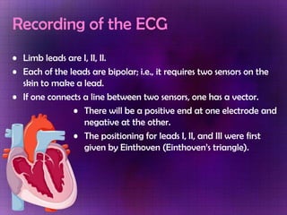
The document discusses electrocardiography (ECG), providing details on the standard 12-lead ECG procedure, what each lead measures, and ECG paper formatting. Common cardiac arrhythmias and conduction abnormalities that can be detected from the ECG are summarized, including sinus bradycardia, atrial flutter, atrial fibrillation, ventricular tachycardia, and Wolff-Parkinson-White syndrome. Characteristics of right and left bundle branch block are also outlined.
ECG Rhythm Interpretation
ST Elevation and non-ST Elevation MIs
ECG Changes
ECG Changes & the Evolving MI
Left Ventricular Hypertrophy
Normal Impulse Conduction
Bundle Branch Blocks
crème de la crème basics to understand electrocardiographic analysis in an easy & simple way with some specifications to its use in Emergency medicine/clinical toxicology practice.
This presentation covers few basic things about ECG, especially for UG Medical students like ECG leads, normal ECG waves, axis of ECG and how to look for common ECG misplacements.
ECG basics for undergraduate
How to identify normal ECG
How to identify abnormal findings in ECG
-Atrial hypertrophy
-Hyperkalaemia
-Atrial fibrillation
-Hypokalaemia
-Atrial flutter
-Pericarditis
-Heart blocks
-Chambers enlargement
-IHD
-AMI
ecg basics made easy, with description of most common ecg types especially in emergency situation.
easy to memorize points and mnemonics included.
approach to ecg diagnosis.
sample ecgs.
How to Make a Field invisible in Odoo 17Celine George
It is possible to hide or invisible some fields in odoo. Commonly using “invisible” attribute in the field definition to invisible the fields. This slide will show how to make a field invisible in odoo 17.
Macroeconomics- Movie Location
This will be used as part of your Personal Professional Portfolio once graded.
Objective:
Prepare a presentation or a paper using research, basic comparative analysis, data organization and application of economic information. You will make an informed assessment of an economic climate outside of the United States to accomplish an entertainment industry objective.
Biological screening of herbal drugs: Introduction and Need for
Phyto-Pharmacological Screening, New Strategies for evaluating
Natural Products, In vitro evaluation techniques for Antioxidants, Antimicrobial and Anticancer drugs. In vivo evaluation techniques
for Anti-inflammatory, Antiulcer, Anticancer, Wound healing, Antidiabetic, Hepatoprotective, Cardio protective, Diuretics and
Antifertility, Toxicity studies as per OECD guidelines
A review of the growth of the Israel Genealogy Research Association Database Collection for the last 12 months. Our collection is now passed the 3 million mark and still growing. See which archives have contributed the most. See the different types of records we have, and which years have had records added. You can also see what we have for the future.
2024.06.01 Introducing a competency framework for languag learning materials ...Sandy Millin
http://sandymillin.wordpress.com/iateflwebinar2024
Published classroom materials form the basis of syllabuses, drive teacher professional development, and have a potentially huge influence on learners, teachers and education systems. All teachers also create their own materials, whether a few sentences on a blackboard, a highly-structured fully-realised online course, or anything in between. Despite this, the knowledge and skills needed to create effective language learning materials are rarely part of teacher training, and are mostly learnt by trial and error.
Knowledge and skills frameworks, generally called competency frameworks, for ELT teachers, trainers and managers have existed for a few years now. However, until I created one for my MA dissertation, there wasn’t one drawing together what we need to know and do to be able to effectively produce language learning materials.
This webinar will introduce you to my framework, highlighting the key competencies I identified from my research. It will also show how anybody involved in language teaching (any language, not just English!), teacher training, managing schools or developing language learning materials can benefit from using the framework.
This slide is special for master students (MIBS & MIFB) in UUM. Also useful for readers who are interested in the topic of contemporary Islamic banking.
A Strategic Approach: GenAI in EducationPeter Windle
Artificial Intelligence (AI) technologies such as Generative AI, Image Generators and Large Language Models have had a dramatic impact on teaching, learning and assessment over the past 18 months. The most immediate threat AI posed was to Academic Integrity with Higher Education Institutes (HEIs) focusing their efforts on combating the use of GenAI in assessment. Guidelines were developed for staff and students, policies put in place too. Innovative educators have forged paths in the use of Generative AI for teaching, learning and assessments leading to pockets of transformation springing up across HEIs, often with little or no top-down guidance, support or direction.
This Gasta posits a strategic approach to integrating AI into HEIs to prepare staff, students and the curriculum for an evolving world and workplace. We will highlight the advantages of working with these technologies beyond the realm of teaching, learning and assessment by considering prompt engineering skills, industry impact, curriculum changes, and the need for staff upskilling. In contrast, not engaging strategically with Generative AI poses risks, including falling behind peers, missed opportunities and failing to ensure our graduates remain employable. The rapid evolution of AI technologies necessitates a proactive and strategic approach if we are to remain relevant.
2. Electrocardiography A recording of the electrical activity of the heart over time Gold standard for diagnosis of cardiac arrhythmias Helps detect electrolyte disturbances Allows for detection of conduction abnormalities Screening tool for ischemic heart disease (stress tests) Helpful with non-cardiac diseases
3. ECG Leads The standard EKG has 12 leads: 3 Standard Limb Leads 3 Augmented Limb Leads 6 Precordial Leads
4. ECG Limb Leads Leads are electrodes which measure the difference in electrical potential between either: 1. Two different points on the body (bipolar leads) 2. One point on the body and a virtual reference point with zero electrical potential, located in the center of the heart (unipolar leads)
5. Recording of the ECG Limb leads are I, II, II. Each of the leads are bipolar; i.e., it requires two sensors on the skin to make a lead. If one connects a line between two sensors, one has a vector. There will be a positive end at one electrode and negative at the other. The positioning for leads I, II, and III were first given by Einthoven (Einthoven’s triangle).
15. ECG Graph Paper Runs at a paper speed of 25 mm/sec Each small block of ECG paper is 1 mm2 At a paper speed of 25 mm/s, one small block equals 0.04 s Five small blocks make up 1 large block which translates into 0.20 s 5 large blocks per second Voltage: 1 mm = 0.1 mV between each individual block vertically
19. Guide in ECG Reading Rhythm Rate: Atrial and Ventricular Axis P wave: morphology and duration P – R interval QRS complex: morphology and duration ST segment T wave U wave Q – T interval
20. Determining the Rhythm Regular or Irregular Accessing whether the PP intervals and RR intervals are regularly spaced If the rhythm is irregular, determine if: Occasionally irregular Regularly irregular (there is the pattern of irregularity) Irregularly irregular (no pattern of irregularity)
21. Determining the Heart Rate Rule of 300 Take the number of “big boxes” between neighboring QRS complexes, and divide this into 300. The result will be approximately equal to the rate Although fast, this method only works for regular rhythms. 10 Second Rule As most EKGs record 10 seconds of rhythm per page, one can simply count the number of beats present on the EKG and multiply by 6 to get the number of beats per 60 seconds. This method works well for irregular rhythms.
22. The QRS Axis The QRS axis represents the net overall direction of the heart’s electrical activity. By near-consensus, the normal QRS axis is defined as ranging from -30° to +90°. -30° to -90° is referred to as a left axis deviation (LAD) +90° to +180° is referred to as a right axis deviation (RAD)
24. The QRS Axis The Quadrant Approach 1. Examine the QRS complex in leads I and aVF to determine if they are predominantly positive or predominantly negative. The combination should place the axis into one of the 4 quadrants below.
25. The QRS Axis The Quadrant Approach 2. In the event that LAD is present, examine lead II to determine if this deviation is pathologic. If the QRS in II is predominantly positive, the LAD is non-pathologic (in other words, the axis is normal). If it is predominantly negative, it is pathologic.
26. P-wave Depolarization of both atria Relationship between P and QRS helps distinguish various cardiac arrhythmias Shape and duration of P may indicate atrialenlargement
29. PR interval from onset of P wave to onset of QRS Normal duration = 0.12-2.0 sec (120-200 ms) (3-4 horizontal boxes) Represents atria to ventricular conduction time (through His bundle) Prolonged PR interval may indicate a 1st degree heart block
34. QRS complex Its duration, amplitude, and morphology are useful in diagnosing cardiac arrhythmias, ventricular hypertrophy, MI, electrolyte derangement, etc. Q wave greater than 1/3 the height of the R wave, greater than 0.04 sec are abnormal and may represent MI
35. QRS morphology and duration I AVR Positive QRS Negative QRS V3 Biphasic QRS 6
36. ST segment Connects the QRS complex and T wave Duration of 0.08-0.12 sec (80-120 msec)
38. S – T Segment I V1 Normal Elevated V3 Depressed 7
39. T waves Represents repolarization or recovery of ventricles Interval from beginning of QRS to apex of T is referred to as the absolute refractory period
45. Chamber Enlargement Right Atrial Enlargement (RAE) Tall P waves in II, III, AVF > 2.5mm Left Atrial Enlargement (LAE) P wave in I > 0.11 secs Terminal Negativity of P wave in VI = > 1mm Bi-atrial Enlargement RAE + LAE
46. Sinus Bradycardia Heart is slower than 60 beats per minute RR interval is longer P wave followed by QRS complex in 1:1 ratio PR interval slightly prolonged
47. Sinus Tachycardia Sinus rhythm is faster than 100 beats per minute RR interval is shorter, less than 0.6 seconds P wave followed by QRS complex in 1:1 ratio
48. Atrial Flutter atria contract at 200-350 beats per minute F waves are larger than normal P waves and they have a saw-toothed waveform ventricular rate is usually regular but slower than the atrial rate fixed ratio of flutter waves to QRS complexes can be observed, for instance 2:1, 3:1 or 4:1
50. Atrial Fibrillation occurs when the atria depolarize repeatedly and in an irregular uncontrolled manner usually at atatrial rate greater than 350 beats per minute no concerted contraction of the atria No P-waves are observed QRS complexes have normal shape, due to normal ventricular conduction. However the RR intervals vary from beat to beat. The ventricular rate may increase to greater than 150 beats per minute if uncontrolled.
52. Ventricular Tachycardia occurs when electrical impulses originating either from the ventricles cause rapid ventricular depolarization (140-250 beats per minute) QRS complexes are wide and bizarre P-waves may be inverted/may be present but not associated with QRS complexes (AV dissociation) RR intervals are usually regular
54. Ventricular Fibrillation occurs when parts of the ventricles depolarize repeatedly in an erratic, uncoordinated manner random, apparently unrelated waves no recognizable QRS complex almost invariably fatal because the uncoordinated contractions of ventricular myocardium electrical defibrillation restores normal regular rhythm
56. Wolff-Parkinson-White Syndrome presence of an accessory atrioventicular pathway located between the wall of the right or left atria and the ventricles, known as the Bundle of Kent - allows the impulse to bypass the AV node and activate the ventricles prematurely initial slur to the QRS complex, known as a delta wave may be observed QRS complexes are wide, > 0.11 sec PR is shortened, to less than 0.12 sec
58. Right Bundle Branch Block Criteria for right bundle branch block (RBBB) [1] QRS >0,12 sec Slurred S wave in lead I and V6 RSR'-pattern in V1 where R' > R
59. Left Bundle Branch Block QRS duration is 120 msec or greater poor R wave progression in V 1 thru V4 T wave vector is in the opposite direction to the QRS vector (T waves are inverted in I, V5 and V6) conduction abnormality often appears as "rabbit ears" (rsR pattern) on the left side of the chest (V4,5,6)