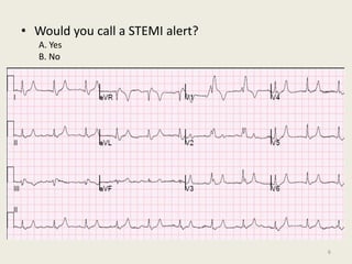
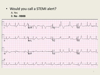
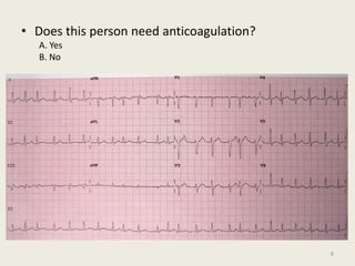
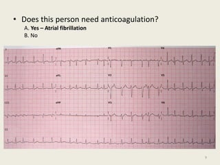
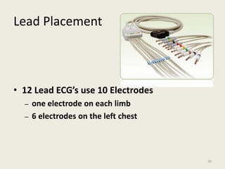
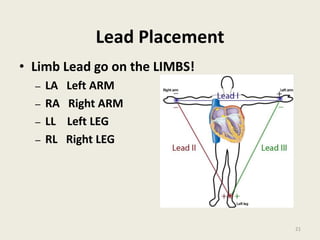
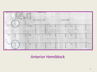
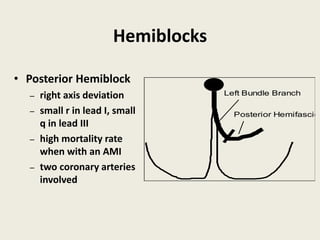
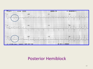
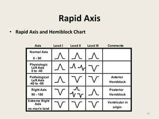
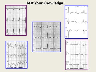
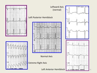
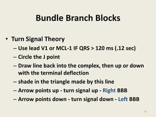
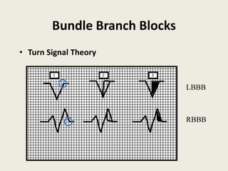
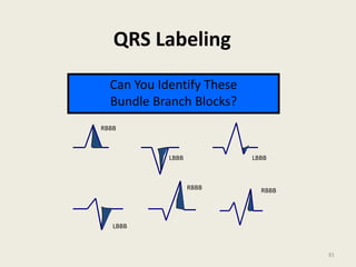
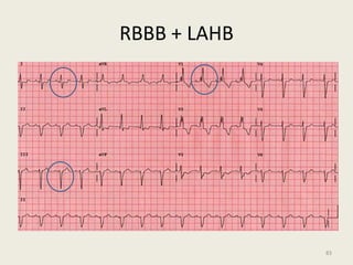
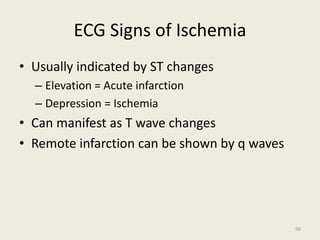
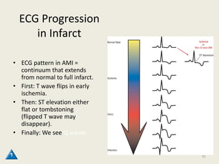
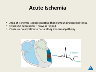
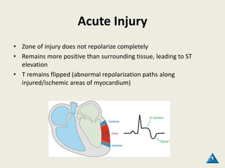
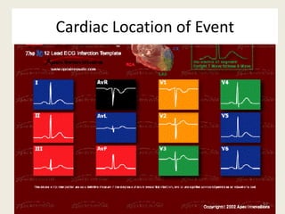
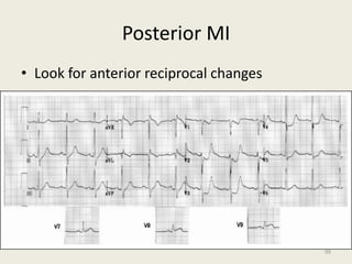
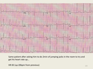
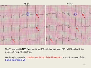
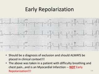
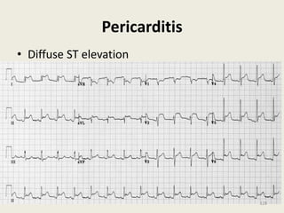
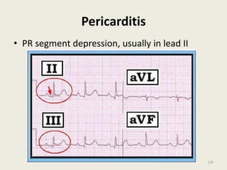
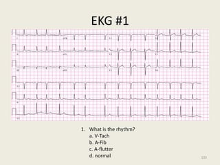
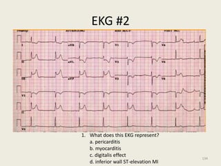
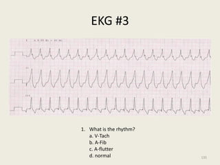
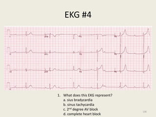
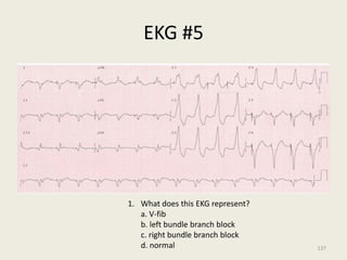
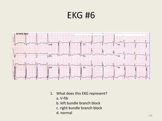
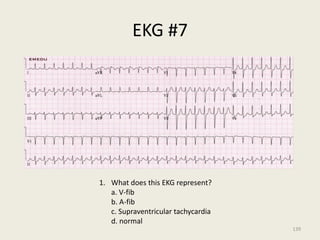
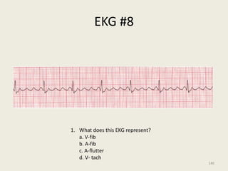
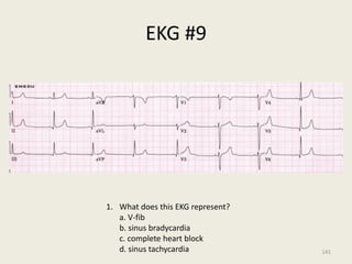
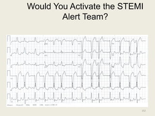
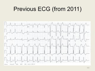
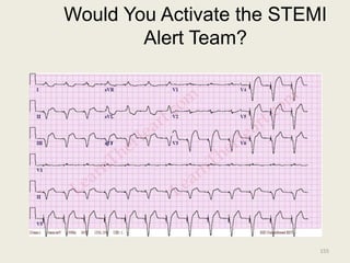
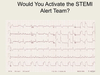
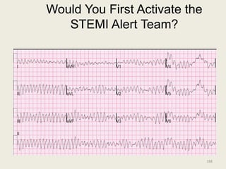
SlideShare a Scribd company logo
1 of 251
Download to read offline
Unraveling the
Mysteries of the
12 Lead EKG
Developed by the
Objectives
• Identify the correct lead placement for
performing a 12 lead EKG
• Identify and interpret heart rhythm and
differing blocks
• Identify extreme axis deviations
• Identify and interpret bundle branch blocks
• Interpret MI location based on ST elevation
2
ECG Pre-test
3
• Is this ECG normal?
A. True
B. False
4
• Is this ECG normal?
A. True
B. False - Wenkebach
5
• Would you call a STEMI alert?
A. Yes
B. No
6
• Would you call a STEMI alert?
A. Yes
B. No - RBBB
7
• Does this person need anticoagulation?
A. Yes
B. No
8
• Does this person need anticoagulation?
A. Yes – Atrial fibrillation
B. No
9
• The initial treatment of choice for this rhythm is
cardioversion.
A. True
B. False
10
• The initial treatment of choice for this rhythm is
cardioversion.
A. True
B. False – SVT (try adenosine first)
11
• This ECG explains the patient’s complaints of
dizziness.
A. True
B. False
12
• This ECG explains the patient’s complaints of
dizziness.
A. True – sinus bradycardia with heart rate of 37 bpm
B. False
13
How did you do?
OK – let’s get started!!
14
Monitoring vs Assessing
• Monitoring – EKG leads can be placed
anywhere
– Allows for identification of VF and Asystole
• Assessing – EKG leads MUST be placed in
specific locations
– Allows for interpretation of changes in the
electrical conduction (depolarization and
repolarization changes) i.e., ischemia.
16
Patient Preparation
• Provide a level of privacy
• Remove the patient’s shirt
• Shave the chest
• Prep the skin
– Remove the dead epithelials
• Electrically non-conductive
• Place the patient in a
hospital gown
YES! – Women Too
• Remove the bra
• Use a sheet to drape the patient
• Diaphoresis
– Dry the chest
– Use alcohol
– Use benzene
Patient Position
• Place the patient in the correct position to
acquire the EKG
– Supine Recommended
– Sitting up is fine
• Ask the patient to hold still
• Keep their hands down by their side
– May need to hold the patient’s hands
19
Lead Placement
• 12 Lead ECG’s use 10 Electrodes
– one electrode on each limb
– 6 electrodes on the left chest
20
Lead Placement
• Limb Lead go on the LIMBS!
– LA Left ARM
– RA Right ARM
– LL Left LEG
– RL Right LEG
21
Left Chest Lead Placement
• Precordial Leads (V leads or MCL leads)
– V1 4th intercostal space, right of sternum
– V2 4th intercostal space, left of the sternum
– V3 between V4 and V2
– V4 5th intercostal space, left of sternum
– V5 5th intercostal space, left of sternum
– V6 5th intercostal space, left of sternum
22
Left Chest EKG
23
The Normal Conduction System
24
Normal ECG
25
Limb Lead Reversal
26
Limb Lead Reversal
27
Waveforms
28
QRS Labeling
Q Waves
First negative deflection after the
P waves in any lead
Q wave
QRS Labeling
R Waves
First positive deflection after the
P waves in any lead
"R"
QRS Labeling
S Wave
Negative deflection below the
baseline after an "R" or "Q" wave
s
QS
s
QRS Labeling
The "J" Point
Also called the" juncture" point.
Where the qrs complex ends
and the ST segment begins
32
QRS Labeling
QRS Morphologies
Can you label these complexes?
R
QS
q
R
s
r
S q
R
r
S
R’
33
Now YOU Do It!
• Video of proper ECG lead placement
34
Interpretation
• Develop a systematic approach to reading
EKGs and use it every time
• The system recommended is:
– Rate
– Rhythm (including intervals and blocks)
– Axis
– Ischemia
35
Rate
• Rule of 300- Divide 300 by the number of
boxes between each QRS = rate
Number of
big boxes
Rate
1 300
2 150
3 100
4 75
5 60
6 50
36
Estimate of Heart Rate
37
What is the heart rate?
(300 / 6) = 50 bpm
www.uptodate.com
38
Rate
• HR of 60-100 per minute is normal
• HR > 100 = tachycardia
• HR < 60 = bradycardia
39
Differential Diagnosis of Tachycardia
Tachycardia Narrow Complex Wide Complex
Regular ST
SVT
Atrial flutter
ST w/ BBB
SVT w/ BBB
VT
Irregular A-fib
A-flutter w/
variable conduction
MAT
A-fib w/ BBB
A-fib w/ WPW
VT
40
Rhythm
• Sinus
– Originating from SA
node
– P wave before
every QRS
– P wave in same
direction as QRS
41
What is this rhythm?
42
Normal Intervals
• PR
– 0.20 sec (less than one
large box)
• QRS
– 0.08 – 0.10 sec (1-2 small
boxes)
• QT
– 450 ms in men, 460 ms in
women
– Based on sex / heart rate
– Half the R-R interval with
normal HR
43
Causes of Prolonged QT
• Causes
– Drugs
– Hypocalcemia,
hypomagnesemia,
hypokalemia
– Hypothermia
– AMI
– Congenital
– Increased ICP
44
Consequences of Prolonged QT
45
Blocks
• AV blocks
– First degree block
• PR interval fixed and > 0.2 sec
– Second degree block, Mobitz type 1
• PR gradually lengthened, then drop QRS
– Second degree block, Mobitz type 2
• PR fixed, but drop QRS randomly
– Type 3 block
• PR and QRS dissociated
46
What is this rhythm?
47
What is this rhythm?
First degree AV block
PR is fixed and longer than 0.2 sec
48
What is this rhythm?
49
What is this rhythm?
Type 1 second degree block (Wenckebach)
50
What is this rhythm?
51
What is this rhythm?
52
What is this rhythm?
53
What is this rhythm?
3rd degree heart block (complete)
54
Section Two
55
Axis
• Axis: predominant
flow of electricity
through the heart
• We look at the QRS
complexes for
ventricular axis
III II
I
NORMAL AXIS
57
Hexaxial Reference System
• Divided into 6 part grid
– Based on the leads
• I
• II
• III
• aVR
• aVF
• aVL
• Degrees of electrical flow
– 0 to +180
– 0 to -180
58
ECG with Normal Axis
59
Extreme Right Axis
-90 to -180 degrees
60
ECG with Extreme Right Axis
61
Differential Diagnosis of Extreme Right
Axis
• Ventricular tachycardia
• Hyperkalemia (acute renal failure)
• Apical MI
• Right Ventricular Hypertrophy
62
Limb Lead Reversal
63
Hemiblocks
• A hemiblock is a block
of one of the fascicles
of the left bundle
branch.
• Hemiblock is an ECG
diagnosis
Left Bundle Branch
Posterior Hemifascicle
Anterior Hemifascicle
Hemiblocks
• Anterior Hemiblock
– pathological left axis
– negative deflection in
leads II and III
– small q in lead I, small
r in lead III
– common block
– 4x higher mortality
rate in AMI
Left Bundle Branch
Anterior Hemifascicle
Anterior Hemiblock
66
Hemiblocks
• Posterior Hemiblock
– right axis deviation
– small r in lead I, small
q in lead III
– high mortality rate
when with an AMI
– two coronary arteries
involved
Left Bundle Branch
Posterior Hemifascicl
Posterior Hemiblock
• Very rare and much more dangerous.
• Posterior hemifascicle has redundant
blood supply from two separate
coronary arteries.
• In setting of an acute MI, two coronary
arteries would have to be occluded
proximally in order to create this
condition.
Posterior Hemiblock
69
Rapid Axis
• Rapid Axis and Hemiblock Chart
Lead I Lead II Lead III
Axis
Normal Axis
0 - 90
Physiologic
Left Axis
0 to -40
Pathological
Left Axis
-40 to -90
Right Axis
90 - 180
Extreme Right
Axis
Anterior
Hemiblock
Hemiblock
Posterior
no man's land
Ventricular in
origin
Comments
70
Test Your Knowledge!
71
Normal Axis
Leftward Axis
(normal)
Left Anterior Hemiblock
Left Posterior Hemiblock
Extreme Right Axis
72
Ventricular Tachycardia
• 12 Lead ECG and VT:
• You may be the only one
to see the rhythm
• A 12 lead ECG of VT is
very helpful to the
cardiologist looking for
the cause
• More benefit and less risk
in knowing for sure
Ventricular Tachycardia
• Rate 110 -250 bpm
• Wide complex (>0.12 – 3 small blocks)
• Regular
• AV dissociation
• Extreme Right Axis Deviation + Upright MCL-1
I II
I II III
MCL-1
I
II
III
EXTREME RIGHT AXIS
ERAD
V1
75
VT
76
Bundle Branch Blocks
• A Bundle Branch Block is a block of one of the
two bundle branches, left or right
• A Bundle Branch is a fascicle of electrical
conduction system cells designed to carry
impulses to the ventricles
• Bundle Branches facilitate “syncytium” or
both ventricles contracting in sync.
77
Bundle Branch Blocks
• Turn Signal Theory
– easy way to
determine left or right
BBB
– use lead V1
– QRS complex must be
at least .12sec (120
ms) or wider (or 3
little squares)
Bundle Branch Blocks
• Turn Signal Theory
– Use lead V1 or MCL-1 IF QRS > 120 ms (.12 sec)
– Circle the J point
– Draw line back into the complex, then up or down
with the terminal deflection
– shade in the triangle made by this line
– Arrow points up - turn signal up - Right BBB
– Arrow points down - turn signal down - Left BBB
79
Bundle Branch Blocks
• Turn Signal Theory
1 2 3
LBBB
RBBB
QRS Labeling
QRS Morphologies
Can you label these complexes?
RBBB
LBBB LBBB
LBBB
RBBB RBBB
Can You Identify These
Bundle Branch Blocks?
81
Right Bundle Branch Block and
Hemiblocks can occur together!
RBBB + Anterior Hemiblock (most commonly seen)
Left Bundle Branch
Anterior Hemifascicle
Right Bundle Branch
RBBB + LAHB
83
Section Three
84
Myocardial Blood Supply
85
AMI
Myocardial Blood Supply
• Right Coronary Artery
• Inferior Wall (LV)
• Posterior Wall (LV)
• Right Ventricle
• SA and AV Node
• Posterior fascicle of LBB
Myocardial Blood Supply
• Left Anterior Descending
• Anterior Wall of LV
• Septal Wall
• Bundle of His and BB
Myocardial Blood Supply
• Circumflex
• Lateral Wall of LV
• Rarely SA and AV nodes
• Posterior Wall of LV
Clinical Manifestations of
Arterial Thrombosis
UA/NSTEMI:
Partially-occlusive thrombus
(primarily platelets)
Intra-plaque
thrombus (platelet
dominated)
Plaque core
ST  MI:
Occlusive thrombus (platelets,
red blood cells, and fibrin)
Intra-plaque
thrombus (platelet
dominated)
Plaque core
SUDDEN DEATH
Adapted from Davies MJ. Circulation. 1990; 82 (supl II): 30-46.
ECG Signs of Ischemia
• Usually indicated by ST changes
– Elevation = Acute infarction
– Depression = Ischemia
• Can manifest as T wave changes
• Remote infarction can be shown by q waves
90
ECG Progression
in Infarct
• ECG pattern in AMI =
continuum that extends
from normal to full infarct.
• First: T wave flips in early
ischemia.
• Then: ST elevation either
flat or tombstoning
(flipped T wave may
disappear).
• Finally: We see Q waves.
91
12 Lead ECG and AMI
• Benefits of 12 Lead ECG’s
– Highly specific (90% + confidence)
– If it shows an MI, there probably is an MI
– Rapid identification of MI in early stages
– Can commit to treat with ECG, history and
physical exam
– Complications can be identified
92
12 Lead ECG and AMI
• Limitations
– Only 46 - 50 % sensitive (may miss 50%)
• Increase sensitivity by looking at the whole heart
– Diagnostic quality necessary
– Training needed to read the 12 leads
– ECG evidence is only one piece of the puzzle
– Some non-MI conditions look like MI’s
93
12 Lead ECG and AMI
A NORMAL 12 LEAD ECG DOES NOT
RULE OUT A MYOCARDIAL
INFARCTION
If there is suspicion for MI, repeat the
ECG
94
Acute Ischemia
• Area of ischemia is more negative than surrounding normal tissue
• Causes ST depression; T wave is flipped
• Causes repolarization to occur along abnormal pathway
95
Acute Injury
• Zone of injury does not repolarize completely
• Remains more positive than surrounding tissue, leading to ST
elevation
• T remains flipped (abnormal repolarization paths along
injured/ischemic areas of myocardium)
96
Cardiac Location of Event
97
Posterior MI
Is there a lead for that?
• You only find what you’re looking for!
– Move V4, V5, V6
– 5th intercostal space
98
Posterior MI
• Look for anterior reciprocal changes
99
What about the right side?
RV infacts
• Move V4 to the
right side same
location
– 5th intercostal
space anterior
axillary
Occur in conjunction with inferior MIs
100
Acute MI with RV involvement
101
Where/What is It?
102
Where/What is It?
103
Where/What is It?
104
Where/What is It?
105
106
Scorecard
• The guidelines call for a 90 minute medical
contact to balloon time.
• Very important to perform immediate or in-
field ECG to make earlier diagnosis to start the
STEMI alert.
107
Interventional Plan for EMS
• Out of hospital 12 lead
• Early notification of
hospital
• O2, NTG, pain control
• ASA, Heparin
• Thrombolytic prescreen
• Transport to PCI Center
Definitive AMI Treatment
Percutaneous Coronary Intervention
When to Consider Thrombolytics
• Acute MI patients in whom first medical
contact to balloon time is like to exceed
2 hours.
• Cath lab is not available.
110
How do thrombolytics or more
appropriately fibrinolytics work?
t-PA
• A naturally occurring blood protein
Plasminogen activates the production of
plasmin – a digestive enzyme
• Presence of a clot causes the endothelia cells
to secrete tissue plasminogen activator which
starts the breakdown of the clot
111
How do fibrinolytics work?
• Fibrinolytics
– Destroy the clot
at the level of the fibrin.
– Activate the production
of plasmin to cause the
digestion of the clot
112
EMS and the AMI:
Making a difference
• Early recognition and treatment
• Early activation of cath lab
• Once infarction begins 500
myocardial cells die each second
• Salvage myocardium
• Decreased incidence of CHF
• Maintain active lifestyles
Infarct Caveats
• Anterior Wall MI
– most lethal (highest mortality)
– can suddenly develop, CHB, VF or VT
– if seen with hemiblocks or BBB, place quick
combo pads on the patient and prepare for
the worst
– can extend to septum (anteroseptal) or lateral
(anterolateral)
– nitrates are great, fluids are spared
114
Infarct Caveats
• Inferior MI
– Most common seen. Can be fatal
– 50% have posterior and right ventricle involved
– Patients may have bradycardia and hypotension
– Could also have 1st degree or Mobitz 1 blocks
– Nausea is common, phenergan or compazine
– Use nitrates with caution, may need fluids
115
Infarct Imitators
• Left Bundle Branch Block
– late depolarization makes ST
elevation difficult to distinguish
– LBBB considered a non-diagnostic
ECG
• Left Ventricular Hypertrophy
– won’t have reciprocal changes
• Early Repolarization
(…but is it really benign?)
Benign early repolarization
Who gets it?
• 2-5% of the general population (Wellens,
2008)
• Usually the young and physically fit
• More prominent in African-Americans
• Generally disappears with advancing age
118
What does it look like?
Red arrows: concave up ST-segment elevation anteriorly
Blue arrows: hyperdynamic, symmetrical, concordant T-waves 119
Classic findings
1. J-point “notching”
2. Concave-up ST segment
(smiley face)
3. ST segment elevation
from baseline in V2-V5,
typically <3mm
4. Large, symmetrically
concordant T-waves in
leads with STE
120
Can we tease it out?
• The degree of ST segment elevation is thought to
be indirectly proportional to the degree of
sympathetic tone
• In other words, the more relaxed the patient, the
more pronounced the ST segment elevation (and
vice versa)
• If you truly want to test your patient, get their
heart rate up and look at the ST segment
121
14yo M w/ palpitations
HR: 64
122
1. Notched J-point
2. Concave down ST
elevation in
precordial leads
123
Same patient after asking him to do 2min of jumping jacks in the room to try and
get his heart rate up…
HR 83 (up 20bpm from previous) 124
HR 64 HR 83
The ST segment is NOT fixed in pts w/ BER and changes from EKG to EKG and with the
degree of sympathetic strain
On the right, note the complete resolution of the ST elevation but maintenance of the
J-point notching in V4
125
Early Repolarization
• Should be a diagnosis of exclusion and should ALWAYS be
placed in clinical context!!!
• The above was taken in a patient with difficulty breathing and
chest pain…and is an Myocardial Infarction -- NOT Early
Repolarization!!! 126
Pericarditis
• Pericarditis is an inflammation of the
pericardium (sac that surrounds the heart).
• This often occurs as a result of a viral
infection.
• However, this can cause severe chest pain and
can lead to ST elevation in all leads.
• Therefore, it is important to distinguish acute
pericarditis from acute myocardial infarction.
127
Pericarditis
• Diffuse ST elevation
128
Pericarditis
• PR segment depression, usually in lead II
129
Pericarditis Treatment
• NSAIDs
• Colchicine
• Occasionally steroids
• Anticoagulation could cause a hemorrhagic
pericardial effusion – life threatening.
130
Section 4
• ECG Tests are next!
131
ECG Quiz
EKG #1
1. What is the rhythm?
a. V-Tach
b. A-Fib
c. A-flutter
d. normal 133
EKG #2
1. What does this EKG represent?
a. pericarditis
b. myocarditis
c. digitalis effect
d. inferior wall ST-elevation MI
134
EKG #3
1. What is the rhythm?
a. V-Tach
b. A-Fib
c. A-flutter
d. normal 135
EKG #4
1. What does this EKG represent?
a. sius bradycardia
b. sinus tachycardia
c. 2nd degree AV block
d. complete heart block
136
EKG #5
1. What does this EKG represent?
a. V-fib
b. left bundle branch block
c. right bundle branch block
d. normal 137
EKG #6
1. What does this EKG represent?
a. V-fib
b. left bundle branch block
c. right bundle branch block
d. normal
138
EKG #7
1. What does this EKG represent?
a. V-fib
b. A-fib
c. Supraventricular tachycardia
d. normal
139
EKG #8
1. What does this EKG represent?
a. V-fib
b. A-fib
c. A-flutter
d. V- tach
140
EKG #9
1. What does this EKG represent?
a. V-fib
b. sinus bradycardia
c. complete heart block
d. sinus tachycardia 141
EKG #10
1. What does this EKG represent?
a. V-fib
b. left bundle branch block
c. right bundle branch block
d. normal
142
EKG #11
1. What diagnostic test would be the best to order next?
a. Echo
b. CTA
c. Cath 143
EKG #12
1. What therapy would be the best to order next?
a. Thrombolytic therapy
b. Emergent cath and PCI
c. Toradol IV 144
STEMI Alerts
Would You Activate the STEMI
Alert Team?
146
YES!
• This is an large anteroseptal, anterior,
and anterolateral MI
147
Would You Activate the STEMI
Alert Team?
148
NO!
• This is Pericarditis – inflammation of the
sac around the heart.
– Diffuse ST elevation
– PR segment depression
– Younger
– Recent viral syndrome
– Hurts worse with deep breaths or lying
down
149
Would You Activate the STEMI
Alert Team?
150
YES!
• This is new-onset Left Bundle Branch
Block
• Also note the lateral ST elevation
151
Would You Activate the STEMI
Alert Team?
152
Previous ECG (from 2011)
153
NO!
• This is a chronic Left Bundle Branch
Block
• Marker of CAD, heart valve disease, as
well as hypertension.
154
Would You Activate the STEMI
Alert Team?
155
NO!
• This is a PACED rhythm!
• No interpretation of the ECG is possible.
156
Would You Activate the STEMI
Alert Team?
157
NO!
• This is Early Repolarization.
– Early repolarization is a common ECG variant,
characterized by either terminal QRS slurring
(the transition from the QRS segment to the ST
segment) or notching (a positive deflection
inscribed on terminal QRS complex) associated
with concave upward ST-segment elevation
and prominent T waves in at least two
contiguous leads.
158
NO!
• This is Early Repolarization.
– This benign ECG phenomenon is noted in 1%
to 2% of the adult population and generally
occurs in the absence of myocardial disease.
– People with this mostly consist of men, young
adults, athletes, and people of African American
heritage
159
160
161
Would You Activate the STEMI
Alert Team?
162
YES!
• This is an inferior – posterior – lateral MI
163
Would You Activate the STEMI
Alert Team?
164
YES!
• This is Ventricular Tachycardia – and
likely is related to MI
165
Would You Activate the STEMI
Alert Team?
166
YES!
• This is an acute Anterior Wall MI with
Ventricular Bigeminy
167
Would You First Activate the
STEMI Alert Team?
168
NO!
• Shock that!
• While MI may be the reason for Vfib, other
reasons also need to be excluded.
• Consider Hypothermia Therapy in route
169
Section 5
170
Review of MHCA Protocols
• STEMI
• Stroke
171
Goals for STEMI
• First Medical Contact (FMC) to PCI < 90 minutes
• Door to ECG time < 10 minutes
• Door In / Door Out Time < 30 minutes
• FMC to Non-PCI hospital to PCI < 120 minutes
EMS specific
• Ideal for all chest pain patients to have in-field
ECG
• Pre-hospital Activation of STEMI network
• Diversion to STEMI hospital
172
EMS Requirements
Equip all ambulances in state with ECG machines by 2012
Ambulance services should obtain EKG within 15 minutes for
 typical chest pain in anyone > 30 years, and
 atypical chest pain in all patients 50 and older
 EMS should interpret and transfer ECG to affiliated ED
 EMS personnel need training / certification in ECG interpretation
of STEMI
173
ECG + Symptoms
• Chest pain,fullness, or
pressure
• Radiation to jaw, teeth,
shoulder, arm, or back
• Shortness of breath
• Epigastric discomfort
• Sweating
• Dizziness
• Cognitive impairment
174
EMS Requirements
 + EKG patients directly to PCI hospital if 90 minutes window
obtainable from first med contact to PCI AND patient is
hemodynamically stable
 + EKG patients directly to PCI hospital if 90 minutes window
obtainable from first med contact to PCI BUT patient is
hemodynamically UNSTABLE
 Go to nearest ED
 Activate Air Transport immediately for transfer to PCI center
175
EMS Requirements
 If no pre-hospital ECG available for a chest pain patient who
arrives at a non-PCI hospital
 Keep the patient on the EMS stretcher until ECG performed
 If EKG results + transfer to PCI hospital with SAME ambulance if
patient hemodynamically stable
176
STEMI Network (24/7) PCI Centers
Jackson
St. Dominic
MBHS
UMMC
CMMC
Hattiesburg
Forrest General Hospital
Wesley
Meridian
Jeff Anderson Hospital
Rush Hospital
Tupelo
North Mississippi Medical Center
Oxford
Baptist Memorial Hospital North
Mississippi
South Haven
Baptist Memorial Hospital Desoto
Corinth
Magnolia Regional Health Center
Vicksburg
River Region Hospital
Greenville
Delta Regional Medical Center
Columbus
Baptist Memorial Hospital Golden
Triangle
Pascagoula
Singing River Health Systems
Gulfport
Gulfport Memorial Hospital
McComb
South West Regional Medical
Center
177
EMS Territorial Boundaries Broken
 It is imperative for EMS to be able to cross county lines
when necessary for reperfusion.
 EMS services should cross-cover for adjacent EMS in
another county.
 A “Heart Attack” should take priority over many non-
life threatening medical conditions.
178
179
Goals for STEMI
• First Medical Contact (FMC) to PCI < 90 minutes
• Door to ECG time < 10 minutes
• Door In / Door Out Time < 30 minutes
• FMC to Non-PCI hospital to PCI < 120 minutes
EMS specific
• Ideal for all chest pain patients to have in-field
ECG
• Pre-hospital Activation of STEMI network
• Diversion to STEMI hospital
180
Phases of EMS Management
of the Stroke Patient
• Activation of 911 system
• EMS response
• On scene assessment and stabilization
• Selection of appropriate destination
• Transport
• Pre-arrival stroke alert to receiving emergency
department (as early as possible)
• Delivery of patient and information
• PI feedback
181
Scene Assessment
• General assessment
– Consider alternative causes of neurologic deficit
• Focused neurologic assessment to include FAST
– Face
– Arm
– Speech
– Time
• Sensitivity 80%/specificity 30%
• Time of onset - may not be available at hospital
182
183
Treatment
• Stabilization
– Standard protocols (check vital signs, ECG,
glucose, hydration and treat as needed)
– Scene time should be minimized but prehospital
care should not be sacrificed for less scene time
184
Select Appropriate Destination
• Transport to the nearest hospital with an
appropriate level of stroke care
– Level may vary as resources change
– Utilize knowledge of local facilities
• Window of opportunity – 4 ½ hours to
completion of fibrinolytic treatment (earlier
more effective than later)
• Useful time – 3 ½ hours until time of arrival at
stroke capable hospital
185
EMS Goals for Stroke
186
1) Initial assessment, transport ASAP:
ABCs ; Obtain time of symptom onset (Last time known well) _______; Source of information
________; Contact information _________.
2) Administer high concentration oxygen, as needed, to maintain O2 Sat >94 percent.
3) Position patient with head/shoulders elevated to 15-30 degrees (unless contraindicated).
4) Maintain NPO.
5) Blood glucose < 60, treat per protocol.
6) Do not treat high blood pressure without physician approval.
7) Perform Stroke Scale – Cincinnati Stroke Scale.
8) Transport patient to the appropriate facility:
a. Transport patient to the closest hospital capable of treating the patient with IV Alteplase
(Stroke Capable or Primary/Comprehensive Stroke Center). Hospitals not able to diagnose
and treat stroke patients (Level 4 hospitals) may be by-passed. EMS may use discretion based
on transport time or other unforeseen factors.
b. Consider transport of the stroke patient with severe symptoms (hemiplegia, aphasia, neglect,
stably intubated) to a Comprehensive Stroke Center if symptom onset to hospital arrival time
is greater than 3 hours and less than 6 hours.
c. Transport patient to the closest appropriate facility if unstable (e.g., cardiac arrest, unstable
airway).
9) IV NS KVO once en route.
10) EKG once en route.
11) Notify receiving facility of estimated arrival time of acute stroke patient, Stroke Scale finding, and
time of onset. 187
Section 6
188
EMS Cardiac
Pharmacology
189
Oxygen
• Indications
– Any suspected cardiopulmonary emergency
– Saturate hemoglobin with oxygen
– Reduce anxiety & further damage
– Note: Pulse oximetry should be monitored
190
Oxygen
• Precautions
– Pulse oximetry inaccurate in:
• Low cardiac output
• Vasoconstriction
• Hypothermia
– NEVER rely on pulse oximetry!
– Too much oxygen can make some patients with
emphysema quit breathing
191
Aspirin
• Indications
– Administer to all patients with ACS, particularly
reperfusion candidates
• Give 325 mg as soon as possible, non-coated preferred
– Blocks formation of thromboxane A2, which
causes platelets to aggregate
192
Anti-Platelet Actions
193
Aspirin
• Precautions
– Many patients are allergic to aspirin – be sure to
ask!
– Does not provide blood thinning effects in all
people (aspirin resistance)
– Relatively contraindicated in patients with active
bleeding
194
Thienopyridines
(Brilinta, Effient,Plavix)
• Indications
– Use as a second anti-platelet agent in patients
with ACS, particularly reperfusion candidates
– Blocks ADP activation of platelets
– Usually given as a bolus dose
• Brilinta – 180 mg (MHCA preferred agent)
• Plavix (clopidogrel) – 600 mg
• Effient – 60 mg
195
Anti-Platelet Actions
196
Thienopyridine
• Precautions
– Plavix does not provide blood thinning effects in
all people (plavix resistance)
– Effient should not be given to patients with
previous stroke or TIA
– Relatively contraindicated in patients with active
bleeding
197
Glycoprotein IIb/IIIa Inhibitors
• Indications
– Inhibit the glycoprotein IIb/IIIa receptor in the
membrane of platelets, inhibiting platelet
aggregation
– Can be used as an early second anti-platelet agent
rather than thienopyridines, especially in those
who can’t swallow or have nausea and vomiting.
198
Anti-Platelet Actions
199
Glycoprotein IIb/IIIa Inhibitors
• Eptifibatide (integrilin)
– Within 10 minutes after bolus, > 90% of platelets
are inhibited
– Platelet function recovers within 4 to 8 hours after
discontinuation
– Dose
• 180 mcg/kg IV bolus, then 2 mcg/kg/min infusion
200
Glycoprotein IIb/IIIa Inhibitors
• Precautions
– Integrilin (eptifibatide) is a derivative of snake
venom
– Use in precaution in those patients with previous
snake bites
201
Heparin
• Indications
– Inhibits thrombin generation by factor Xa
inhibition and also inhibit thrombin indirectly by
formation of a complex with antithrombin III
– Exists in two forms
• Unfractionated
• Low molecular weight
202
Unfractionated Heparin
• Dosing
– Initial bolus 60 IU/kg
• Maximum bolus: 4000 IU
• Check efficacy of dose with ACT
• Not always effective
– Continuous infusion at 800-1200 units/hour
203
Low Molecular Weight Heparin
Lovenox (enoxaparin)
• Dosing in ACS in those proceeding to PCI or to
receive thrombolytics
– 30 mg IV
• Bolus is active for 3 hours
• Initial dosing in medically treated patients
– 1 mg/kg SQ
• Dose is active for 12 hours
204
Heparins
• Precautions
– Contraindications: active bleeding; recent
intracranial, intraspinal or eye surgery; severe
hypertension; bleeding disorders;
gastroinintestinal bleeding
– DO NOT use if platelet count is below 100 000
205
Nitroglycerin
• Indications
– Chest pain of suspected cardiac origin
– Unstable angina
– Complications of AMI, including congestive heart
failure, left ventricular failure
– Hypertensive crisis or urgency with chest pain
206
Nitroglycerin
• What it does…
– Decreases pain of ischemia
– Increases venous dilation
– Decreases venous blood return to heart
– Decreases preload and cardiac
oxygen consumption
– Dilates coronary arteries
– Increases cardiac collateral flow
207
Nitroglycerin
• What it does NOT do…
– Prevent heart attacks
– Save lives
– Limit infarct size
208
Nitroglycerin
• Dosing
– Sublingual Route
• 0.3 to 0.4 mg; repeat every 5 minutes
– Aerosol Spray
• Spray for 0.5 to 1.0 second at 5 minute intervals
– IV Infusion
• Infuse at 10 to 20 µg/min
• Route of choice for emergencies
• Titrate to effect
209
Nitroglycerin
• Precautions
– Use extreme caution if systolic BP <90 mm Hg
– Use extreme caution in Inferior and/or RV infarctions
– Suspect RV infarction with inferior ST changes
– Limit BP drop to 10% if patient is normotensive
– Limit BP drop to 30% if patient is hypertensive
– Watch for headache, drop in BP, syncope, tachycardia
– Tell patient to sit or lie down during administration
210
Morphine Sulfate
• Indications
– Chest pain and anxiety associated with AMI or
cardiac ischemia
– Acute cardiogenic pulmonary edema (if blood
pressure is adequate)
211
Morphine Sulfate
• Dosing
– 1 to 4 mg IV (over 1 to 5 minutes) every 5 to 10
minutes as needed
212
Morphine Sulfate
• Precautions
– Administer slowly and titrate to effect
– May compromise respiration; therefore use with
caution in acute pulmonary edema
– Causes hypotension in volume-depleted patients
213
Fibrinolytics
• Indications
– For AMI in adults
• ST elevation or new or presumably new LBBB; strongly
suspicious for injury
• Time of onset of symptoms < 12 hours
– For strokes in adelts
• Time of onset of symptoms< 4.5 hours
214
Fibrinolytics
• Dosing
– For fibrinolytic use, all patients should have 2
peripheral IV lines
• 1 line exclusively for fibrinolytic administration
215
Fibrinolytics
• Dosing for AMI Patients
– Tenecteplase (TNKase)
• Bolus 30 to 50 mg
– Alteplase, recombinant (tPA)
• Accelerated Infusion
– 15 mg IV bolus
– Then 0.75 mg/kg over the next 30 minutes
» Not to exceed 50 mg
– Then 0.5 mg/kg over the next 60 minutes
» Not to exceed 35 mg
216
Fibrinolytics
• Dosing for Acute Ischemic Stroke
– Alteplase, recombinant (tPA)
• Give 0.9 mg/kg (maximum 90 mg) infused over 60
minutes
– Give 10% of total dose as an initial IV bolus over 1 minute
– Give the remaining 90% over the next 60 minutes
– Alteplase is the only agent approved for use in
Ischemic Stroke patients
217
Fibrinolytics
• Precautions
– Specific Exclusion Criteria
• Active internal bleeding (except mensus) within 21 days
• History of CVA, intracranial, or intraspinal within 3
months
• Major trauma or serious injury within 14 days
• Aortic dissection
• Severe uncontrolled hypertension
218
Fibrinolytics
• Precautions
– Specific Exclusion Criteria
• Known bleeding disorders
• Prolonged CPR with evidence of thoracic trauma
• Lumbar puncture within 7 days
• Recent arterial puncture at noncompressible site
• During the first 24 hours of fibrinolytic therapy for
ischemic stroke, do not give aspirin or heparin
219
Amiodarone
• Indications
– Powerful anti-arrhythmic with activity in both
atria and ventricles; so that, this drug can be used
for atrial fibrillation and VT
– Can be used to prevent recurrent VF
220
Amiodarone
• Dosing
– 150 mg bolus dose
• May repeat x 1
– Can also use continual IV infusion
• 1 mg/min x 6 hours, then
• 0.5 mg/min
221
Amiodarone
• Precautions
– May produce vasodilation & hypotension
– May have negative inotropic effects
– Terminal elimination
• IV half-life lasts hours
• Oral half-life lasts up to 40 days
222
Lidocaine
• Indications
– VT
– Vfib
– Frequent PVCs
223
Lidocaine
• Bolus Dosing
– Initial dose: 1.0 to 1.5 mg/kg bolus IV
– May repeat bolus x 1 for refractory VF
– May also be given down ET tube
• Maintenance Infusion
– 2 to 4 mg/min IV continuous infusion
224
Lidocaine
• Precautions
– Reduce maintenance dose (not loading dose) in
presence of impaired liver function or left
ventricular dysfunction
– Discontinue infusion immediately if signs of
toxicity (seizures, confusion) develop
225
Atropine Sulfate
• Indications
– Should only be used for bradycardia
• Relative or Absolute
– Used to increase heart rate
226
Atropine Sulfate
• Dosing
– 1 mg IV push
– Repeat every 3 to 5 minutes
– May give via ET tube (2 to 2.5 mg) diluted in 10
mL of NS
227
Atropine Sulfate
• Precautions
– Increases myocardial oxygen demand
– May result in unwanted tachycardia or
dysrhythmia
– When given in low doses (<0.4 mg), can cause a
paradoxical bradycardia
228
Dopamine
• Indications
– Second drug for symptomatic bradycardia (after
atropine)
– Use for hypotension (systolic BP 70 to 100 mm Hg)
with S/S of shock
229
Dopamine
• Dosing
– IV Infusions (Titrate to Effect)
• Low Dose “Renal Dose"
– 1 to 5 µg/kg per minute
• Moderate Dose “Cardiac Dose"
– 5 to 10 µg/kg per minute
• High Dose “Vasopressor Dose"
– 10 to 20 µg/kg per minute
230
Dopamine
• Precautions
– May use in patients with hypovolemia but only after
volume replacement
– May cause tachyarrhythmias, excessive vasoconstriction
– DO NOT mix with sodium bicarbonate
231
Epinephrine
• Indications
– Increases:
• Heart rate
• Force of contraction
• Conduction velocity
– Peripheral vasoconstriction (raises blood pressure)
– Bronchial dilation
232
Epinephrine
• Dosing
– 1 mg IV push; may repeat every 3 to 5 minutes
– May use higher doses (0.2 mg/kg) if lower dose is
not effective
– Endotracheal Route
• 2.0 to 2.5 mg diluted in 10 mL normal saline
– Profound Bradycardia
• 2 to 10 µg/min infusion (add 1 mg of 1:1000 to 500 mL
normal saline; infuse at 1 to 5 mL/min)
233
Epinephrine
• Precautions
– Raising blood pressure and increasing heart rate
may cause myocardial ischemia, angina, and
increased myocardial oxygen demand
– Do not mix or give with alkaline solutions
– Higher doses have not improved outcome & may
cause myocardial dysfunction
234
Diltiazem
• Indications
– To control ventricular rate in atrial fibrillation and
atrial flutter
– Use after adenosine to treat refractory PSVT in
patients with narrow QRS complex and adequate
blood pressure
235
Diltiazem
• Dosing
– Acute Rate Control
• 10 to 20 mg (0.25 mg/kg) IV over 2 minutes
• May repeat in 15 minutes at 20 to 25 mg (0.35 mg/kg)
over 2 minutes
– Maintenance Infusion
• 5 to 15 mg/hour, titrated to heart rate
236
Diltiazem
• Precautions
– Do not use calcium channel blockers for tachycardias of
uncertain origin
– Avoid calcium channel blockers in patients with Wolff-
Parkinson-White syndrome, in patients with sick sinus
syndrome, or in patients with AV block without a
pacemaker
– Expect blood pressure drop resulting from peripheral
vasodilation
– Concurrent IV administration with IV ß-blockers can cause
severe hypotension or heart block
237
Question 1
• Which of the following is an adverse reaction
to nitroglycerin?
A) Hypertension
B) Hypotension
C) Lacrimation
D) Arrhythmias
238
Question 1
• Which of the following is an adverse reaction
to nitroglycerin?
A) Hypertension
B) Hypotension
C) Lacrimation
D) Arrhythmias
239
Question 2
• Which of the following must be given within
4.5 hours of the beginning of a stroke?
A. Thrombolytics
B. Anti-platelets
C. Heparin
240
Question 2
• Which of the following must be given within
4.5 hours of the beginning of a stroke?
A. Thrombolytics
B. Anti-platelets
C. Heparin
241
Question 3
• Which of the following agents is most
efficacious in the conversion of acute AF into
sinus rhythm?
a. Metoprolol
b. Digoxin
c. Amiodarone
d. Diltiazem
e. Esmolol
242
Question 3
• Which of the following agents is most
efficacious in the conversion of acute AF into
sinus rhythm?
a. Metoprolol
b. Digoxin
c. Amiodarone
d. Diltiazem
e. Esmolol
243
Question 4
• The following are true for aspirin, except:
a. Aspirin is indicated in combination with
warfarin in patients at high risk for mechanical
valve thrombosis
b. Clopidogrel should be administered to aspirin-
intolerant patients acutely with an STEMI
c. Aspirin is indicated in acute thrombotic stroke
d. Aspirin is FDA approved for primary
prevention of MI.
244
Question 4
• The following are true for aspirin, except:
a. Aspirin is indicated in combination with
warfarin in patients at high risk for mechanical
valve thrombosis
b. Clopidogrel should be administered to aspirin-
intolerant patients acutely with an STEMI
c. Aspirin is indicated in acute thrombotic stroke
d. Aspirin is FDA approved for primary
prevention of MI.
245
Question 5
• Appropriate upfront medical therapy in a
previously healthy 51 year old man having a
STEMI includes all of the following except:
a. Aspirin
b. Heparin
c. Lipitor
d. Brilinta
246
Question 5
• Appropriate upfront medical therapy in a
previously healthy 51 year old man having a
STEMI includes all of the following except:
a. Aspirin
b. Heparin
c. Lipitor
d. Brilinta
247
CONCLUSIONS
• Be constantly alert—patients can change in
seconds
• Know your drugs---use resources
• Remember that every drug, even OTC drugs,
have the potential to result in a serious
adverse reaction
CONCLUSIONS
• Never leave the sending facility unless you feel
thoroughly comfortable with your patient and
with the medications you are being asked to
administer or monitor
• Make sure that you are thoroughly prepared
for any complication
• Know where possible diversion hospitals are
located
• Use your EMS medical director whenever
necessary
CONCLUSIONS
Questions?
The End
• Thank you for your time today in learning the
interpretation of ECGs.
• With your new knowledge and proficiencies,
patients of Mississippi are in better hands.
252

More Related Content

What's hot

hemodynamic in cath lab: aortic stenosis and hocm
hemodynamic in cath lab: aortic stenosis and hocmhemodynamic in cath lab: aortic stenosis and hocm
hemodynamic in cath lab: aortic stenosis and hocmrahul arora
 
Angulated views in coronary angiography,an introductory lecture for cath lab ...
Angulated views in coronary angiography,an introductory lecture for cath lab ...Angulated views in coronary angiography,an introductory lecture for cath lab ...
Angulated views in coronary angiography,an introductory lecture for cath lab ...LPS Institute of Cardiology Kanpur UP India
 
Basics of echo & principles of doppler echocardiography
Basics of echo & principles of doppler echocardiographyBasics of echo & principles of doppler echocardiography
Basics of echo & principles of doppler echocardiographyabrahahailu
 
Echo assessment of lv systolic function and swma
Echo assessment of lv systolic function and swmaEcho assessment of lv systolic function and swma
Echo assessment of lv systolic function and swmaFuad Farooq
 
Localization of WPW( accessory Pathway) by surface ECG
Localization of WPW( accessory Pathway) by surface ECGLocalization of WPW( accessory Pathway) by surface ECG
Localization of WPW( accessory Pathway) by surface ECGRaghu Kishore Galla
 
PRESSURE MEASUREMENT by Cardiac catheterisation_Dr Amol Patil.pptx
PRESSURE MEASUREMENT by Cardiac catheterisation_Dr Amol Patil.pptxPRESSURE MEASUREMENT by Cardiac catheterisation_Dr Amol Patil.pptx
PRESSURE MEASUREMENT by Cardiac catheterisation_Dr Amol Patil.pptxAshishSharma907946
 
Electrophysiology study protocol
Electrophysiology study protocolElectrophysiology study protocol
Electrophysiology study protocolSatyam Rajvanshi
 

What's hot (20)

Junctional arrhythmias
Junctional arrhythmiasJunctional arrhythmias
Junctional arrhythmias
 
cath Lab Hemoduhynamic
cath Lab Hemoduhynamiccath Lab Hemoduhynamic
cath Lab Hemoduhynamic
 
Echo base optimization of crt therapy
Echo base optimization of crt therapyEcho base optimization of crt therapy
Echo base optimization of crt therapy
 
hemodynamic in cath lab: aortic stenosis and hocm
hemodynamic in cath lab: aortic stenosis and hocmhemodynamic in cath lab: aortic stenosis and hocm
hemodynamic in cath lab: aortic stenosis and hocm
 
Ecg !
Ecg !Ecg !
Ecg !
 
EPS 的基本設定_20120916_南區
EPS 的基本設定_20120916_南區EPS 的基本設定_20120916_南區
EPS 的基本設定_20120916_南區
 
Contrast echocardiography
Contrast echocardiography Contrast echocardiography
Contrast echocardiography
 
Angulated views in coronary angiography,an introductory lecture for cath lab ...
Angulated views in coronary angiography,an introductory lecture for cath lab ...Angulated views in coronary angiography,an introductory lecture for cath lab ...
Angulated views in coronary angiography,an introductory lecture for cath lab ...
 
Atrial tachycardia
Atrial tachycardiaAtrial tachycardia
Atrial tachycardia
 
Basics of echo & principles of doppler echocardiography
Basics of echo & principles of doppler echocardiographyBasics of echo & principles of doppler echocardiography
Basics of echo & principles of doppler echocardiography
 
Echo assessment of lv systolic function and swma
Echo assessment of lv systolic function and swmaEcho assessment of lv systolic function and swma
Echo assessment of lv systolic function and swma
 
Cardiac Anatomy_20120916_南區
Cardiac Anatomy_20120916_南區Cardiac Anatomy_20120916_南區
Cardiac Anatomy_20120916_南區
 
Atrial enlargement
Atrial enlargementAtrial enlargement
Atrial enlargement
 
Localization of WPW( accessory Pathway) by surface ECG
Localization of WPW( accessory Pathway) by surface ECGLocalization of WPW( accessory Pathway) by surface ECG
Localization of WPW( accessory Pathway) by surface ECG
 
EKG PA Class 2017
EKG PA Class 2017EKG PA Class 2017
EKG PA Class 2017
 
PRESSURE MEASUREMENT by Cardiac catheterisation_Dr Amol Patil.pptx
PRESSURE MEASUREMENT by Cardiac catheterisation_Dr Amol Patil.pptxPRESSURE MEASUREMENT by Cardiac catheterisation_Dr Amol Patil.pptx
PRESSURE MEASUREMENT by Cardiac catheterisation_Dr Amol Patil.pptx
 
Restrictive cardiomyopathy
Restrictive cardiomyopathyRestrictive cardiomyopathy
Restrictive cardiomyopathy
 
Cath hemodynamics vir
Cath hemodynamics virCath hemodynamics vir
Cath hemodynamics vir
 
The secret of ECG
The secret of ECG The secret of ECG
The secret of ECG
 
Electrophysiology study protocol
Electrophysiology study protocolElectrophysiology study protocol
Electrophysiology study protocol
 

Similar to 12 Lead EKG Interpretation ( PDFDrive ).pdf

Similar to 12 Lead EKG Interpretation ( PDFDrive ).pdf (20)

Stemi or no stemi
Stemi or no stemi Stemi or no stemi
Stemi or no stemi
 
Presentation on basics of ECG............
Presentation on basics of ECG............Presentation on basics of ECG............
Presentation on basics of ECG............
 
Basic of ecg_dr nazmun
Basic of ecg_dr nazmunBasic of ecg_dr nazmun
Basic of ecg_dr nazmun
 
Ecg
EcgEcg
Ecg
 
Ecg in children
Ecg in childrenEcg in children
Ecg in children
 
Basic ECG Interpretation.pptx
Basic ECG Interpretation.pptxBasic ECG Interpretation.pptx
Basic ECG Interpretation.pptx
 
The electrocardiogram (ecg)
The electrocardiogram (ecg)The electrocardiogram (ecg)
The electrocardiogram (ecg)
 
Ecg fundamentals
Ecg fundamentalsEcg fundamentals
Ecg fundamentals
 
Ecg final the best
Ecg final the bestEcg final the best
Ecg final the best
 
Electrocardiography
ElectrocardiographyElectrocardiography
Electrocardiography
 
ECG-2 RAMA.pptx
ECG-2 RAMA.pptxECG-2 RAMA.pptx
ECG-2 RAMA.pptx
 
Cardio 2
Cardio 2Cardio 2
Cardio 2
 
Smackslidecom ecg-ayzixy-5ec31bc4bd35f
Smackslidecom ecg-ayzixy-5ec31bc4bd35fSmackslidecom ecg-ayzixy-5ec31bc4bd35f
Smackslidecom ecg-ayzixy-5ec31bc4bd35f
 
Speedy ECG for medical student
Speedy ECG for medical studentSpeedy ECG for medical student
Speedy ECG for medical student
 
12 lead interpretation
12 lead interpretation12 lead interpretation
12 lead interpretation
 
Electrocardiography
ElectrocardiographyElectrocardiography
Electrocardiography
 
Зүрхний цахилгаан бичлэг
Зүрхний цахилгаан бичлэг Зүрхний цахилгаан бичлэг
Зүрхний цахилгаан бичлэг
 
ECG
ECGECG
ECG
 
base-110816084037-phpapp02.pdf
base-110816084037-phpapp02.pdfbase-110816084037-phpapp02.pdf
base-110816084037-phpapp02.pdf
 
Ecg basics
Ecg basicsEcg basics
Ecg basics
 

Recently uploaded

Call Girls Jabalpur Just Call 9907093804 Top Class Call Girl Service Available
Call Girls Jabalpur Just Call 9907093804 Top Class Call Girl Service AvailableCall Girls Jabalpur Just Call 9907093804 Top Class Call Girl Service Available
Call Girls Jabalpur Just Call 9907093804 Top Class Call Girl Service AvailableDipal Arora
 
Premium Call Girls Cottonpet Whatsapp 7001035870 Independent Escort Service
Premium Call Girls Cottonpet Whatsapp 7001035870 Independent Escort ServicePremium Call Girls Cottonpet Whatsapp 7001035870 Independent Escort Service
Premium Call Girls Cottonpet Whatsapp 7001035870 Independent Escort Servicevidya singh
 
(👑VVIP ISHAAN ) Russian Call Girls Service Navi Mumbai🖕9920874524🖕Independent...
(👑VVIP ISHAAN ) Russian Call Girls Service Navi Mumbai🖕9920874524🖕Independent...(👑VVIP ISHAAN ) Russian Call Girls Service Navi Mumbai🖕9920874524🖕Independent...
(👑VVIP ISHAAN ) Russian Call Girls Service Navi Mumbai🖕9920874524🖕Independent...Taniya Sharma
 
Call Girls Ludhiana Just Call 9907093804 Top Class Call Girl Service Available
Call Girls Ludhiana Just Call 9907093804 Top Class Call Girl Service AvailableCall Girls Ludhiana Just Call 9907093804 Top Class Call Girl Service Available
Call Girls Ludhiana Just Call 9907093804 Top Class Call Girl Service AvailableDipal Arora
 
Bangalore Call Girls Nelamangala Number 7001035870 Meetin With Bangalore Esc...
Bangalore Call Girls Nelamangala Number 7001035870  Meetin With Bangalore Esc...Bangalore Call Girls Nelamangala Number 7001035870  Meetin With Bangalore Esc...
Bangalore Call Girls Nelamangala Number 7001035870 Meetin With Bangalore Esc...narwatsonia7
 
Call Girls Siliguri Just Call 9907093804 Top Class Call Girl Service Available
Call Girls Siliguri Just Call 9907093804 Top Class Call Girl Service AvailableCall Girls Siliguri Just Call 9907093804 Top Class Call Girl Service Available
Call Girls Siliguri Just Call 9907093804 Top Class Call Girl Service AvailableDipal Arora
 
(Rocky) Jaipur Call Girl - 09521753030 Escorts Service 50% Off with Cash ON D...
(Rocky) Jaipur Call Girl - 09521753030 Escorts Service 50% Off with Cash ON D...(Rocky) Jaipur Call Girl - 09521753030 Escorts Service 50% Off with Cash ON D...
(Rocky) Jaipur Call Girl - 09521753030 Escorts Service 50% Off with Cash ON D...indiancallgirl4rent
 
Top Rated Bangalore Call Girls Richmond Circle ⟟ 8250192130 ⟟ Call Me For Gen...
Top Rated Bangalore Call Girls Richmond Circle ⟟ 8250192130 ⟟ Call Me For Gen...Top Rated Bangalore Call Girls Richmond Circle ⟟ 8250192130 ⟟ Call Me For Gen...
Top Rated Bangalore Call Girls Richmond Circle ⟟ 8250192130 ⟟ Call Me For Gen...narwatsonia7
 
Call Girls Nagpur Just Call 9907093804 Top Class Call Girl Service Available
Call Girls Nagpur Just Call 9907093804 Top Class Call Girl Service AvailableCall Girls Nagpur Just Call 9907093804 Top Class Call Girl Service Available
Call Girls Nagpur Just Call 9907093804 Top Class Call Girl Service AvailableDipal Arora
 
All Time Service Available Call Girls Marine Drive 📳 9820252231 For 18+ VIP C...
All Time Service Available Call Girls Marine Drive 📳 9820252231 For 18+ VIP C...All Time Service Available Call Girls Marine Drive 📳 9820252231 For 18+ VIP C...
All Time Service Available Call Girls Marine Drive 📳 9820252231 For 18+ VIP C...Arohi Goyal
 
VIP Call Girls Tirunelveli Aaradhya 8250192130 Independent Escort Service Tir...
VIP Call Girls Tirunelveli Aaradhya 8250192130 Independent Escort Service Tir...VIP Call Girls Tirunelveli Aaradhya 8250192130 Independent Escort Service Tir...
VIP Call Girls Tirunelveli Aaradhya 8250192130 Independent Escort Service Tir...narwatsonia7
 
Call Girls Coimbatore Just Call 9907093804 Top Class Call Girl Service Available
Call Girls Coimbatore Just Call 9907093804 Top Class Call Girl Service AvailableCall Girls Coimbatore Just Call 9907093804 Top Class Call Girl Service Available
Call Girls Coimbatore Just Call 9907093804 Top Class Call Girl Service AvailableDipal Arora
 
Call Girls Dehradun Just Call 9907093804 Top Class Call Girl Service Available
Call Girls Dehradun Just Call 9907093804 Top Class Call Girl Service AvailableCall Girls Dehradun Just Call 9907093804 Top Class Call Girl Service Available
Call Girls Dehradun Just Call 9907093804 Top Class Call Girl Service AvailableDipal Arora
 
Lucknow Call girls - 8800925952 - 24x7 service with hotel room
Lucknow Call girls - 8800925952 - 24x7 service with hotel roomLucknow Call girls - 8800925952 - 24x7 service with hotel room
Lucknow Call girls - 8800925952 - 24x7 service with hotel roomdiscovermytutordmt
 
Night 7k to 12k Chennai City Center Call Girls 👉👉 7427069034⭐⭐ 100% Genuine E...
Night 7k to 12k Chennai City Center Call Girls 👉👉 7427069034⭐⭐ 100% Genuine E...Night 7k to 12k Chennai City Center Call Girls 👉👉 7427069034⭐⭐ 100% Genuine E...
Night 7k to 12k Chennai City Center Call Girls 👉👉 7427069034⭐⭐ 100% Genuine E...hotbabesbook
 
The Most Attractive Hyderabad Call Girls Kothapet 𖠋 6297143586 𖠋 Will You Mis...
The Most Attractive Hyderabad Call Girls Kothapet 𖠋 6297143586 𖠋 Will You Mis...The Most Attractive Hyderabad Call Girls Kothapet 𖠋 6297143586 𖠋 Will You Mis...
The Most Attractive Hyderabad Call Girls Kothapet 𖠋 6297143586 𖠋 Will You Mis...chandars293
 
Best Rate (Hyderabad) Call Girls Jahanuma ⟟ 8250192130 ⟟ High Class Call Girl...
Best Rate (Hyderabad) Call Girls Jahanuma ⟟ 8250192130 ⟟ High Class Call Girl...Best Rate (Hyderabad) Call Girls Jahanuma ⟟ 8250192130 ⟟ High Class Call Girl...
Best Rate (Hyderabad) Call Girls Jahanuma ⟟ 8250192130 ⟟ High Class Call Girl...astropune
 
Call Girls Service Surat Samaira ❤️🍑 8250192130 👄 Independent Escort Service ...
Call Girls Service Surat Samaira ❤️🍑 8250192130 👄 Independent Escort Service ...Call Girls Service Surat Samaira ❤️🍑 8250192130 👄 Independent Escort Service ...
Call Girls Service Surat Samaira ❤️🍑 8250192130 👄 Independent Escort Service ...CALL GIRLS
 
Book Paid Powai Call Girls Mumbai 𖠋 9930245274 𖠋Low Budget Full Independent H...
Book Paid Powai Call Girls Mumbai 𖠋 9930245274 𖠋Low Budget Full Independent H...Book Paid Powai Call Girls Mumbai 𖠋 9930245274 𖠋Low Budget Full Independent H...
Book Paid Powai Call Girls Mumbai 𖠋 9930245274 𖠋Low Budget Full Independent H...Call Girls in Nagpur High Profile
 
Call Girls Bareilly Just Call 9907093804 Top Class Call Girl Service Available
Call Girls Bareilly Just Call 9907093804 Top Class Call Girl Service AvailableCall Girls Bareilly Just Call 9907093804 Top Class Call Girl Service Available
Call Girls Bareilly Just Call 9907093804 Top Class Call Girl Service AvailableDipal Arora
 

Recently uploaded (20)

Call Girls Jabalpur Just Call 9907093804 Top Class Call Girl Service Available
Call Girls Jabalpur Just Call 9907093804 Top Class Call Girl Service AvailableCall Girls Jabalpur Just Call 9907093804 Top Class Call Girl Service Available
Call Girls Jabalpur Just Call 9907093804 Top Class Call Girl Service Available
 
Premium Call Girls Cottonpet Whatsapp 7001035870 Independent Escort Service
Premium Call Girls Cottonpet Whatsapp 7001035870 Independent Escort ServicePremium Call Girls Cottonpet Whatsapp 7001035870 Independent Escort Service
Premium Call Girls Cottonpet Whatsapp 7001035870 Independent Escort Service
 
(👑VVIP ISHAAN ) Russian Call Girls Service Navi Mumbai🖕9920874524🖕Independent...
(👑VVIP ISHAAN ) Russian Call Girls Service Navi Mumbai🖕9920874524🖕Independent...(👑VVIP ISHAAN ) Russian Call Girls Service Navi Mumbai🖕9920874524🖕Independent...
(👑VVIP ISHAAN ) Russian Call Girls Service Navi Mumbai🖕9920874524🖕Independent...
 
Call Girls Ludhiana Just Call 9907093804 Top Class Call Girl Service Available
Call Girls Ludhiana Just Call 9907093804 Top Class Call Girl Service AvailableCall Girls Ludhiana Just Call 9907093804 Top Class Call Girl Service Available
Call Girls Ludhiana Just Call 9907093804 Top Class Call Girl Service Available
 
Bangalore Call Girls Nelamangala Number 7001035870 Meetin With Bangalore Esc...
Bangalore Call Girls Nelamangala Number 7001035870  Meetin With Bangalore Esc...Bangalore Call Girls Nelamangala Number 7001035870  Meetin With Bangalore Esc...
Bangalore Call Girls Nelamangala Number 7001035870 Meetin With Bangalore Esc...
 
Call Girls Siliguri Just Call 9907093804 Top Class Call Girl Service Available
Call Girls Siliguri Just Call 9907093804 Top Class Call Girl Service AvailableCall Girls Siliguri Just Call 9907093804 Top Class Call Girl Service Available
Call Girls Siliguri Just Call 9907093804 Top Class Call Girl Service Available
 
(Rocky) Jaipur Call Girl - 09521753030 Escorts Service 50% Off with Cash ON D...
(Rocky) Jaipur Call Girl - 09521753030 Escorts Service 50% Off with Cash ON D...(Rocky) Jaipur Call Girl - 09521753030 Escorts Service 50% Off with Cash ON D...
(Rocky) Jaipur Call Girl - 09521753030 Escorts Service 50% Off with Cash ON D...
 
Top Rated Bangalore Call Girls Richmond Circle ⟟ 8250192130 ⟟ Call Me For Gen...
Top Rated Bangalore Call Girls Richmond Circle ⟟ 8250192130 ⟟ Call Me For Gen...Top Rated Bangalore Call Girls Richmond Circle ⟟ 8250192130 ⟟ Call Me For Gen...
Top Rated Bangalore Call Girls Richmond Circle ⟟ 8250192130 ⟟ Call Me For Gen...
 
Call Girls Nagpur Just Call 9907093804 Top Class Call Girl Service Available
Call Girls Nagpur Just Call 9907093804 Top Class Call Girl Service AvailableCall Girls Nagpur Just Call 9907093804 Top Class Call Girl Service Available
Call Girls Nagpur Just Call 9907093804 Top Class Call Girl Service Available
 
All Time Service Available Call Girls Marine Drive 📳 9820252231 For 18+ VIP C...
All Time Service Available Call Girls Marine Drive 📳 9820252231 For 18+ VIP C...All Time Service Available Call Girls Marine Drive 📳 9820252231 For 18+ VIP C...
All Time Service Available Call Girls Marine Drive 📳 9820252231 For 18+ VIP C...
 
VIP Call Girls Tirunelveli Aaradhya 8250192130 Independent Escort Service Tir...
VIP Call Girls Tirunelveli Aaradhya 8250192130 Independent Escort Service Tir...VIP Call Girls Tirunelveli Aaradhya 8250192130 Independent Escort Service Tir...
VIP Call Girls Tirunelveli Aaradhya 8250192130 Independent Escort Service Tir...
 
Call Girls Coimbatore Just Call 9907093804 Top Class Call Girl Service Available
Call Girls Coimbatore Just Call 9907093804 Top Class Call Girl Service AvailableCall Girls Coimbatore Just Call 9907093804 Top Class Call Girl Service Available
Call Girls Coimbatore Just Call 9907093804 Top Class Call Girl Service Available
 
Call Girls Dehradun Just Call 9907093804 Top Class Call Girl Service Available
Call Girls Dehradun Just Call 9907093804 Top Class Call Girl Service AvailableCall Girls Dehradun Just Call 9907093804 Top Class Call Girl Service Available
Call Girls Dehradun Just Call 9907093804 Top Class Call Girl Service Available
 
Lucknow Call girls - 8800925952 - 24x7 service with hotel room
Lucknow Call girls - 8800925952 - 24x7 service with hotel roomLucknow Call girls - 8800925952 - 24x7 service with hotel room
Lucknow Call girls - 8800925952 - 24x7 service with hotel room
 
Night 7k to 12k Chennai City Center Call Girls 👉👉 7427069034⭐⭐ 100% Genuine E...
Night 7k to 12k Chennai City Center Call Girls 👉👉 7427069034⭐⭐ 100% Genuine E...Night 7k to 12k Chennai City Center Call Girls 👉👉 7427069034⭐⭐ 100% Genuine E...
Night 7k to 12k Chennai City Center Call Girls 👉👉 7427069034⭐⭐ 100% Genuine E...
 
The Most Attractive Hyderabad Call Girls Kothapet 𖠋 6297143586 𖠋 Will You Mis...
The Most Attractive Hyderabad Call Girls Kothapet 𖠋 6297143586 𖠋 Will You Mis...The Most Attractive Hyderabad Call Girls Kothapet 𖠋 6297143586 𖠋 Will You Mis...
The Most Attractive Hyderabad Call Girls Kothapet 𖠋 6297143586 𖠋 Will You Mis...
 
Best Rate (Hyderabad) Call Girls Jahanuma ⟟ 8250192130 ⟟ High Class Call Girl...
Best Rate (Hyderabad) Call Girls Jahanuma ⟟ 8250192130 ⟟ High Class Call Girl...Best Rate (Hyderabad) Call Girls Jahanuma ⟟ 8250192130 ⟟ High Class Call Girl...
Best Rate (Hyderabad) Call Girls Jahanuma ⟟ 8250192130 ⟟ High Class Call Girl...
 
Call Girls Service Surat Samaira ❤️🍑 8250192130 👄 Independent Escort Service ...
Call Girls Service Surat Samaira ❤️🍑 8250192130 👄 Independent Escort Service ...Call Girls Service Surat Samaira ❤️🍑 8250192130 👄 Independent Escort Service ...
Call Girls Service Surat Samaira ❤️🍑 8250192130 👄 Independent Escort Service ...
 
Book Paid Powai Call Girls Mumbai 𖠋 9930245274 𖠋Low Budget Full Independent H...
Book Paid Powai Call Girls Mumbai 𖠋 9930245274 𖠋Low Budget Full Independent H...Book Paid Powai Call Girls Mumbai 𖠋 9930245274 𖠋Low Budget Full Independent H...
Book Paid Powai Call Girls Mumbai 𖠋 9930245274 𖠋Low Budget Full Independent H...
 
Call Girls Bareilly Just Call 9907093804 Top Class Call Girl Service Available
Call Girls Bareilly Just Call 9907093804 Top Class Call Girl Service AvailableCall Girls Bareilly Just Call 9907093804 Top Class Call Girl Service Available
Call Girls Bareilly Just Call 9907093804 Top Class Call Girl Service Available
 

12 Lead EKG Interpretation ( PDFDrive ).pdf

  • 1. Unraveling the Mysteries of the 12 Lead EKG Developed by the
  • 2. Objectives • Identify the correct lead placement for performing a 12 lead EKG • Identify and interpret heart rhythm and differing blocks • Identify extreme axis deviations • Identify and interpret bundle branch blocks • Interpret MI location based on ST elevation 2
  • 4. • Is this ECG normal? A. True B. False 4
  • 5. • Is this ECG normal? A. True B. False - Wenkebach 5
  • 6. • Would you call a STEMI alert? A. Yes B. No 6
  • 7. • Would you call a STEMI alert? A. Yes B. No - RBBB 7
  • 8. • Does this person need anticoagulation? A. Yes B. No 8
  • 9. • Does this person need anticoagulation? A. Yes – Atrial fibrillation B. No 9
  • 10. • The initial treatment of choice for this rhythm is cardioversion. A. True B. False 10
  • 11. • The initial treatment of choice for this rhythm is cardioversion. A. True B. False – SVT (try adenosine first) 11
  • 12. • This ECG explains the patient’s complaints of dizziness. A. True B. False 12
  • 13. • This ECG explains the patient’s complaints of dizziness. A. True – sinus bradycardia with heart rate of 37 bpm B. False 13
  • 14. How did you do? OK – let’s get started!! 14
  • 15. Monitoring vs Assessing • Monitoring – EKG leads can be placed anywhere – Allows for identification of VF and Asystole • Assessing – EKG leads MUST be placed in specific locations – Allows for interpretation of changes in the electrical conduction (depolarization and repolarization changes) i.e., ischemia. 16
  • 16. Patient Preparation • Provide a level of privacy • Remove the patient’s shirt • Shave the chest • Prep the skin – Remove the dead epithelials • Electrically non-conductive • Place the patient in a hospital gown
  • 17. YES! – Women Too • Remove the bra • Use a sheet to drape the patient • Diaphoresis – Dry the chest – Use alcohol – Use benzene
  • 18. Patient Position • Place the patient in the correct position to acquire the EKG – Supine Recommended – Sitting up is fine • Ask the patient to hold still • Keep their hands down by their side – May need to hold the patient’s hands 19
  • 19. Lead Placement • 12 Lead ECG’s use 10 Electrodes – one electrode on each limb – 6 electrodes on the left chest 20
  • 20. Lead Placement • Limb Lead go on the LIMBS! – LA Left ARM – RA Right ARM – LL Left LEG – RL Right LEG 21
  • 21. Left Chest Lead Placement • Precordial Leads (V leads or MCL leads) – V1 4th intercostal space, right of sternum – V2 4th intercostal space, left of the sternum – V3 between V4 and V2 – V4 5th intercostal space, left of sternum – V5 5th intercostal space, left of sternum – V6 5th intercostal space, left of sternum 22
  • 28. QRS Labeling Q Waves First negative deflection after the P waves in any lead Q wave
  • 29. QRS Labeling R Waves First positive deflection after the P waves in any lead "R"
  • 30. QRS Labeling S Wave Negative deflection below the baseline after an "R" or "Q" wave s QS s
  • 31. QRS Labeling The "J" Point Also called the" juncture" point. Where the qrs complex ends and the ST segment begins 32
  • 32. QRS Labeling QRS Morphologies Can you label these complexes? R QS q R s r S q R r S R’ 33
  • 33. Now YOU Do It! • Video of proper ECG lead placement 34
  • 34. Interpretation • Develop a systematic approach to reading EKGs and use it every time • The system recommended is: – Rate – Rhythm (including intervals and blocks) – Axis – Ischemia 35
  • 35. Rate • Rule of 300- Divide 300 by the number of boxes between each QRS = rate Number of big boxes Rate 1 300 2 150 3 100 4 75 5 60 6 50 36
  • 36. Estimate of Heart Rate 37
  • 37. What is the heart rate? (300 / 6) = 50 bpm www.uptodate.com 38
  • 38. Rate • HR of 60-100 per minute is normal • HR > 100 = tachycardia • HR < 60 = bradycardia 39
  • 39. Differential Diagnosis of Tachycardia Tachycardia Narrow Complex Wide Complex Regular ST SVT Atrial flutter ST w/ BBB SVT w/ BBB VT Irregular A-fib A-flutter w/ variable conduction MAT A-fib w/ BBB A-fib w/ WPW VT 40
  • 40. Rhythm • Sinus – Originating from SA node – P wave before every QRS – P wave in same direction as QRS 41
  • 41. What is this rhythm? 42
  • 42. Normal Intervals • PR – 0.20 sec (less than one large box) • QRS – 0.08 – 0.10 sec (1-2 small boxes) • QT – 450 ms in men, 460 ms in women – Based on sex / heart rate – Half the R-R interval with normal HR 43
  • 43. Causes of Prolonged QT • Causes – Drugs – Hypocalcemia, hypomagnesemia, hypokalemia – Hypothermia – AMI – Congenital – Increased ICP 44
  • 45. Blocks • AV blocks – First degree block • PR interval fixed and > 0.2 sec – Second degree block, Mobitz type 1 • PR gradually lengthened, then drop QRS – Second degree block, Mobitz type 2 • PR fixed, but drop QRS randomly – Type 3 block • PR and QRS dissociated 46
  • 46. What is this rhythm? 47
  • 47. What is this rhythm? First degree AV block PR is fixed and longer than 0.2 sec 48
  • 48. What is this rhythm? 49
  • 49. What is this rhythm? Type 1 second degree block (Wenckebach) 50
  • 50. What is this rhythm? 51
  • 51. What is this rhythm? 52
  • 52. What is this rhythm? 53
  • 53. What is this rhythm? 3rd degree heart block (complete) 54
  • 55. Axis • Axis: predominant flow of electricity through the heart • We look at the QRS complexes for ventricular axis
  • 57. Hexaxial Reference System • Divided into 6 part grid – Based on the leads • I • II • III • aVR • aVF • aVL • Degrees of electrical flow – 0 to +180 – 0 to -180 58
  • 58. ECG with Normal Axis 59
  • 59. Extreme Right Axis -90 to -180 degrees 60
  • 60. ECG with Extreme Right Axis 61
  • 61. Differential Diagnosis of Extreme Right Axis • Ventricular tachycardia • Hyperkalemia (acute renal failure) • Apical MI • Right Ventricular Hypertrophy 62
  • 63. Hemiblocks • A hemiblock is a block of one of the fascicles of the left bundle branch. • Hemiblock is an ECG diagnosis Left Bundle Branch Posterior Hemifascicle Anterior Hemifascicle
  • 64. Hemiblocks • Anterior Hemiblock – pathological left axis – negative deflection in leads II and III – small q in lead I, small r in lead III – common block – 4x higher mortality rate in AMI Left Bundle Branch Anterior Hemifascicle
  • 66. Hemiblocks • Posterior Hemiblock – right axis deviation – small r in lead I, small q in lead III – high mortality rate when with an AMI – two coronary arteries involved Left Bundle Branch Posterior Hemifascicl
  • 67. Posterior Hemiblock • Very rare and much more dangerous. • Posterior hemifascicle has redundant blood supply from two separate coronary arteries. • In setting of an acute MI, two coronary arteries would have to be occluded proximally in order to create this condition.
  • 69. Rapid Axis • Rapid Axis and Hemiblock Chart Lead I Lead II Lead III Axis Normal Axis 0 - 90 Physiologic Left Axis 0 to -40 Pathological Left Axis -40 to -90 Right Axis 90 - 180 Extreme Right Axis Anterior Hemiblock Hemiblock Posterior no man's land Ventricular in origin Comments 70
  • 71. Normal Axis Leftward Axis (normal) Left Anterior Hemiblock Left Posterior Hemiblock Extreme Right Axis 72
  • 72. Ventricular Tachycardia • 12 Lead ECG and VT: • You may be the only one to see the rhythm • A 12 lead ECG of VT is very helpful to the cardiologist looking for the cause • More benefit and less risk in knowing for sure
  • 73. Ventricular Tachycardia • Rate 110 -250 bpm • Wide complex (>0.12 – 3 small blocks) • Regular • AV dissociation • Extreme Right Axis Deviation + Upright MCL-1 I II I II III MCL-1
  • 75. VT 76
  • 76. Bundle Branch Blocks • A Bundle Branch Block is a block of one of the two bundle branches, left or right • A Bundle Branch is a fascicle of electrical conduction system cells designed to carry impulses to the ventricles • Bundle Branches facilitate “syncytium” or both ventricles contracting in sync. 77
  • 77. Bundle Branch Blocks • Turn Signal Theory – easy way to determine left or right BBB – use lead V1 – QRS complex must be at least .12sec (120 ms) or wider (or 3 little squares)
  • 78. Bundle Branch Blocks • Turn Signal Theory – Use lead V1 or MCL-1 IF QRS > 120 ms (.12 sec) – Circle the J point – Draw line back into the complex, then up or down with the terminal deflection – shade in the triangle made by this line – Arrow points up - turn signal up - Right BBB – Arrow points down - turn signal down - Left BBB 79
  • 79. Bundle Branch Blocks • Turn Signal Theory 1 2 3 LBBB RBBB
  • 80. QRS Labeling QRS Morphologies Can you label these complexes? RBBB LBBB LBBB LBBB RBBB RBBB Can You Identify These Bundle Branch Blocks? 81
  • 81. Right Bundle Branch Block and Hemiblocks can occur together! RBBB + Anterior Hemiblock (most commonly seen) Left Bundle Branch Anterior Hemifascicle Right Bundle Branch
  • 85. AMI Myocardial Blood Supply • Right Coronary Artery • Inferior Wall (LV) • Posterior Wall (LV) • Right Ventricle • SA and AV Node • Posterior fascicle of LBB
  • 86. Myocardial Blood Supply • Left Anterior Descending • Anterior Wall of LV • Septal Wall • Bundle of His and BB
  • 87. Myocardial Blood Supply • Circumflex • Lateral Wall of LV • Rarely SA and AV nodes • Posterior Wall of LV
  • 88. Clinical Manifestations of Arterial Thrombosis UA/NSTEMI: Partially-occlusive thrombus (primarily platelets) Intra-plaque thrombus (platelet dominated) Plaque core ST  MI: Occlusive thrombus (platelets, red blood cells, and fibrin) Intra-plaque thrombus (platelet dominated) Plaque core SUDDEN DEATH Adapted from Davies MJ. Circulation. 1990; 82 (supl II): 30-46.
  • 89. ECG Signs of Ischemia • Usually indicated by ST changes – Elevation = Acute infarction – Depression = Ischemia • Can manifest as T wave changes • Remote infarction can be shown by q waves 90
  • 90. ECG Progression in Infarct • ECG pattern in AMI = continuum that extends from normal to full infarct. • First: T wave flips in early ischemia. • Then: ST elevation either flat or tombstoning (flipped T wave may disappear). • Finally: We see Q waves. 91
  • 91. 12 Lead ECG and AMI • Benefits of 12 Lead ECG’s – Highly specific (90% + confidence) – If it shows an MI, there probably is an MI – Rapid identification of MI in early stages – Can commit to treat with ECG, history and physical exam – Complications can be identified 92
  • 92. 12 Lead ECG and AMI • Limitations – Only 46 - 50 % sensitive (may miss 50%) • Increase sensitivity by looking at the whole heart – Diagnostic quality necessary – Training needed to read the 12 leads – ECG evidence is only one piece of the puzzle – Some non-MI conditions look like MI’s 93
  • 93. 12 Lead ECG and AMI A NORMAL 12 LEAD ECG DOES NOT RULE OUT A MYOCARDIAL INFARCTION If there is suspicion for MI, repeat the ECG 94
  • 94. Acute Ischemia • Area of ischemia is more negative than surrounding normal tissue • Causes ST depression; T wave is flipped • Causes repolarization to occur along abnormal pathway 95
  • 95. Acute Injury • Zone of injury does not repolarize completely • Remains more positive than surrounding tissue, leading to ST elevation • T remains flipped (abnormal repolarization paths along injured/ischemic areas of myocardium) 96
  • 97. Posterior MI Is there a lead for that? • You only find what you’re looking for! – Move V4, V5, V6 – 5th intercostal space 98
  • 98. Posterior MI • Look for anterior reciprocal changes 99
  • 99. What about the right side? RV infacts • Move V4 to the right side same location – 5th intercostal space anterior axillary Occur in conjunction with inferior MIs 100
  • 100. Acute MI with RV involvement 101
  • 105. 106
  • 106. Scorecard • The guidelines call for a 90 minute medical contact to balloon time. • Very important to perform immediate or in- field ECG to make earlier diagnosis to start the STEMI alert. 107
  • 107. Interventional Plan for EMS • Out of hospital 12 lead • Early notification of hospital • O2, NTG, pain control • ASA, Heparin • Thrombolytic prescreen • Transport to PCI Center
  • 108. Definitive AMI Treatment Percutaneous Coronary Intervention
  • 109. When to Consider Thrombolytics • Acute MI patients in whom first medical contact to balloon time is like to exceed 2 hours. • Cath lab is not available. 110
  • 110. How do thrombolytics or more appropriately fibrinolytics work? t-PA • A naturally occurring blood protein Plasminogen activates the production of plasmin – a digestive enzyme • Presence of a clot causes the endothelia cells to secrete tissue plasminogen activator which starts the breakdown of the clot 111
  • 111. How do fibrinolytics work? • Fibrinolytics – Destroy the clot at the level of the fibrin. – Activate the production of plasmin to cause the digestion of the clot 112
  • 112. EMS and the AMI: Making a difference • Early recognition and treatment • Early activation of cath lab • Once infarction begins 500 myocardial cells die each second • Salvage myocardium • Decreased incidence of CHF • Maintain active lifestyles
  • 113. Infarct Caveats • Anterior Wall MI – most lethal (highest mortality) – can suddenly develop, CHB, VF or VT – if seen with hemiblocks or BBB, place quick combo pads on the patient and prepare for the worst – can extend to septum (anteroseptal) or lateral (anterolateral) – nitrates are great, fluids are spared 114
  • 114. Infarct Caveats • Inferior MI – Most common seen. Can be fatal – 50% have posterior and right ventricle involved – Patients may have bradycardia and hypotension – Could also have 1st degree or Mobitz 1 blocks – Nausea is common, phenergan or compazine – Use nitrates with caution, may need fluids 115
  • 115. Infarct Imitators • Left Bundle Branch Block – late depolarization makes ST elevation difficult to distinguish – LBBB considered a non-diagnostic ECG • Left Ventricular Hypertrophy – won’t have reciprocal changes • Early Repolarization
  • 116. (…but is it really benign?) Benign early repolarization
  • 117. Who gets it? • 2-5% of the general population (Wellens, 2008) • Usually the young and physically fit • More prominent in African-Americans • Generally disappears with advancing age 118
  • 118. What does it look like? Red arrows: concave up ST-segment elevation anteriorly Blue arrows: hyperdynamic, symmetrical, concordant T-waves 119
  • 119. Classic findings 1. J-point “notching” 2. Concave-up ST segment (smiley face) 3. ST segment elevation from baseline in V2-V5, typically <3mm 4. Large, symmetrically concordant T-waves in leads with STE 120
  • 120. Can we tease it out? • The degree of ST segment elevation is thought to be indirectly proportional to the degree of sympathetic tone • In other words, the more relaxed the patient, the more pronounced the ST segment elevation (and vice versa) • If you truly want to test your patient, get their heart rate up and look at the ST segment 121
  • 121. 14yo M w/ palpitations HR: 64 122
  • 122. 1. Notched J-point 2. Concave down ST elevation in precordial leads 123
  • 123. Same patient after asking him to do 2min of jumping jacks in the room to try and get his heart rate up… HR 83 (up 20bpm from previous) 124
  • 124. HR 64 HR 83 The ST segment is NOT fixed in pts w/ BER and changes from EKG to EKG and with the degree of sympathetic strain On the right, note the complete resolution of the ST elevation but maintenance of the J-point notching in V4 125
  • 125. Early Repolarization • Should be a diagnosis of exclusion and should ALWAYS be placed in clinical context!!! • The above was taken in a patient with difficulty breathing and chest pain…and is an Myocardial Infarction -- NOT Early Repolarization!!! 126
  • 126. Pericarditis • Pericarditis is an inflammation of the pericardium (sac that surrounds the heart). • This often occurs as a result of a viral infection. • However, this can cause severe chest pain and can lead to ST elevation in all leads. • Therefore, it is important to distinguish acute pericarditis from acute myocardial infarction. 127
  • 127. Pericarditis • Diffuse ST elevation 128
  • 128. Pericarditis • PR segment depression, usually in lead II 129
  • 129. Pericarditis Treatment • NSAIDs • Colchicine • Occasionally steroids • Anticoagulation could cause a hemorrhagic pericardial effusion – life threatening. 130
  • 130. Section 4 • ECG Tests are next! 131
  • 132. EKG #1 1. What is the rhythm? a. V-Tach b. A-Fib c. A-flutter d. normal 133
  • 133. EKG #2 1. What does this EKG represent? a. pericarditis b. myocarditis c. digitalis effect d. inferior wall ST-elevation MI 134
  • 134. EKG #3 1. What is the rhythm? a. V-Tach b. A-Fib c. A-flutter d. normal 135
  • 135. EKG #4 1. What does this EKG represent? a. sius bradycardia b. sinus tachycardia c. 2nd degree AV block d. complete heart block 136
  • 136. EKG #5 1. What does this EKG represent? a. V-fib b. left bundle branch block c. right bundle branch block d. normal 137
  • 137. EKG #6 1. What does this EKG represent? a. V-fib b. left bundle branch block c. right bundle branch block d. normal 138
  • 138. EKG #7 1. What does this EKG represent? a. V-fib b. A-fib c. Supraventricular tachycardia d. normal 139
  • 139. EKG #8 1. What does this EKG represent? a. V-fib b. A-fib c. A-flutter d. V- tach 140
  • 140. EKG #9 1. What does this EKG represent? a. V-fib b. sinus bradycardia c. complete heart block d. sinus tachycardia 141
  • 141. EKG #10 1. What does this EKG represent? a. V-fib b. left bundle branch block c. right bundle branch block d. normal 142
  • 142. EKG #11 1. What diagnostic test would be the best to order next? a. Echo b. CTA c. Cath 143
  • 143. EKG #12 1. What therapy would be the best to order next? a. Thrombolytic therapy b. Emergent cath and PCI c. Toradol IV 144
  • 145. Would You Activate the STEMI Alert Team? 146
  • 146. YES! • This is an large anteroseptal, anterior, and anterolateral MI 147
  • 147. Would You Activate the STEMI Alert Team? 148
  • 148. NO! • This is Pericarditis – inflammation of the sac around the heart. – Diffuse ST elevation – PR segment depression – Younger – Recent viral syndrome – Hurts worse with deep breaths or lying down 149
  • 149. Would You Activate the STEMI Alert Team? 150
  • 150. YES! • This is new-onset Left Bundle Branch Block • Also note the lateral ST elevation 151
  • 151. Would You Activate the STEMI Alert Team? 152
  • 152. Previous ECG (from 2011) 153
  • 153. NO! • This is a chronic Left Bundle Branch Block • Marker of CAD, heart valve disease, as well as hypertension. 154
  • 154. Would You Activate the STEMI Alert Team? 155
  • 155. NO! • This is a PACED rhythm! • No interpretation of the ECG is possible. 156
  • 156. Would You Activate the STEMI Alert Team? 157
  • 157. NO! • This is Early Repolarization. – Early repolarization is a common ECG variant, characterized by either terminal QRS slurring (the transition from the QRS segment to the ST segment) or notching (a positive deflection inscribed on terminal QRS complex) associated with concave upward ST-segment elevation and prominent T waves in at least two contiguous leads. 158
  • 158. NO! • This is Early Repolarization. – This benign ECG phenomenon is noted in 1% to 2% of the adult population and generally occurs in the absence of myocardial disease. – People with this mostly consist of men, young adults, athletes, and people of African American heritage 159
  • 159. 160
  • 160. 161
  • 161. Would You Activate the STEMI Alert Team? 162
  • 162. YES! • This is an inferior – posterior – lateral MI 163
  • 163. Would You Activate the STEMI Alert Team? 164
  • 164. YES! • This is Ventricular Tachycardia – and likely is related to MI 165
  • 165. Would You Activate the STEMI Alert Team? 166
  • 166. YES! • This is an acute Anterior Wall MI with Ventricular Bigeminy 167
  • 167. Would You First Activate the STEMI Alert Team? 168
  • 168. NO! • Shock that! • While MI may be the reason for Vfib, other reasons also need to be excluded. • Consider Hypothermia Therapy in route 169
  • 170. Review of MHCA Protocols • STEMI • Stroke 171
  • 171. Goals for STEMI • First Medical Contact (FMC) to PCI < 90 minutes • Door to ECG time < 10 minutes • Door In / Door Out Time < 30 minutes • FMC to Non-PCI hospital to PCI < 120 minutes EMS specific • Ideal for all chest pain patients to have in-field ECG • Pre-hospital Activation of STEMI network • Diversion to STEMI hospital 172
  • 172. EMS Requirements Equip all ambulances in state with ECG machines by 2012 Ambulance services should obtain EKG within 15 minutes for  typical chest pain in anyone > 30 years, and  atypical chest pain in all patients 50 and older  EMS should interpret and transfer ECG to affiliated ED  EMS personnel need training / certification in ECG interpretation of STEMI 173
  • 173. ECG + Symptoms • Chest pain,fullness, or pressure • Radiation to jaw, teeth, shoulder, arm, or back • Shortness of breath • Epigastric discomfort • Sweating • Dizziness • Cognitive impairment 174
  • 174. EMS Requirements  + EKG patients directly to PCI hospital if 90 minutes window obtainable from first med contact to PCI AND patient is hemodynamically stable  + EKG patients directly to PCI hospital if 90 minutes window obtainable from first med contact to PCI BUT patient is hemodynamically UNSTABLE  Go to nearest ED  Activate Air Transport immediately for transfer to PCI center 175
  • 175. EMS Requirements  If no pre-hospital ECG available for a chest pain patient who arrives at a non-PCI hospital  Keep the patient on the EMS stretcher until ECG performed  If EKG results + transfer to PCI hospital with SAME ambulance if patient hemodynamically stable 176
  • 176. STEMI Network (24/7) PCI Centers Jackson St. Dominic MBHS UMMC CMMC Hattiesburg Forrest General Hospital Wesley Meridian Jeff Anderson Hospital Rush Hospital Tupelo North Mississippi Medical Center Oxford Baptist Memorial Hospital North Mississippi South Haven Baptist Memorial Hospital Desoto Corinth Magnolia Regional Health Center Vicksburg River Region Hospital Greenville Delta Regional Medical Center Columbus Baptist Memorial Hospital Golden Triangle Pascagoula Singing River Health Systems Gulfport Gulfport Memorial Hospital McComb South West Regional Medical Center 177
  • 177. EMS Territorial Boundaries Broken  It is imperative for EMS to be able to cross county lines when necessary for reperfusion.  EMS services should cross-cover for adjacent EMS in another county.  A “Heart Attack” should take priority over many non- life threatening medical conditions. 178
  • 178. 179
  • 179. Goals for STEMI • First Medical Contact (FMC) to PCI < 90 minutes • Door to ECG time < 10 minutes • Door In / Door Out Time < 30 minutes • FMC to Non-PCI hospital to PCI < 120 minutes EMS specific • Ideal for all chest pain patients to have in-field ECG • Pre-hospital Activation of STEMI network • Diversion to STEMI hospital 180
  • 180. Phases of EMS Management of the Stroke Patient • Activation of 911 system • EMS response • On scene assessment and stabilization • Selection of appropriate destination • Transport • Pre-arrival stroke alert to receiving emergency department (as early as possible) • Delivery of patient and information • PI feedback 181
  • 181. Scene Assessment • General assessment – Consider alternative causes of neurologic deficit • Focused neurologic assessment to include FAST – Face – Arm – Speech – Time • Sensitivity 80%/specificity 30% • Time of onset - may not be available at hospital 182
  • 182. 183
  • 183. Treatment • Stabilization – Standard protocols (check vital signs, ECG, glucose, hydration and treat as needed) – Scene time should be minimized but prehospital care should not be sacrificed for less scene time 184
  • 184. Select Appropriate Destination • Transport to the nearest hospital with an appropriate level of stroke care – Level may vary as resources change – Utilize knowledge of local facilities • Window of opportunity – 4 ½ hours to completion of fibrinolytic treatment (earlier more effective than later) • Useful time – 3 ½ hours until time of arrival at stroke capable hospital 185
  • 185. EMS Goals for Stroke 186
  • 186. 1) Initial assessment, transport ASAP: ABCs ; Obtain time of symptom onset (Last time known well) _______; Source of information ________; Contact information _________. 2) Administer high concentration oxygen, as needed, to maintain O2 Sat >94 percent. 3) Position patient with head/shoulders elevated to 15-30 degrees (unless contraindicated). 4) Maintain NPO. 5) Blood glucose < 60, treat per protocol. 6) Do not treat high blood pressure without physician approval. 7) Perform Stroke Scale – Cincinnati Stroke Scale. 8) Transport patient to the appropriate facility: a. Transport patient to the closest hospital capable of treating the patient with IV Alteplase (Stroke Capable or Primary/Comprehensive Stroke Center). Hospitals not able to diagnose and treat stroke patients (Level 4 hospitals) may be by-passed. EMS may use discretion based on transport time or other unforeseen factors. b. Consider transport of the stroke patient with severe symptoms (hemiplegia, aphasia, neglect, stably intubated) to a Comprehensive Stroke Center if symptom onset to hospital arrival time is greater than 3 hours and less than 6 hours. c. Transport patient to the closest appropriate facility if unstable (e.g., cardiac arrest, unstable airway). 9) IV NS KVO once en route. 10) EKG once en route. 11) Notify receiving facility of estimated arrival time of acute stroke patient, Stroke Scale finding, and time of onset. 187
  • 189. Oxygen • Indications – Any suspected cardiopulmonary emergency – Saturate hemoglobin with oxygen – Reduce anxiety & further damage – Note: Pulse oximetry should be monitored 190
  • 190. Oxygen • Precautions – Pulse oximetry inaccurate in: • Low cardiac output • Vasoconstriction • Hypothermia – NEVER rely on pulse oximetry! – Too much oxygen can make some patients with emphysema quit breathing 191
  • 191. Aspirin • Indications – Administer to all patients with ACS, particularly reperfusion candidates • Give 325 mg as soon as possible, non-coated preferred – Blocks formation of thromboxane A2, which causes platelets to aggregate 192
  • 193. Aspirin • Precautions – Many patients are allergic to aspirin – be sure to ask! – Does not provide blood thinning effects in all people (aspirin resistance) – Relatively contraindicated in patients with active bleeding 194
  • 194. Thienopyridines (Brilinta, Effient,Plavix) • Indications – Use as a second anti-platelet agent in patients with ACS, particularly reperfusion candidates – Blocks ADP activation of platelets – Usually given as a bolus dose • Brilinta – 180 mg (MHCA preferred agent) • Plavix (clopidogrel) – 600 mg • Effient – 60 mg 195
  • 196. Thienopyridine • Precautions – Plavix does not provide blood thinning effects in all people (plavix resistance) – Effient should not be given to patients with previous stroke or TIA – Relatively contraindicated in patients with active bleeding 197
  • 197. Glycoprotein IIb/IIIa Inhibitors • Indications – Inhibit the glycoprotein IIb/IIIa receptor in the membrane of platelets, inhibiting platelet aggregation – Can be used as an early second anti-platelet agent rather than thienopyridines, especially in those who can’t swallow or have nausea and vomiting. 198
  • 199. Glycoprotein IIb/IIIa Inhibitors • Eptifibatide (integrilin) – Within 10 minutes after bolus, > 90% of platelets are inhibited – Platelet function recovers within 4 to 8 hours after discontinuation – Dose • 180 mcg/kg IV bolus, then 2 mcg/kg/min infusion 200
  • 200. Glycoprotein IIb/IIIa Inhibitors • Precautions – Integrilin (eptifibatide) is a derivative of snake venom – Use in precaution in those patients with previous snake bites 201
  • 201. Heparin • Indications – Inhibits thrombin generation by factor Xa inhibition and also inhibit thrombin indirectly by formation of a complex with antithrombin III – Exists in two forms • Unfractionated • Low molecular weight 202
  • 202. Unfractionated Heparin • Dosing – Initial bolus 60 IU/kg • Maximum bolus: 4000 IU • Check efficacy of dose with ACT • Not always effective – Continuous infusion at 800-1200 units/hour 203
  • 203. Low Molecular Weight Heparin Lovenox (enoxaparin) • Dosing in ACS in those proceeding to PCI or to receive thrombolytics – 30 mg IV • Bolus is active for 3 hours • Initial dosing in medically treated patients – 1 mg/kg SQ • Dose is active for 12 hours 204
  • 204. Heparins • Precautions – Contraindications: active bleeding; recent intracranial, intraspinal or eye surgery; severe hypertension; bleeding disorders; gastroinintestinal bleeding – DO NOT use if platelet count is below 100 000 205
  • 205. Nitroglycerin • Indications – Chest pain of suspected cardiac origin – Unstable angina – Complications of AMI, including congestive heart failure, left ventricular failure – Hypertensive crisis or urgency with chest pain 206
  • 206. Nitroglycerin • What it does… – Decreases pain of ischemia – Increases venous dilation – Decreases venous blood return to heart – Decreases preload and cardiac oxygen consumption – Dilates coronary arteries – Increases cardiac collateral flow 207
  • 207. Nitroglycerin • What it does NOT do… – Prevent heart attacks – Save lives – Limit infarct size 208
  • 208. Nitroglycerin • Dosing – Sublingual Route • 0.3 to 0.4 mg; repeat every 5 minutes – Aerosol Spray • Spray for 0.5 to 1.0 second at 5 minute intervals – IV Infusion • Infuse at 10 to 20 µg/min • Route of choice for emergencies • Titrate to effect 209
  • 209. Nitroglycerin • Precautions – Use extreme caution if systolic BP <90 mm Hg – Use extreme caution in Inferior and/or RV infarctions – Suspect RV infarction with inferior ST changes – Limit BP drop to 10% if patient is normotensive – Limit BP drop to 30% if patient is hypertensive – Watch for headache, drop in BP, syncope, tachycardia – Tell patient to sit or lie down during administration 210
  • 210. Morphine Sulfate • Indications – Chest pain and anxiety associated with AMI or cardiac ischemia – Acute cardiogenic pulmonary edema (if blood pressure is adequate) 211
  • 211. Morphine Sulfate • Dosing – 1 to 4 mg IV (over 1 to 5 minutes) every 5 to 10 minutes as needed 212
  • 212. Morphine Sulfate • Precautions – Administer slowly and titrate to effect – May compromise respiration; therefore use with caution in acute pulmonary edema – Causes hypotension in volume-depleted patients 213
  • 213. Fibrinolytics • Indications – For AMI in adults • ST elevation or new or presumably new LBBB; strongly suspicious for injury • Time of onset of symptoms < 12 hours – For strokes in adelts • Time of onset of symptoms< 4.5 hours 214
  • 214. Fibrinolytics • Dosing – For fibrinolytic use, all patients should have 2 peripheral IV lines • 1 line exclusively for fibrinolytic administration 215
  • 215. Fibrinolytics • Dosing for AMI Patients – Tenecteplase (TNKase) • Bolus 30 to 50 mg – Alteplase, recombinant (tPA) • Accelerated Infusion – 15 mg IV bolus – Then 0.75 mg/kg over the next 30 minutes » Not to exceed 50 mg – Then 0.5 mg/kg over the next 60 minutes » Not to exceed 35 mg 216
  • 216. Fibrinolytics • Dosing for Acute Ischemic Stroke – Alteplase, recombinant (tPA) • Give 0.9 mg/kg (maximum 90 mg) infused over 60 minutes – Give 10% of total dose as an initial IV bolus over 1 minute – Give the remaining 90% over the next 60 minutes – Alteplase is the only agent approved for use in Ischemic Stroke patients 217
  • 217. Fibrinolytics • Precautions – Specific Exclusion Criteria • Active internal bleeding (except mensus) within 21 days • History of CVA, intracranial, or intraspinal within 3 months • Major trauma or serious injury within 14 days • Aortic dissection • Severe uncontrolled hypertension 218
  • 218. Fibrinolytics • Precautions – Specific Exclusion Criteria • Known bleeding disorders • Prolonged CPR with evidence of thoracic trauma • Lumbar puncture within 7 days • Recent arterial puncture at noncompressible site • During the first 24 hours of fibrinolytic therapy for ischemic stroke, do not give aspirin or heparin 219
  • 219. Amiodarone • Indications – Powerful anti-arrhythmic with activity in both atria and ventricles; so that, this drug can be used for atrial fibrillation and VT – Can be used to prevent recurrent VF 220
  • 220. Amiodarone • Dosing – 150 mg bolus dose • May repeat x 1 – Can also use continual IV infusion • 1 mg/min x 6 hours, then • 0.5 mg/min 221
  • 221. Amiodarone • Precautions – May produce vasodilation & hypotension – May have negative inotropic effects – Terminal elimination • IV half-life lasts hours • Oral half-life lasts up to 40 days 222
  • 222. Lidocaine • Indications – VT – Vfib – Frequent PVCs 223
  • 223. Lidocaine • Bolus Dosing – Initial dose: 1.0 to 1.5 mg/kg bolus IV – May repeat bolus x 1 for refractory VF – May also be given down ET tube • Maintenance Infusion – 2 to 4 mg/min IV continuous infusion 224
  • 224. Lidocaine • Precautions – Reduce maintenance dose (not loading dose) in presence of impaired liver function or left ventricular dysfunction – Discontinue infusion immediately if signs of toxicity (seizures, confusion) develop 225
  • 225. Atropine Sulfate • Indications – Should only be used for bradycardia • Relative or Absolute – Used to increase heart rate 226
  • 226. Atropine Sulfate • Dosing – 1 mg IV push – Repeat every 3 to 5 minutes – May give via ET tube (2 to 2.5 mg) diluted in 10 mL of NS 227
  • 227. Atropine Sulfate • Precautions – Increases myocardial oxygen demand – May result in unwanted tachycardia or dysrhythmia – When given in low doses (<0.4 mg), can cause a paradoxical bradycardia 228
  • 228. Dopamine • Indications – Second drug for symptomatic bradycardia (after atropine) – Use for hypotension (systolic BP 70 to 100 mm Hg) with S/S of shock 229
  • 229. Dopamine • Dosing – IV Infusions (Titrate to Effect) • Low Dose “Renal Dose" – 1 to 5 µg/kg per minute • Moderate Dose “Cardiac Dose" – 5 to 10 µg/kg per minute • High Dose “Vasopressor Dose" – 10 to 20 µg/kg per minute 230
  • 230. Dopamine • Precautions – May use in patients with hypovolemia but only after volume replacement – May cause tachyarrhythmias, excessive vasoconstriction – DO NOT mix with sodium bicarbonate 231
  • 231. Epinephrine • Indications – Increases: • Heart rate • Force of contraction • Conduction velocity – Peripheral vasoconstriction (raises blood pressure) – Bronchial dilation 232
  • 232. Epinephrine • Dosing – 1 mg IV push; may repeat every 3 to 5 minutes – May use higher doses (0.2 mg/kg) if lower dose is not effective – Endotracheal Route • 2.0 to 2.5 mg diluted in 10 mL normal saline – Profound Bradycardia • 2 to 10 µg/min infusion (add 1 mg of 1:1000 to 500 mL normal saline; infuse at 1 to 5 mL/min) 233
  • 233. Epinephrine • Precautions – Raising blood pressure and increasing heart rate may cause myocardial ischemia, angina, and increased myocardial oxygen demand – Do not mix or give with alkaline solutions – Higher doses have not improved outcome & may cause myocardial dysfunction 234
  • 234. Diltiazem • Indications – To control ventricular rate in atrial fibrillation and atrial flutter – Use after adenosine to treat refractory PSVT in patients with narrow QRS complex and adequate blood pressure 235
  • 235. Diltiazem • Dosing – Acute Rate Control • 10 to 20 mg (0.25 mg/kg) IV over 2 minutes • May repeat in 15 minutes at 20 to 25 mg (0.35 mg/kg) over 2 minutes – Maintenance Infusion • 5 to 15 mg/hour, titrated to heart rate 236
  • 236. Diltiazem • Precautions – Do not use calcium channel blockers for tachycardias of uncertain origin – Avoid calcium channel blockers in patients with Wolff- Parkinson-White syndrome, in patients with sick sinus syndrome, or in patients with AV block without a pacemaker – Expect blood pressure drop resulting from peripheral vasodilation – Concurrent IV administration with IV ß-blockers can cause severe hypotension or heart block 237
  • 237. Question 1 • Which of the following is an adverse reaction to nitroglycerin? A) Hypertension B) Hypotension C) Lacrimation D) Arrhythmias 238
  • 238. Question 1 • Which of the following is an adverse reaction to nitroglycerin? A) Hypertension B) Hypotension C) Lacrimation D) Arrhythmias 239
  • 239. Question 2 • Which of the following must be given within 4.5 hours of the beginning of a stroke? A. Thrombolytics B. Anti-platelets C. Heparin 240
  • 240. Question 2 • Which of the following must be given within 4.5 hours of the beginning of a stroke? A. Thrombolytics B. Anti-platelets C. Heparin 241
  • 241. Question 3 • Which of the following agents is most efficacious in the conversion of acute AF into sinus rhythm? a. Metoprolol b. Digoxin c. Amiodarone d. Diltiazem e. Esmolol 242
  • 242. Question 3 • Which of the following agents is most efficacious in the conversion of acute AF into sinus rhythm? a. Metoprolol b. Digoxin c. Amiodarone d. Diltiazem e. Esmolol 243
  • 243. Question 4 • The following are true for aspirin, except: a. Aspirin is indicated in combination with warfarin in patients at high risk for mechanical valve thrombosis b. Clopidogrel should be administered to aspirin- intolerant patients acutely with an STEMI c. Aspirin is indicated in acute thrombotic stroke d. Aspirin is FDA approved for primary prevention of MI. 244
  • 244. Question 4 • The following are true for aspirin, except: a. Aspirin is indicated in combination with warfarin in patients at high risk for mechanical valve thrombosis b. Clopidogrel should be administered to aspirin- intolerant patients acutely with an STEMI c. Aspirin is indicated in acute thrombotic stroke d. Aspirin is FDA approved for primary prevention of MI. 245
  • 245. Question 5 • Appropriate upfront medical therapy in a previously healthy 51 year old man having a STEMI includes all of the following except: a. Aspirin b. Heparin c. Lipitor d. Brilinta 246
  • 246. Question 5 • Appropriate upfront medical therapy in a previously healthy 51 year old man having a STEMI includes all of the following except: a. Aspirin b. Heparin c. Lipitor d. Brilinta 247
  • 247. CONCLUSIONS • Be constantly alert—patients can change in seconds • Know your drugs---use resources • Remember that every drug, even OTC drugs, have the potential to result in a serious adverse reaction
  • 248. CONCLUSIONS • Never leave the sending facility unless you feel thoroughly comfortable with your patient and with the medications you are being asked to administer or monitor
  • 249. • Make sure that you are thoroughly prepared for any complication • Know where possible diversion hospitals are located • Use your EMS medical director whenever necessary CONCLUSIONS
  • 251. The End • Thank you for your time today in learning the interpretation of ECGs. • With your new knowledge and proficiencies, patients of Mississippi are in better hands. 252