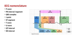
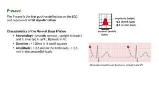
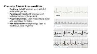
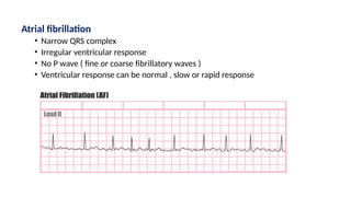
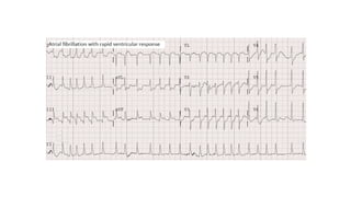
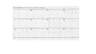
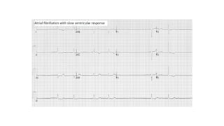
Electrocardiogram
‘ECG’
Zaid kamel Alhusaini, MD
Cardiology fellow
2023
What is ECG ?
it'sused to record the electrical
activity of the heart from different
angles to both identity and locate
pathology
Electrode are placed on different
parts of a patient’slimb and chest
ECG Electrodes vs ECG leads
ECG Electrodes is a conductive pad that is
attached to the skin to record electrical activity
ECG leads (= views) is a graphical representation
of the heart’s electrical activity
ECG leads
Limb leads
• Standard leads : I,II,III
• Augmented leads : aVR, aVF, aVL
Chest leads
V1-V2 : Septal view of the heart
V3-V4 : Anterior view of the heart
V5-V6 : Lateral view of heart
ECG paper is divided by heavy vertical and
horizontal lines into big squares , each big square
is subdivided into 5 small squares.
Calibration : Duration (Time) X Voltage(Amplitude)
Small square : 1mm X 1mm (40ms X 0.1mV)
Large square : 5mm X 5 mm ( 200ms X 0.5 mV )
• 5 large square = 1 second
• 300 large square = 60 second (1 minute )
• Before you interpret an ECG , check the
standard setting of calibration:
• Speed : 25mm/sec
• Voltage : 2 big squares = one mV = 10mm
• We can change ECG setting (speed and
voltage) according to the situation
ECG nomenclature
• P wave
• PR interval /segment
• QRS complex
• J point
• ST segment
• T wave
• U wave
• QT interval
• RR interval
P-wave
The P wave is the first positive deflection on the ECG
and represents atrial depolarization
Characteristics of the Normal Sinus P Wave
• Morphology : Smooth contour , upright in leads I
and II, inverted in aVR , Biphasic in V1
• Duration : < 120ms or 3 small squares
• Amplitude : < 2.5 mm in the limb leads , < 1.5
mm in the precordial leads
Common P Wave Abnormalities
• P mitrale (bifid P waves), seen with left
atrial enlargement
• P pulmonale (peaked P waves), seen
with right atrial enlargement
• P wave inversion, seen with ectopic atrial
and junctional rhythms
• Variable P wave morphology, seen in
multifocal atrial rhythms
PR interval
• Normal duration is 120-200 ms ( 3-5 small
square )
• Abnormally short/ long/ varying as due Heart
blocks
QRS complex
• Represent ventricular depolarization
• Duration(width): < 2.5 small square , wide if > 3 small
square
• Amplitude(height): Lead I+ Lead II+ Lead III > 15 small
square , can be normal/ low voltage/ alternans.
• Component :
• Q wave : First negative deflection
• R wave : First positive deflection , it show normal
progression from V1 to V5 , transitional zone (R = S) in V3 or
V4
• S wave : First positive after R deflection
• R’ wave : Second positive deflection
• QRS could be monophasic or biphasic or triphasic ,
not every QRS contain Q ,R,S .
Q wave
• Any negative deflection that precedes an R
wave
• Q waves in different leads
• Small Q waves are normal in most leads ,
typically seen in the lateral leads
• Deeper Q waves (>2 mm) may be seen in
leads III and aVR as a normal variant
• Normally Q waves are not seen in V1-3
• Pathological Q Waves
• Usually indicate current or prior
myocardial infarction .
• Q waves are considered pathological
if:
• > 1 mm wide
• > 2 mm deep
• > 25% of depth of QRS complex
• Seen in leads V1-3
R wave
• Normally R wave progress from V1-V5
• Normal Transition in V3-4
• Poor R Wave Progression is defined by R
wave height ≤ 3 mm in V3
QRS complex comment should include :
• Duration: Normal < 2.5 small square , wide if > 3 small square
• Amplitude: normal/ low voltage/ alternans
• Axis : Normal axis or Abnormal (LAD,RAD,NW axis )
• Q wave : Detect any pathological Q waves
• R wave: Normal or Poor R progression
ST Segment
• Isoelectric line between the end of the S wave
(J point) and the beginning of the T wave
• Displacement: elevation/ depression
• Contour: horizontal /upsloping / downsloping
T wave
• Represent Ventricular repolarization
• Upright in all leads except aVR and V1
• Amplitude
< 1 large square in limb leads
< 2 large square in chest leads
• T wave abnormalities :
Peaked T waves , Hyperacute T waves ,
Inverted T waves , Biphasic T waves ,
Flattened T waves
T wave abnormalities
• Inverted: ischemia
• Peaked: Early ischemia, hyperkalemia (↑K)
• Flat/U waves: hypokalemia (↓K)
QT Interval
• Normal QT is 350-450 msec
• QT interval is less than half the
preceding RR interval
• QT interval should be adjusted for
heart rate (QTc)
• QTc is prolonged if > 450 ms
• QTc short if < 350 ms
Rate and Rhythm
• Normal heart rate : 60 – 100 beat per min
• Rate can be Normal /Tachycardia
/Bradycardia
• Rhythm can be Regular , regularly irregular ,
irregularly irregular
• How to calculate HR ?
• If the rhythm is regular :
• 300/RR interval in big squares.
• 1500/RR interval in small squares
• If the rhythm is irregular :
• Number of QRS complexes in 30 big square x
10.
Cardiac axis
• Normally the electrical activity begin at the SAN to
both atrium then spread to AVN down to His
bundle , right bundle and left bundle to Purkinje
fiber to cause ventricular depolarization.
• Whenever the direction of the electrical activity
move toward the lead cause positive deflection ,
move away the lead cause negative deflection ,
move parapedicular to the lead make biphasic
wave.
• Cardiac axis give us an idea of the overall direction
of electrical activity .
Normal Axis = QRS axis between -30° and +90°.
Left Axis Deviation = QRS axis between -30° and -90° .
Right Axis Deviation = QRS axis between +90° and
180.
Extreme Axis Deviation = QRS axis between -90° and
180° (AKA “Northwest Axis”).
Assessment for cardiac axis :
• look for lead I,III (or aVF ) and examine the QRS
complex in each lead and determine if it is Positive or
Negative
• Another method is by find Biphasic QRS and calculate
QRS axis , axis is 90 degree to biphasic lead .
How to write an ECG report
I. Rate
II. Rhythm
III. Waves
• P wave
• PR interval
• QRS complex ( Axis , width , amplitude , waves )
• ST segment
• T wave
• QT interval
IV : Final diagnosis
ECG Report
Rate : 300/3.5 = 85 beat per minute
Rhythm: Regular, normal sinus rhythm
waves :
• P wave: Normal P wave morphology and
axis , each P wave followed by QRS.
• PR interval : Normal , constant .
• QRS complex ( Axis , width , amplitude ,
waves ) : Normal
• T wave : Normal
• ST segment : Normal , isoelectric
• QT interval : Normal
Final diagnosis : Normal ECG
Common ECG abnormality
• Tachyarrhythmias
• Bradyarrhythmias
• Bundle branch blocks
• Myocardial ischemia and infarction
• Chamber Enlargement
• Miscellaneous
Tachyarrhythmias and Bradyarrhythmias
• The key for identification of the rhythm is the
identification of the pacemaker of the heart
• To be able to reach the final diagnosis you will
need to answer three questions : rate , rhythm,
and the pacemaker
• Normal pacemaker of the heart is SA node ,in
arrhythmia the pacemaker can be atrial focus ,
AVN or ventricular
Characteristic features of different types of pacemaker
• SA Node : P wave upright in lead I,II, inverted in aVR
• Normal sinus rhythm
• Sinus tachycardia
• Sinus bradycardia
• Sinus arrhythmia
• Sinus pause
• Atrial ectopic focus : P wave is small , inverted or replaced by fibrillatory or
flutter wave or there more than three P wave morphologies
• Atrial tachycardia
• Atrial flutter
• Atrial fibrillation
• Multifocal atrial tachycardia
• Wandering atrial pacemaker
• Junctional in AVN: P wave is absent or retrograde (negative in lead II , upright
in aVR , P wave just before or inside or after QRS)
• Junctional tachycardia
• Accelerated junctional rhythm
• Escape junctional Rhythm
• Ventricular ectopic focus : wide QRS complex , T wave axis opposite to QRS axis
and AV dissociation (no constant relation between P wave and QRS)
• Ventricular tachycardia
• Accelerated Ventricular rhythm
• Escape idioventricular rhythm
• Ventricular fibrillation
Sinus Rhythms
Regular narrow complex QRS with sinus
pacemaker (P wave upright in lead I,II,
inverted in aVR )
Heart rate :
• 60-100: Normal sinus rhythm
• >100 : Sinus tachycardia
• <60: Sinus bradycardia
• Sinus arrhythmia :changing sinus
node rate with respiratory cycle lead
to irregular sinus rhythm
• Sinus pause : SAN transiently ceases
to generate the electrical impulses ,
sinus pause lasting > 2-3 seconds is
called sinus arrest
Atrial Rhythms
Narrow complex QRS with atrial pacemaker , can be regular or
irregular according to type of arrhythmia
Regular
• Non sinus P-wave : Atrial tachycardia
• Flutter wave : Atrial flutter with fixed block
Irregular
• Fibrillatory wave : Atrial fibrillation
• Flutter wave : Atrial flutter with variable block
• 3 or more different P wave :
• Multifocal atrial tachycardia (rate > 100 )
• Wandering atrial pacemaker (rate < 100)
Atrial tachycardia
• Narrow QRS complex
• Tachycardia
• Regular
• Ectopic atrial focus : Non-sinus P wave ( small, Biphasic , inverted , .. )
Atrial fibrillation
• Narrow QRS complex
• Irregular ventricular response
• No P wave ( fine or coarse fibrillatory waves )
• Ventricular response can be normal , slow or rapid response
Atrial flutter
• Narrow QRS complex
• Flutter wave ( saw teeth appearance)
• Regular ventricular response , irregular if with variable AV block
Multifocal atrial tachycardia (MAT) ,
Wandering atrial pacemaker (WAP)
• Narrow QRS complex
• Irregular
• 3 or more different P wave
• Heart rate :
• <100 : Wandering atrial pacemaker
• > 100: Multifocal atrial tachycardia
Junctional Rhythms
Regular narrow complex QRS with
Junctional pacemaker (P wave come
just before or inside or after QRS)
Heart rate :
• > 100 : Junctional tachycardia
• 60-100 : Accelerated junctional rhythm
• 40-60 : Escape junctional Rhythm
SVT is any tachyarrhythmia arising from above the level of the Bundle of His,
It is often used synonymously with AV nodal re-entry tachycardia (AVNRT)
Classification of SVT :
Atrial :
Regular Atrial
• Sinus tachycardia
• Atrial tachycardia
• Atrial flutter
Irregular Atrial
• Atrial fibrillation
• Atrial flutter (variable block)
• Multifocal atrial tachycardia
AV node
• AVRT
• AVNRT
• junctional tachycardia
ECG features of AVNRT
• Regular tachycardia (150 -
200)
• Narrow QRS complexes
• P waves is absent , if visible
exhibit retrograde
conduction may be buried
within or after or before the
QRS complex
Ventricular Rhythms
Regular wide complex QRS ( > 120 ms )
with Ventricular pacemaker
• wide QRS complex
• T wave axis opposite to QRS axis
• AV dissociation (no constant relation
between P wave and QRS)
Heart rate :
• > 100 : Ventricular tachycardia
• 40-100 : Accelerated Ventricular rhythm
• <40 : Escape idioventricular rhythm
Ventricular tachycardia types:
• Monomorphic VT and Polymorphic VT
• Sustained VT and non-sustained VT
• Torsades de pointes (TdP) is a specific form of PVT occurring in the
context of QT prolongation , it has a characteristic morphology in
which the QRS complexes “twist” around the isoelectric line.
• Ventricular Fibrillation (VF) : Chaotic irregular deflections of varying
amplitude with no identifiable P waves, QRS complexes, or T waves.
Tachyarrhythmia
Narrow complex tachycardia
Regular
• Sinus tachycardia : P wave upright in lead I,II, inverted in aVR
• Atrial tachycardia ( focal ) : P wave is small or inverted
• Atrial flutter with fixed block : P wave is replaced by flutter wave
• Supraventricular tachycardia ( AVNRT ): P wave is absent or retrograde
• Junctional tachycardia : P wave is absent or retrograde
Irregular
• Atrial fibrillation : P wave is replaced by fibrillatory wave or absent P wave
• Atrial flutter with variable block : P wave is replaced by flutter wave
• Multifocal atrial tachycardia ( MAT ) : Three or more different P wave morphology
Wide complex tachycardia
Regular
• Ventricular tachycardia ( Monomorphic ) : Wide QRS complex , QRS are same morphology.
• Any regular SVT with aberrancy or preexcitation.
Irregular
• Ventricular tachycardia ( Polymorphic ) : Wide QRS complex with different morphology
• Ventricular fibrillation
• Any irregular SVT with aberrancy or preexcitation
Premature beats
• Premature Atrial Complex (PAC)
• Premature Junctional Complex (PJC)
• Premature Ventricular Complex (PVC)
Premature Atrial Complex (PAC)
• A premature beat arising from ectopic tissue within the atria. There is an
abnormal P wave, usually followed by a normal QRS complex.
• PACs may also be conducted aberrantly or not conducted at all.
Premature Junctional Complex (PJC)
• A premature beat arising from an ectopic focus within the Atrioventricular
(AV) junction.
• Narrow QRS complex, either without a preceding P wave or with a retrograde
P wave which may appear before, during, or after the QRS complex
Premature Ventricular Complex (PVC)
A premature beat arising from an ectopic focus within the ventricles.
Broad QRS complex with abnormal morphology
• Premature : occurs earlier than would be expected.
• Discordant ST segment and T wave changes.
• Usually followed by a full compensatory pause.
• Retrograde capture of the atria may or may not occur.
Heart block ( AV block)
• First degree HB
• Second degree HB ( Mobitz 1 , Mobitz 2 )
• Third degree HB ( Complete Heart block )
First degree HB
Prolonged PR interval > 5 small square
Mobitz I block (Wenckebach phenomenon)
Progressive PR lengthening followed by drop beat due to
malfunctioning AV nodal cells
Mobitz II block
intermittent non-conducted P waves without progressive
prolongation of the PR interval usually due to failure of
conduction at the level of the His-Purkinje system (i.e. below
the AV node)
Third degree heart block ( CHB )
• Severe bradycardia due to absence of AV conduction (Complete failure impulse conduct
from atrium to ventricle
• The ECG demonstrates bradycardia with complete AV dissociation (QRS rate < P rate)
• Regular P-P interval
• Regular R-R interval
• More P waves are present than QRS complexes ( AV dissociation )
• Bradycardia
Bundle branch block
•RBBB
•LBBB
•Hemiblock
•Bifascicular block
•Trifascicular block
RBBB
• Wide QRS > 120 ms (3 square)
• rSR’ pattern or broad monophasic R wave in V1-3
with 2ry T wave inversion
• Wide, slurred S wave in the lateral leads
LBBB
• Wide QRS > 120 ms (3 square)
• Dominant S wave in V1 (with ST and T
wave opposite QRS)
• Broad monophasic R wave in lateral leads
Intraventricular conduction delay
• Wide QRS but the pattern is not consistent with RBBB or LBBB .
• Can be due to fascicular block , Cardiomyopathy , LVH , Electrolyte abnormally , drugs as TCA
or Non-specific IVCD ( not due to any of the cause )
Hemiblock
Left anterior hemiblock (LAHB ,LAFB)
• Pathology in left anterior fascicle branch of left bundle
• ECG : LAD and deep S in inferior leads
Left posterior hemiblock (LPHB ,LPFB )
• Pathology in posterior fascicle branch of left bundle
• ECG : RAD and deep S in lateral leads
Bifascicular block
RBBB + LAHB or LPHB
Trifascicular block
Bifascicular block +
First degree AV block
Myocardial ischemia and infarction
ECG changes in myocardial ischemia
• Certain ECG findings suggest myocardial ischemia without total obstruction
• ECG findings should be in at least two successive leads in the same wall
• ECG changes :
• ST depression ≥ 0.5 mm
• T wave flattening / inversion
ECG changes in myocardial infarction (STEMI)
• ECG findings should be in at least two successive leads in the same wall
• ECG changes :
• ST elevation ( injury ) : ≥ 1mm in limb lead and ≥2mm in chest lead
• T wave changes ( Peripheral ischemia ) : Hyperacute T wave , Biphasic then
inverted
• Pathological Q (Central necrosis ) :Wide and deep Q wave
• Poor R progression
• Reciprocal ST depression : Presences STD in leads other than leads
showing STE , with inferior MI look for reciprocal in lateral leads , with
anterior MI looks for reciprocal STD in inferior leads
Stages of MI
 Hyperacute MI : Hyperacute T wave +- STE ( No pathological Q wave )
 Acute MI : STE , Pathological Q may present or not
 Recent MI ( subacute , evolving ) : Pathological Q wave , STD return to baseline , Inverted T wave
 Old MI ( chronic ) : Pathological Q or Poor R progression with Normal ST , T wave (Persistent STE in
old MI if with LV aneurysm )
Chamber Enlargement
• Left atrium enlargement
• Right atrium enlargement
• Left Ventricular Hypertrophy (LVH)
• Right Ventricular Hypertrophy (RVH)
LA enlargement :
Lead II : P mitrale, broad +- notched P wave
V1 : Negative deflection of P wave > 1*1mm
RA enlargement :
Lead II: P pulmonale ,Tall and peaked P wave
V1 : Positive deflection of P wave is tall > 1 mm
Left Ventricular Hypertrophy (LVH)
Voltage Criteria
• R wave in V5 or V6 > 25 mm ( 5 large square )
• R wave in V5 or V6 plus S wave in V1 or V2 > 35
mm (7 large square )
• R wave in aVL > 11 mm
Non Voltage Criteria (Strain pattern)
• ST depression +- T wave inversion in the lateral
leads
• ST elevation in V1-3
Additional ECG changes seen in LVH
• Left axis deviation
• IVCD
Typical appearance of LVH:
• S(V1 or V2) + R(V5 or V6) > 35mm
• Strain Pattern : ST Elevation V1-4 , ST Depression / Inverted T waves V5 and V6
Right Ventricular Hypertrophy (RVH)
• Dominant R wave in V1 (> 7mm tall or R/S ratio > 1)
• Dominant S wave in V5 or V6 (> 7mm deep or R/S ratio < 1)
• QRS duration < 120ms (changes not due to RBBB)
• Right axis deviation
• RV strain pattern : STD / T wave inversion in V1-4 and inferior leads.
Typical appearance of RVH:
• Dominant R wave in V1
• Dominant S wave in V6
• Right axis deviation
• Right ventricular strain pattern with ST depression and T-wave inversion in V1-4
Miscellaneous
• WPW syndrome
• Long QT syndrome
• Pericarditis
• Pacemaker ECG
• Low Voltage ECG
• Electrical alternans
Wolff-Parkinson-White (WPW) Syndrome
Presence of a congenital accessory pathway (AP)
that can result in symptomatic and life-
threatening arrhythmias.
ECG features of WPW :
• PR interval < 120ms
• Delta wave: slurring slow rise of initial
portion of the QRS
• Wide QRS
• QT interval usually measured in lead II or V5,V6 , however the
lead with longest measurement should be used
• QT interval is less than half the preceding RR interval
• QT interval is inversely proportional to heart rate , QT interval
shortens at faster rate and lengthens at slower rates so QT
interval should be corrected for heart rate ( corrected QT
interval , QTc )
• QTc estimated QT interval at standard heart rate of 60 bpm
Long QT syndrome
• Causes of a prolonged QTc :
• Congenital long QT syndrome
• Hypokalemia
• Hypomagnesaemia
• Hypocalcemia
• Myocardial ischemia
• Drugs
• Raised intracranial pressure
• QTc prolonged if > 440 ms in men , and > 460 ms in women
• Short QT if < 350ms
Pericarditis
• Widespread concave STE and PR depression
• Stages of ECG Findings in Pericarditis
Stage I - widespread ST elevation
Stage II - resolution of ST elevation, T wave flattening
Stage III - T wave inversion
Stage IV - resolution of T wave inversion
DD : Benign early repolarization (BER)
• usually benign ECG pattern producing widespread ST segment elevation that is commonly seen in young
• It is generally thought to be a normal variant that is not indicative of underlying cardiac disease
• Ecg : Widespread concave ST elevation , Notching or slurring at the J point
Pacemaker ECG
• Spike proceeding P wave or QRS or both
Low Voltage ECG
• Normal QRS amplitude : lead I + II + III > 15 small squares
• Low voltage criteria :QRS amplitude less than 5 mm in the limb leads and/or less than 10 mm
in the precordial leads.
• Causes of low voltage : Pleural effusion , Pericardial effusion, Emphysema, Obesity,
Myxedema , Infiltrative diseases as amyloid cardiomyopathy.
Electrical alternans
• Beat to beat variability in QRS voltage
• Due to massive pericardial effusion (swinging heart)
Massive pericardial effusion produces a characteristic ECG triad of:
• Low QRS voltage
• Tachycardia
• Electrical alternans
1. The P wave represents_________.
2. The QRS complex represent_________.
3. The T wave represents__________.
4. The normal PR interval in adult is ________ ms.
5. In Normal Sinus rhythm the P wave negative in lead_____
and positive in lead _______.
6. A wide QRS with deep S wave in lead V1 and monophasic
R wave in V6 is seen in ______.
7. Tall and peaked P wave suggestive of _______.
8. Wide P wave ( >120 ms) in lead II suggest __________.
9. Normal QRS width is less than_____.
10. Widespread STE with PR depression seen in______.
11. Lateral leads are lead________.
12. A premature beat with abnormal P wave followed with a normal
QRS complex is called __________.
13. Irregular narrow QRS complex with 3 or more different P wave and
normal heart rate is called _______.
14. Beat to beat variability in QRS voltage suggest________.
15. Progressive PR lengthening followed by drop beat seen in _______.
16. Normal QRS axis is between_______.
17. A delta wave is seen in what syndrome ?
18. Name 3 causes that may prolong QT interval ?
19. Define Ventricular tachycardia ?
20. Define Atrial fibrillation ?
21. Name the LVH and RVH criteria ?
22. How to calculate heart rate in irregular rhythm ?
23. Name 3 causes of low voltage QRS complexes ?
‘Work hard in
silence ,let
success make
the noise’

ECG.pptx sdsadsadsadsadasdsadsadsadsadsad

  • 1.
  • 2.
    What is ECG? it'sused to record the electrical activity of the heart from different angles to both identity and locate pathology Electrode are placed on different parts of a patient’slimb and chest
  • 3.
    ECG Electrodes vsECG leads ECG Electrodes is a conductive pad that is attached to the skin to record electrical activity ECG leads (= views) is a graphical representation of the heart’s electrical activity
  • 4.
    ECG leads Limb leads •Standard leads : I,II,III • Augmented leads : aVR, aVF, aVL Chest leads V1-V2 : Septal view of the heart V3-V4 : Anterior view of the heart V5-V6 : Lateral view of heart
  • 7.
    ECG paper isdivided by heavy vertical and horizontal lines into big squares , each big square is subdivided into 5 small squares. Calibration : Duration (Time) X Voltage(Amplitude) Small square : 1mm X 1mm (40ms X 0.1mV) Large square : 5mm X 5 mm ( 200ms X 0.5 mV ) • 5 large square = 1 second • 300 large square = 60 second (1 minute )
  • 8.
    • Before youinterpret an ECG , check the standard setting of calibration: • Speed : 25mm/sec • Voltage : 2 big squares = one mV = 10mm • We can change ECG setting (speed and voltage) according to the situation
  • 10.
    ECG nomenclature • Pwave • PR interval /segment • QRS complex • J point • ST segment • T wave • U wave • QT interval • RR interval
  • 11.
    P-wave The P waveis the first positive deflection on the ECG and represents atrial depolarization Characteristics of the Normal Sinus P Wave • Morphology : Smooth contour , upright in leads I and II, inverted in aVR , Biphasic in V1 • Duration : < 120ms or 3 small squares • Amplitude : < 2.5 mm in the limb leads , < 1.5 mm in the precordial leads
  • 12.
    Common P WaveAbnormalities • P mitrale (bifid P waves), seen with left atrial enlargement • P pulmonale (peaked P waves), seen with right atrial enlargement • P wave inversion, seen with ectopic atrial and junctional rhythms • Variable P wave morphology, seen in multifocal atrial rhythms
  • 13.
    PR interval • Normalduration is 120-200 ms ( 3-5 small square ) • Abnormally short/ long/ varying as due Heart blocks
  • 14.
    QRS complex • Representventricular depolarization • Duration(width): < 2.5 small square , wide if > 3 small square • Amplitude(height): Lead I+ Lead II+ Lead III > 15 small square , can be normal/ low voltage/ alternans. • Component : • Q wave : First negative deflection • R wave : First positive deflection , it show normal progression from V1 to V5 , transitional zone (R = S) in V3 or V4 • S wave : First positive after R deflection • R’ wave : Second positive deflection • QRS could be monophasic or biphasic or triphasic , not every QRS contain Q ,R,S .
  • 15.
    Q wave • Anynegative deflection that precedes an R wave • Q waves in different leads • Small Q waves are normal in most leads , typically seen in the lateral leads • Deeper Q waves (>2 mm) may be seen in leads III and aVR as a normal variant • Normally Q waves are not seen in V1-3 • Pathological Q Waves • Usually indicate current or prior myocardial infarction . • Q waves are considered pathological if: • > 1 mm wide • > 2 mm deep • > 25% of depth of QRS complex • Seen in leads V1-3
  • 16.
    R wave • NormallyR wave progress from V1-V5 • Normal Transition in V3-4 • Poor R Wave Progression is defined by R wave height ≤ 3 mm in V3
  • 17.
    QRS complex commentshould include : • Duration: Normal < 2.5 small square , wide if > 3 small square • Amplitude: normal/ low voltage/ alternans • Axis : Normal axis or Abnormal (LAD,RAD,NW axis ) • Q wave : Detect any pathological Q waves • R wave: Normal or Poor R progression
  • 18.
    ST Segment • Isoelectricline between the end of the S wave (J point) and the beginning of the T wave • Displacement: elevation/ depression • Contour: horizontal /upsloping / downsloping
  • 19.
    T wave • RepresentVentricular repolarization • Upright in all leads except aVR and V1 • Amplitude < 1 large square in limb leads < 2 large square in chest leads • T wave abnormalities : Peaked T waves , Hyperacute T waves , Inverted T waves , Biphasic T waves , Flattened T waves
  • 20.
    T wave abnormalities •Inverted: ischemia • Peaked: Early ischemia, hyperkalemia (↑K) • Flat/U waves: hypokalemia (↓K)
  • 21.
    QT Interval • NormalQT is 350-450 msec • QT interval is less than half the preceding RR interval • QT interval should be adjusted for heart rate (QTc) • QTc is prolonged if > 450 ms • QTc short if < 350 ms
  • 22.
    Rate and Rhythm •Normal heart rate : 60 – 100 beat per min • Rate can be Normal /Tachycardia /Bradycardia • Rhythm can be Regular , regularly irregular , irregularly irregular • How to calculate HR ? • If the rhythm is regular : • 300/RR interval in big squares. • 1500/RR interval in small squares • If the rhythm is irregular : • Number of QRS complexes in 30 big square x 10.
  • 23.
    Cardiac axis • Normallythe electrical activity begin at the SAN to both atrium then spread to AVN down to His bundle , right bundle and left bundle to Purkinje fiber to cause ventricular depolarization. • Whenever the direction of the electrical activity move toward the lead cause positive deflection , move away the lead cause negative deflection , move parapedicular to the lead make biphasic wave. • Cardiac axis give us an idea of the overall direction of electrical activity .
  • 24.
    Normal Axis =QRS axis between -30° and +90°. Left Axis Deviation = QRS axis between -30° and -90° . Right Axis Deviation = QRS axis between +90° and 180. Extreme Axis Deviation = QRS axis between -90° and 180° (AKA “Northwest Axis”). Assessment for cardiac axis : • look for lead I,III (or aVF ) and examine the QRS complex in each lead and determine if it is Positive or Negative • Another method is by find Biphasic QRS and calculate QRS axis , axis is 90 degree to biphasic lead .
  • 30.
    How to writean ECG report I. Rate II. Rhythm III. Waves • P wave • PR interval • QRS complex ( Axis , width , amplitude , waves ) • ST segment • T wave • QT interval IV : Final diagnosis
  • 31.
    ECG Report Rate :300/3.5 = 85 beat per minute Rhythm: Regular, normal sinus rhythm waves : • P wave: Normal P wave morphology and axis , each P wave followed by QRS. • PR interval : Normal , constant . • QRS complex ( Axis , width , amplitude , waves ) : Normal • T wave : Normal • ST segment : Normal , isoelectric • QT interval : Normal Final diagnosis : Normal ECG
  • 33.
    Common ECG abnormality •Tachyarrhythmias • Bradyarrhythmias • Bundle branch blocks • Myocardial ischemia and infarction • Chamber Enlargement • Miscellaneous
  • 34.
    Tachyarrhythmias and Bradyarrhythmias •The key for identification of the rhythm is the identification of the pacemaker of the heart • To be able to reach the final diagnosis you will need to answer three questions : rate , rhythm, and the pacemaker • Normal pacemaker of the heart is SA node ,in arrhythmia the pacemaker can be atrial focus , AVN or ventricular
  • 35.
    Characteristic features ofdifferent types of pacemaker • SA Node : P wave upright in lead I,II, inverted in aVR • Normal sinus rhythm • Sinus tachycardia • Sinus bradycardia • Sinus arrhythmia • Sinus pause • Atrial ectopic focus : P wave is small , inverted or replaced by fibrillatory or flutter wave or there more than three P wave morphologies • Atrial tachycardia • Atrial flutter • Atrial fibrillation • Multifocal atrial tachycardia • Wandering atrial pacemaker
  • 36.
    • Junctional inAVN: P wave is absent or retrograde (negative in lead II , upright in aVR , P wave just before or inside or after QRS) • Junctional tachycardia • Accelerated junctional rhythm • Escape junctional Rhythm • Ventricular ectopic focus : wide QRS complex , T wave axis opposite to QRS axis and AV dissociation (no constant relation between P wave and QRS) • Ventricular tachycardia • Accelerated Ventricular rhythm • Escape idioventricular rhythm • Ventricular fibrillation
  • 37.
    Sinus Rhythms Regular narrowcomplex QRS with sinus pacemaker (P wave upright in lead I,II, inverted in aVR ) Heart rate : • 60-100: Normal sinus rhythm • >100 : Sinus tachycardia • <60: Sinus bradycardia
  • 38.
    • Sinus arrhythmia:changing sinus node rate with respiratory cycle lead to irregular sinus rhythm • Sinus pause : SAN transiently ceases to generate the electrical impulses , sinus pause lasting > 2-3 seconds is called sinus arrest
  • 39.
    Atrial Rhythms Narrow complexQRS with atrial pacemaker , can be regular or irregular according to type of arrhythmia Regular • Non sinus P-wave : Atrial tachycardia • Flutter wave : Atrial flutter with fixed block Irregular • Fibrillatory wave : Atrial fibrillation • Flutter wave : Atrial flutter with variable block • 3 or more different P wave : • Multifocal atrial tachycardia (rate > 100 ) • Wandering atrial pacemaker (rate < 100)
  • 40.
    Atrial tachycardia • NarrowQRS complex • Tachycardia • Regular • Ectopic atrial focus : Non-sinus P wave ( small, Biphasic , inverted , .. )
  • 42.
    Atrial fibrillation • NarrowQRS complex • Irregular ventricular response • No P wave ( fine or coarse fibrillatory waves ) • Ventricular response can be normal , slow or rapid response
  • 46.
    Atrial flutter • NarrowQRS complex • Flutter wave ( saw teeth appearance) • Regular ventricular response , irregular if with variable AV block
  • 49.
    Multifocal atrial tachycardia(MAT) , Wandering atrial pacemaker (WAP) • Narrow QRS complex • Irregular • 3 or more different P wave • Heart rate : • <100 : Wandering atrial pacemaker • > 100: Multifocal atrial tachycardia
  • 50.
    Junctional Rhythms Regular narrowcomplex QRS with Junctional pacemaker (P wave come just before or inside or after QRS) Heart rate : • > 100 : Junctional tachycardia • 60-100 : Accelerated junctional rhythm • 40-60 : Escape junctional Rhythm
  • 51.
    SVT is anytachyarrhythmia arising from above the level of the Bundle of His, It is often used synonymously with AV nodal re-entry tachycardia (AVNRT) Classification of SVT : Atrial : Regular Atrial • Sinus tachycardia • Atrial tachycardia • Atrial flutter Irregular Atrial • Atrial fibrillation • Atrial flutter (variable block) • Multifocal atrial tachycardia AV node • AVRT • AVNRT • junctional tachycardia
  • 53.
    ECG features ofAVNRT • Regular tachycardia (150 - 200) • Narrow QRS complexes • P waves is absent , if visible exhibit retrograde conduction may be buried within or after or before the QRS complex
  • 55.
    Ventricular Rhythms Regular widecomplex QRS ( > 120 ms ) with Ventricular pacemaker • wide QRS complex • T wave axis opposite to QRS axis • AV dissociation (no constant relation between P wave and QRS) Heart rate : • > 100 : Ventricular tachycardia • 40-100 : Accelerated Ventricular rhythm • <40 : Escape idioventricular rhythm
  • 56.
    Ventricular tachycardia types: •Monomorphic VT and Polymorphic VT • Sustained VT and non-sustained VT
  • 58.
    • Torsades depointes (TdP) is a specific form of PVT occurring in the context of QT prolongation , it has a characteristic morphology in which the QRS complexes “twist” around the isoelectric line. • Ventricular Fibrillation (VF) : Chaotic irregular deflections of varying amplitude with no identifiable P waves, QRS complexes, or T waves.
  • 60.
    Tachyarrhythmia Narrow complex tachycardia Regular •Sinus tachycardia : P wave upright in lead I,II, inverted in aVR • Atrial tachycardia ( focal ) : P wave is small or inverted • Atrial flutter with fixed block : P wave is replaced by flutter wave • Supraventricular tachycardia ( AVNRT ): P wave is absent or retrograde • Junctional tachycardia : P wave is absent or retrograde Irregular • Atrial fibrillation : P wave is replaced by fibrillatory wave or absent P wave • Atrial flutter with variable block : P wave is replaced by flutter wave • Multifocal atrial tachycardia ( MAT ) : Three or more different P wave morphology
  • 61.
    Wide complex tachycardia Regular •Ventricular tachycardia ( Monomorphic ) : Wide QRS complex , QRS are same morphology. • Any regular SVT with aberrancy or preexcitation. Irregular • Ventricular tachycardia ( Polymorphic ) : Wide QRS complex with different morphology • Ventricular fibrillation • Any irregular SVT with aberrancy or preexcitation
  • 62.
    Premature beats • PrematureAtrial Complex (PAC) • Premature Junctional Complex (PJC) • Premature Ventricular Complex (PVC)
  • 63.
    Premature Atrial Complex(PAC) • A premature beat arising from ectopic tissue within the atria. There is an abnormal P wave, usually followed by a normal QRS complex. • PACs may also be conducted aberrantly or not conducted at all.
  • 64.
    Premature Junctional Complex(PJC) • A premature beat arising from an ectopic focus within the Atrioventricular (AV) junction. • Narrow QRS complex, either without a preceding P wave or with a retrograde P wave which may appear before, during, or after the QRS complex
  • 65.
    Premature Ventricular Complex(PVC) A premature beat arising from an ectopic focus within the ventricles. Broad QRS complex with abnormal morphology • Premature : occurs earlier than would be expected. • Discordant ST segment and T wave changes. • Usually followed by a full compensatory pause. • Retrograde capture of the atria may or may not occur.
  • 66.
    Heart block (AV block) • First degree HB • Second degree HB ( Mobitz 1 , Mobitz 2 ) • Third degree HB ( Complete Heart block )
  • 67.
    First degree HB ProlongedPR interval > 5 small square
  • 69.
    Mobitz I block(Wenckebach phenomenon) Progressive PR lengthening followed by drop beat due to malfunctioning AV nodal cells
  • 70.
    Mobitz II block intermittentnon-conducted P waves without progressive prolongation of the PR interval usually due to failure of conduction at the level of the His-Purkinje system (i.e. below the AV node)
  • 71.
    Third degree heartblock ( CHB ) • Severe bradycardia due to absence of AV conduction (Complete failure impulse conduct from atrium to ventricle • The ECG demonstrates bradycardia with complete AV dissociation (QRS rate < P rate) • Regular P-P interval • Regular R-R interval • More P waves are present than QRS complexes ( AV dissociation ) • Bradycardia
  • 72.
  • 73.
    RBBB • Wide QRS> 120 ms (3 square) • rSR’ pattern or broad monophasic R wave in V1-3 with 2ry T wave inversion • Wide, slurred S wave in the lateral leads
  • 75.
    LBBB • Wide QRS> 120 ms (3 square) • Dominant S wave in V1 (with ST and T wave opposite QRS) • Broad monophasic R wave in lateral leads
  • 78.
    Intraventricular conduction delay •Wide QRS but the pattern is not consistent with RBBB or LBBB . • Can be due to fascicular block , Cardiomyopathy , LVH , Electrolyte abnormally , drugs as TCA or Non-specific IVCD ( not due to any of the cause )
  • 79.
    Hemiblock Left anterior hemiblock(LAHB ,LAFB) • Pathology in left anterior fascicle branch of left bundle • ECG : LAD and deep S in inferior leads Left posterior hemiblock (LPHB ,LPFB ) • Pathology in posterior fascicle branch of left bundle • ECG : RAD and deep S in lateral leads
  • 82.
    Bifascicular block RBBB +LAHB or LPHB Trifascicular block Bifascicular block + First degree AV block
  • 85.
  • 86.
    ECG changes inmyocardial ischemia • Certain ECG findings suggest myocardial ischemia without total obstruction • ECG findings should be in at least two successive leads in the same wall • ECG changes : • ST depression ≥ 0.5 mm • T wave flattening / inversion
  • 87.
    ECG changes inmyocardial infarction (STEMI) • ECG findings should be in at least two successive leads in the same wall • ECG changes : • ST elevation ( injury ) : ≥ 1mm in limb lead and ≥2mm in chest lead • T wave changes ( Peripheral ischemia ) : Hyperacute T wave , Biphasic then inverted • Pathological Q (Central necrosis ) :Wide and deep Q wave • Poor R progression • Reciprocal ST depression : Presences STD in leads other than leads showing STE , with inferior MI look for reciprocal in lateral leads , with anterior MI looks for reciprocal STD in inferior leads
  • 88.
    Stages of MI Hyperacute MI : Hyperacute T wave +- STE ( No pathological Q wave )  Acute MI : STE , Pathological Q may present or not  Recent MI ( subacute , evolving ) : Pathological Q wave , STD return to baseline , Inverted T wave  Old MI ( chronic ) : Pathological Q or Poor R progression with Normal ST , T wave (Persistent STE in old MI if with LV aneurysm )
  • 93.
    Chamber Enlargement • Leftatrium enlargement • Right atrium enlargement • Left Ventricular Hypertrophy (LVH) • Right Ventricular Hypertrophy (RVH)
  • 94.
    LA enlargement : LeadII : P mitrale, broad +- notched P wave V1 : Negative deflection of P wave > 1*1mm RA enlargement : Lead II: P pulmonale ,Tall and peaked P wave V1 : Positive deflection of P wave is tall > 1 mm
  • 95.
    Left Ventricular Hypertrophy(LVH) Voltage Criteria • R wave in V5 or V6 > 25 mm ( 5 large square ) • R wave in V5 or V6 plus S wave in V1 or V2 > 35 mm (7 large square ) • R wave in aVL > 11 mm Non Voltage Criteria (Strain pattern) • ST depression +- T wave inversion in the lateral leads • ST elevation in V1-3 Additional ECG changes seen in LVH • Left axis deviation • IVCD
  • 96.
    Typical appearance ofLVH: • S(V1 or V2) + R(V5 or V6) > 35mm • Strain Pattern : ST Elevation V1-4 , ST Depression / Inverted T waves V5 and V6
  • 97.
    Right Ventricular Hypertrophy(RVH) • Dominant R wave in V1 (> 7mm tall or R/S ratio > 1) • Dominant S wave in V5 or V6 (> 7mm deep or R/S ratio < 1) • QRS duration < 120ms (changes not due to RBBB) • Right axis deviation • RV strain pattern : STD / T wave inversion in V1-4 and inferior leads.
  • 98.
    Typical appearance ofRVH: • Dominant R wave in V1 • Dominant S wave in V6 • Right axis deviation • Right ventricular strain pattern with ST depression and T-wave inversion in V1-4
  • 100.
    Miscellaneous • WPW syndrome •Long QT syndrome • Pericarditis • Pacemaker ECG • Low Voltage ECG • Electrical alternans
  • 101.
    Wolff-Parkinson-White (WPW) Syndrome Presenceof a congenital accessory pathway (AP) that can result in symptomatic and life- threatening arrhythmias. ECG features of WPW : • PR interval < 120ms • Delta wave: slurring slow rise of initial portion of the QRS • Wide QRS
  • 103.
    • QT intervalusually measured in lead II or V5,V6 , however the lead with longest measurement should be used • QT interval is less than half the preceding RR interval • QT interval is inversely proportional to heart rate , QT interval shortens at faster rate and lengthens at slower rates so QT interval should be corrected for heart rate ( corrected QT interval , QTc ) • QTc estimated QT interval at standard heart rate of 60 bpm Long QT syndrome
  • 104.
    • Causes ofa prolonged QTc : • Congenital long QT syndrome • Hypokalemia • Hypomagnesaemia • Hypocalcemia • Myocardial ischemia • Drugs • Raised intracranial pressure • QTc prolonged if > 440 ms in men , and > 460 ms in women • Short QT if < 350ms
  • 106.
    Pericarditis • Widespread concaveSTE and PR depression • Stages of ECG Findings in Pericarditis Stage I - widespread ST elevation Stage II - resolution of ST elevation, T wave flattening Stage III - T wave inversion Stage IV - resolution of T wave inversion
  • 108.
    DD : Benignearly repolarization (BER) • usually benign ECG pattern producing widespread ST segment elevation that is commonly seen in young • It is generally thought to be a normal variant that is not indicative of underlying cardiac disease • Ecg : Widespread concave ST elevation , Notching or slurring at the J point
  • 109.
    Pacemaker ECG • Spikeproceeding P wave or QRS or both
  • 111.
    Low Voltage ECG •Normal QRS amplitude : lead I + II + III > 15 small squares • Low voltage criteria :QRS amplitude less than 5 mm in the limb leads and/or less than 10 mm in the precordial leads. • Causes of low voltage : Pleural effusion , Pericardial effusion, Emphysema, Obesity, Myxedema , Infiltrative diseases as amyloid cardiomyopathy.
  • 112.
    Electrical alternans • Beatto beat variability in QRS voltage • Due to massive pericardial effusion (swinging heart)
  • 113.
    Massive pericardial effusionproduces a characteristic ECG triad of: • Low QRS voltage • Tachycardia • Electrical alternans
  • 116.
    1. The Pwave represents_________. 2. The QRS complex represent_________. 3. The T wave represents__________. 4. The normal PR interval in adult is ________ ms. 5. In Normal Sinus rhythm the P wave negative in lead_____ and positive in lead _______. 6. A wide QRS with deep S wave in lead V1 and monophasic R wave in V6 is seen in ______. 7. Tall and peaked P wave suggestive of _______. 8. Wide P wave ( >120 ms) in lead II suggest __________. 9. Normal QRS width is less than_____.
  • 117.
    10. Widespread STEwith PR depression seen in______. 11. Lateral leads are lead________. 12. A premature beat with abnormal P wave followed with a normal QRS complex is called __________. 13. Irregular narrow QRS complex with 3 or more different P wave and normal heart rate is called _______. 14. Beat to beat variability in QRS voltage suggest________. 15. Progressive PR lengthening followed by drop beat seen in _______. 16. Normal QRS axis is between_______.
  • 118.
    17. A deltawave is seen in what syndrome ? 18. Name 3 causes that may prolong QT interval ? 19. Define Ventricular tachycardia ? 20. Define Atrial fibrillation ? 21. Name the LVH and RVH criteria ? 22. How to calculate heart rate in irregular rhythm ? 23. Name 3 causes of low voltage QRS complexes ?
  • 131.
    ‘Work hard in silence,let success make the noise’