Download to read offline
![Efficacy of use of a-Si EPID as imaging device in IMRT QA
DOI: 10.9790/4861-07132743 www.iosrjournals.org 28 | Page
W. (2006) developed a method for rapid evaluation of IMRT plans, using portal images for reconstruction of the
dose delivered to a virtual 3D phantom.
II. Materials And Methods
Elekta Precise (Crawley, UK) linear accelerator, which support IMRT deliver, was used in this study
(figure 2.1a). This linear accelerator (LINAC) produces photon energies of 6 and 15MV, also it produces
electrons with energies of 6, 8, 10, 12 and 15 Mev. Only 6 MV photon beam is used in this study. The LINAC is
equipped with MLC. The MLC consists of 40 leaf pairs with 1 cm leaf width at the isocenter.The type of EPID
used throughout this study is an amorphous silicon flat panel-type imager (Elekta iViewGT, release 3.2).The
detector panel is a PerkinElmer Amorphous Silicon (a-Si) detector, and provides a resolution of 1024 × 1024
16-bit pixel images, with a detector panel size of 41 × 41 cm2
(approximately 26 × 26 cm2
at isocenter).Display
pixel factor information;When images are acquired by iViewGTTM
, the pixel values are re-normalized or
averaged before the image data is saved to the database. At the time of acquisition, the pixel scaling factor is
saved with the images in the database. This feature allows us to determine the original accumulated pixel value
by dividing the pixel value by the pixel scaling factor.Polymethyl methacrylate (PMMA), also known as
acrylic. Trade names are Lucite, Plexiglas or Perspex. It consists of 1 cm for each sheet of density 1.19 g/cm3
and area 30 х 30 cm2
.GAFCHROMIC® EBT2 Dosimetry Film (8x10 in size) was used. The film spatial
resolution >5,000 dpi (dots per inch) with dose range 1cGy – 10Gy (in red color channel).Omni ProTM
IMRT is
software used for complete dosimetric verification and QA of IMRT treatment cycle. This software function is
to import and compare calculated doses fromTPS planned data with measured dose distributions from films that
were exposed in the IMRT phantom. It includes 1D profile, 2D isodose profiles as well as fully automated
comparisons using analysis tools such as Gamma index method.A computerized welhÖfer WP 700 water
phantom version 3.5 was used in the present study for acquiring the beam data.XiO treatment planning system
(TPS) (from CMS Inc., USA) version 4.2.2 implemented superposition algorithm is used in this study.
Calibration of the EPID Several steps are necessary to reconstruct the dose in the phantom or patient from the
pixel values of the EPID. All measurements of this study were performed at gantry and collimator angle 0°.
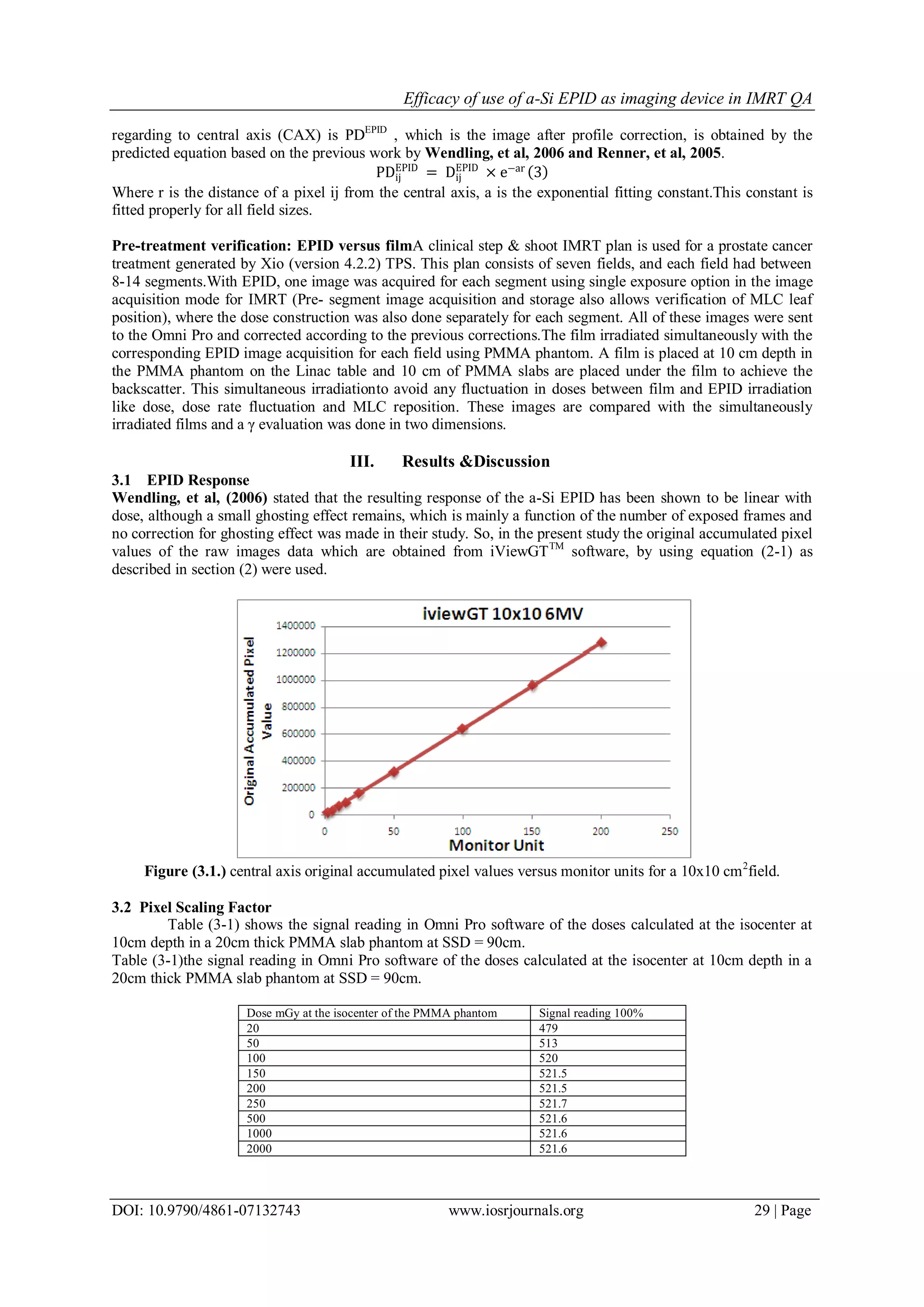
A- Calibration of EPID response to different doses:EPID images of reference field size (typically 10x10
cm2
) were recorded for different MU (2, 5, 10, 15, 25, 50, 100, 150 and 200 MU).The acquired images by
iViewGTTM
are re-normalized (automatically by the iViewGTTM
) before saving process; which means that
all images will appear with the same optical density (OD) for all different doses images. Sofor each image
the pixel scaling factor (PSF) is recorded to determine the original accumulated pixel value by dividing the
pixel value by this factor
Originalaccumulatedpixelvalue=
Pixelvalue
PixelScalingFactor (PSF )
(1)
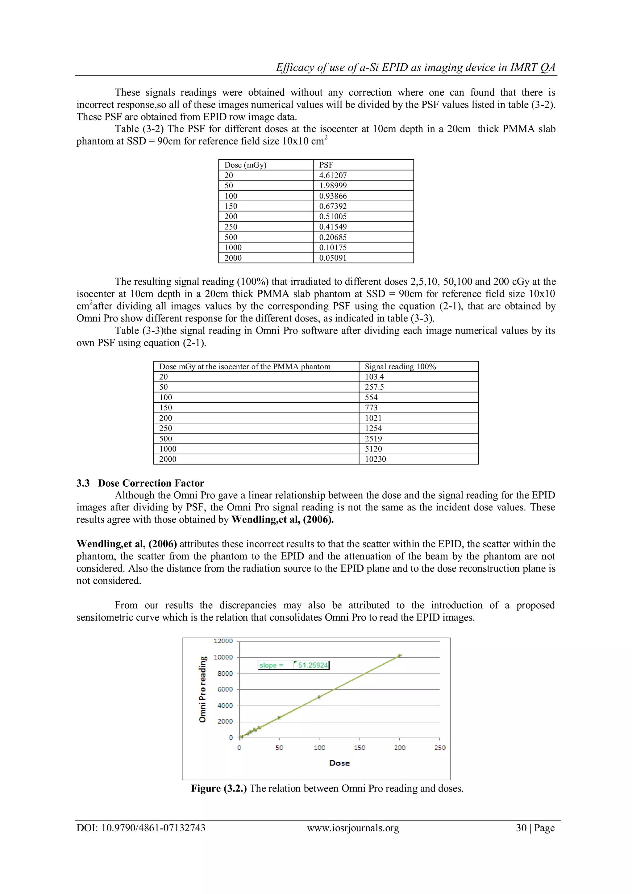
To convert these images into doses, all images were exported to Omni Pro system.The Omni Pro
software can read the Optical Density OD of the image.Each EPID image imported to Omni Pro will be
corrected for its own PSF.even after dividing each image by its corresponding PSF it will still incorrect because
of several reasons:The scattered radiation from EPID;The scatter within the phantom;The scatter from the
phantom to the EPID;The attenuation of the beam by the phantom; andThe distance from the radiation source to
the EPID plane and to the dose reconstruction plane. (Wendling M., et al, 2006).Record the Omni Pro reading
value for each EPID dose, and get the Dose correction factor (DF), which is the Omni Pro reading value for a
specific dose divided by the Actual irradiated dose at isocenter
DF =
OmniProreadingvalueforaspecificdose
Actualirradiateddose
(2)
B- Calibration of the EPID profile
In order to determine the necessary parameters for correcting the dose profile reconstruction, i.e.,
relating pixel values in the EPID images with absolute dose values in the phantom, the EPID images at different
square field sizes are recorded with PMMA slab phantom of thickness 20 cm(an average thickness to encompass
the phantom (or patients) thickness at 6 MV photon beam). For all phantom measurements an isocentric setup
[SAD (source –axis distance) setup] was used. Dose profiles for the same field sizeswere obtained using
semiflex 0.147 ion chamber in the full-scatter water phantom [Source-Skin Distance (SSD) = 88.6cm] were
measured with ion chamber at 11.4cm depth.Where 11.4 cm water is equivalent to 10 cm of Perspex. These dose
profiles will be the reference for EPID dose profile correction. The EPID images were taken and corrected by
PSF & DF. After the conversion of EPID pixel values into dose values according to the dose response relation,
the resulting image is called dose image DEPID
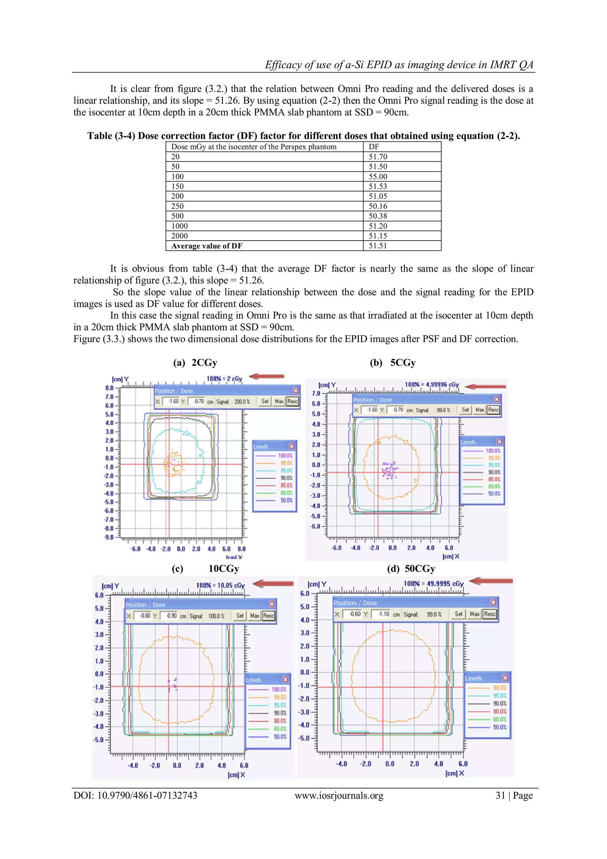
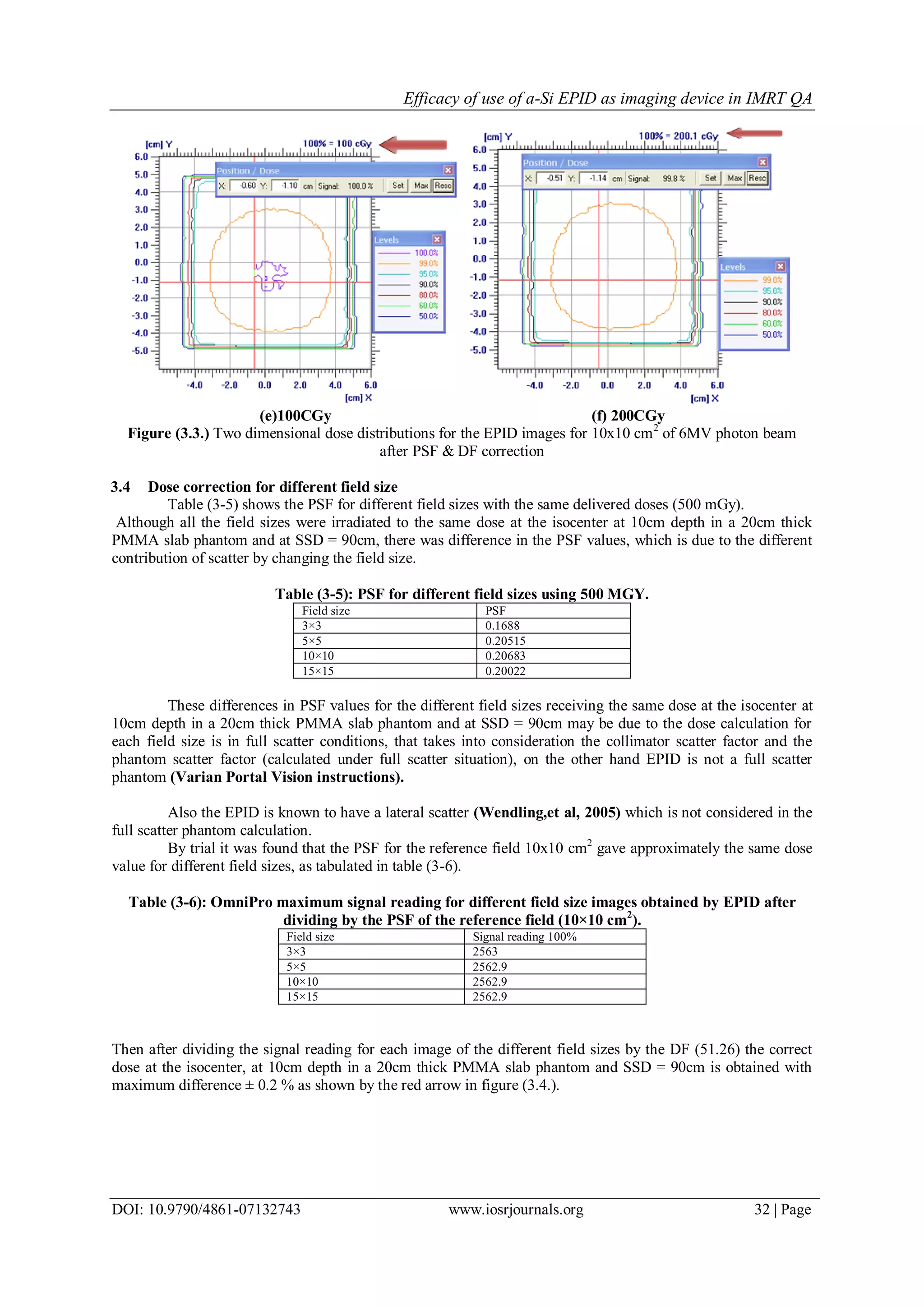
. The corrected portal pixel dose according to its position](https://image.slidesharecdn.com/g07132743-151121105604-lva1-app6891/75/Efficacy-of-Use-of-A-Si-EPID-as-Imaging-Device-in-IMRT-QA-2-2048.jpg)
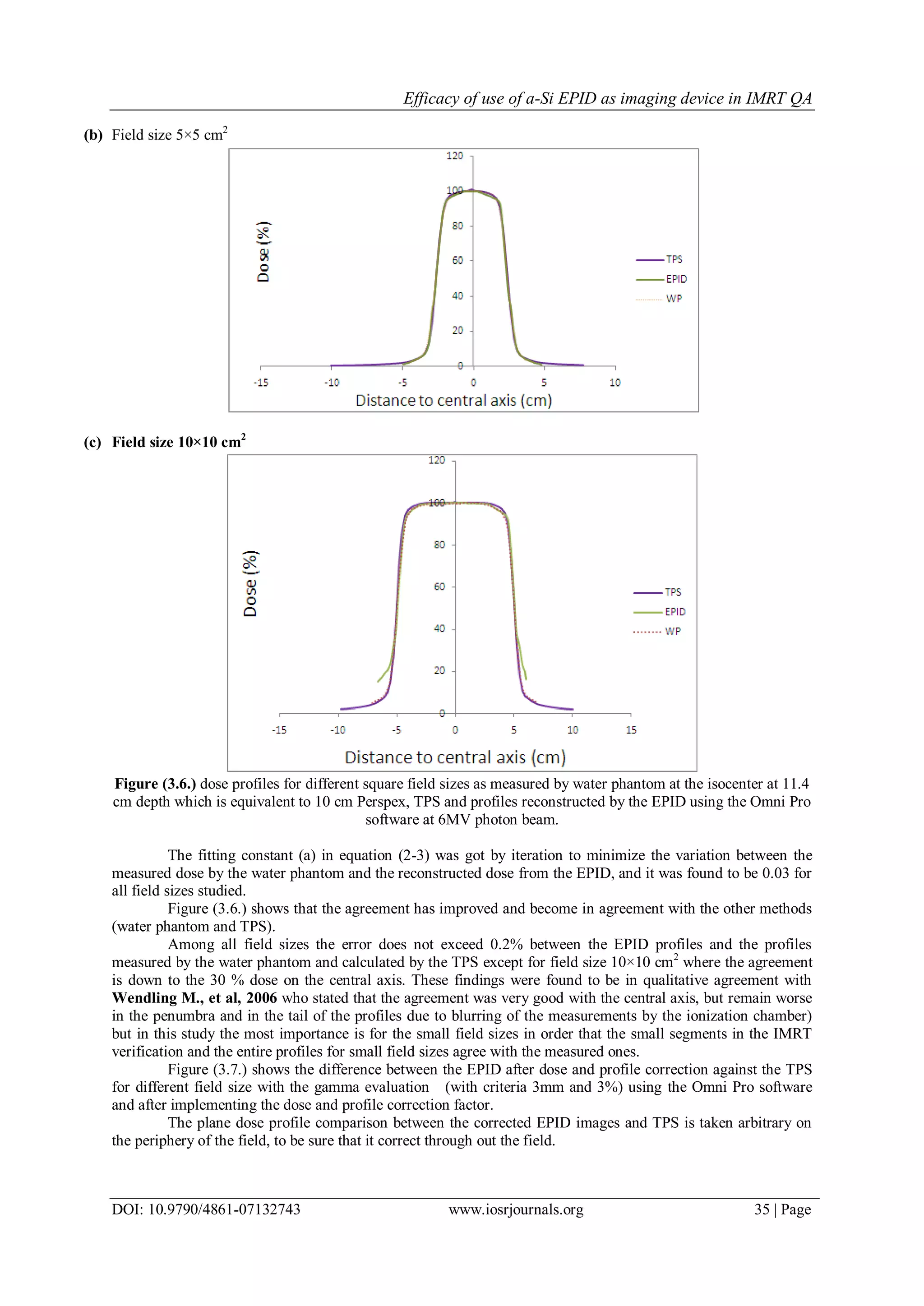
![Efficacy of use of a-Si EPID as imaging device in IMRT QA
DOI: 10.9790/4861-07132743 www.iosrjournals.org 34 | Page
(c) Field size 10×10 cm2
Figure (3.5.) dose profile as measured by water phantom and from TPS against the reconstructed dose
profile from the EPID images without profile correction (a) 3×3 cm2 field size. (b) 5×5 cm2field size. (c)
10×10 cm2 field size. All profiles are normalized to 100% of the measured dose.
Figure (3.5.) shows a difference between water phantom measurements and the reconstructed profiles from the
EPID images for the studied field sizes.
Table (3-7) show the maximum percent of error between profiles measured and t
he EPID at different field sizes.
Table (3-7) (% error) between EPID and the water phantom dose profiles.
Field size(cm2
) Error (%)
3×3 3%
5×5 3.5%
10×10 3%
It is clear from figure (3.5.) and table (3-7); that the profile behaviour along the central axis region by
the three methods is good, and became worse in the penumbra region and the tails. This is because within the
EPID mainly lateral X-ray scatters and optical photon scatter occurs, resulting in more flattened at the shoulders
more than in water phantom &TPS. These findings are in agreement with the results of Wendling, et al, 2006.
The fit parameter [a] in equation (2-3) correct for these behaviour where it is obtained from the water phantom
measurements.
Figure (3.6.) shows the dose profiles of square fields for different sizes obtained by EPID after correction using
equation (2-3) and water phantom and TPS.
(a) Field size 3×3 cm2](https://image.slidesharecdn.com/g07132743-151121105604-lva1-app6891/75/Efficacy-of-Use-of-A-Si-EPID-as-Imaging-Device-in-IMRT-QA-8-2048.jpg)
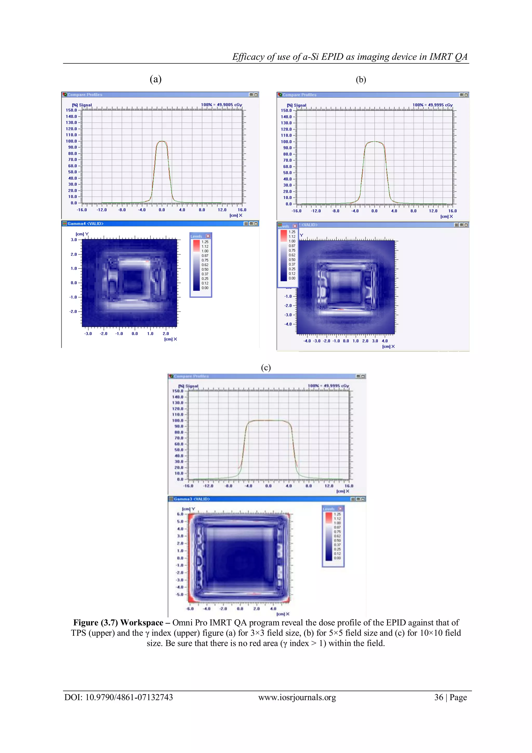


The document summarizes research into using an amorphous silicon electronic portal imaging device (EPID) for intensity modulated radiation therapy (IMRT) quality assurance. It describes calibrating the EPID to correctly relate pixel values to dose. Measurements were made with the EPID and with film in a phantom to verify that the EPID provides accurate dose distributions for an IMRT plan compared to the treatment planning system and film measurements. The study shows the EPID can accurately verify IMRT field doses in a homogeneous phantom and replace film for pretreatment dose verification when used with the appropriate calibration and correction procedures.


![[IJCT-V3I2P37] Authors: Amritpal Singh, Prithvipal Singh](https://cdn.slidesharecdn.com/ss_thumbnails/ijct-v3i2p37-160609073616-thumbnail.jpg?width=640&height=640&fit=bounds)