Downloaded 13 times
![International Research Journal of Engineering and Technology (IRJET) e-ISSN: 2395-0056
Volume: 04 Issue: 12 | Dec-2017 www.irjet.net p-ISSN: 2395-0072
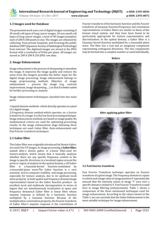
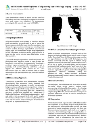
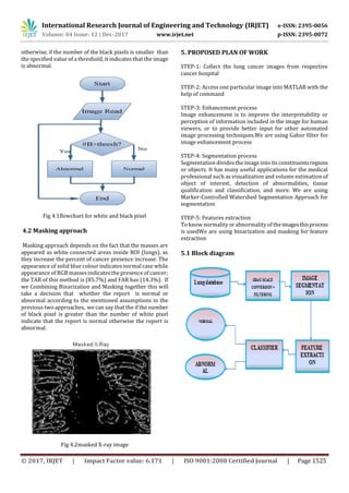
© 2017, IRJET | Impact Factor value: 6.171 | ISO 9001:2008 Certified Journal | Page 1526
Fig 5.1result for detected image
Fig 5.2 result for normal image
6. Advantages
(a) Early detection of cancer greatly increases the chances
for successful treatment.
(b) With the use of this treatment is often simpler and more
likely to be effective.
(c)The proposed systems are more efficient and give the
better result.
(d)Provides better image quality and accuracy
7. Applications
[1]It is widely used in many medical areasforearlydetection
of cancer .so the the proper treatment will beprovidedtothe
patient.
[1] Febr Mokhled S. AL-TARAWNEH,“LungCancerDetection
Using Image Processing Techniques”, Leonardo Electronic
Journal of Practices and Technologies, June 2012.
[2] Muhammad Usman, Muhammad Shoaib and Mohamad
Rahal, “Lung Cancer Detection Using Digital Image
Processing”, PIERS Proceedings, Stockholm, Sweden, Aug.
12-15, 2013.
[3] Anita Chaudhary and Sonit Sukhraj Singh, “Multi-
resolution Analysis Technique for Lung Cancer Detection in
Computed Tomographic Images”, International Journal of
Research in Engineering & Applied Sciences, IJREAS Volume
2, Issue 2 January 2012
[2]This image processing technique can also be used to
detect other cancer such as breast cancer and tumor in
our body part.
8.Conclusion
An image processing technique is built to detect diseases at
early stage of cancer so the patient can take the treatment at
early stages.The time factor is major factor to discover the
abnormal tissue in target x-ray images.The accuracy andthe
quality of image is one the major core factor of this
research.Image quality as well as image enhancement stage
were adopted as low pre-processing techniques based on
gabor filter.This technique is efficient forsegmentationstage
so the region of interest for feature extraction obtaining.on
the basis of general features a normality and abnormality
comparison is made.the main feature for detection of
accurate image comparison are pixel percentage and mask
labeling which gives us the indication that the process of
detection this disease plays a very important and essential
role to avoid serious stages and to reduce its percentage
distribution in the world. To obtain moreaccurateresultswe
three stages: Image Enhancementstage,ImageSegmentation
stage and Features Extraction stage.
ACKNOWLEDGEMENT
The success of any work depends on efforts of many
individuals. We would like to take this opportunity to
express our deep gratitude to those who extended their
support and have guided us to complete this project work.
I liketo thank Prof.Mohd.Nasiruddin (HOD)forprovidingus
the necessary information about topic. I would again like to
thank Prof.Dr. Sajid Anwar, Principal of the College, for
providing us the necessary help and facilities we needed.
I express my thanks to all the staff members of Electronics&
communication Engineering department who have directly
or indirectly extended their kind co-operation in the
completion of my reaserch paper.
REFERENCES](https://image.slidesharecdn.com/irjet-v4i12278-180118083937/85/Lung-Cancer-Detection-using-Image-Processing-Techniques-5-320.jpg)

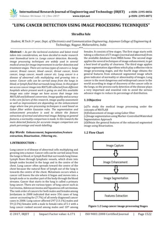
This document presents a technique for detecting lung cancer in x-ray images using image processing. It involves enhancing images using Gabor filtering, segmenting images using marker-controlled watershed segmentation, and extracting features using binarization and masking. The key steps are collecting lung x-ray images, enhancing quality using Gabor filtering, segmenting regions of interest using watershed segmentation, extracting pixel counts and mask features, and classifying images as normal or abnormal based on these features. The goal is to enable early detection of lung cancer through automated analysis of medical images.