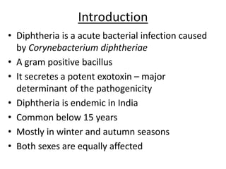
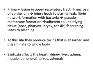
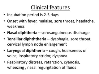
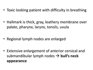
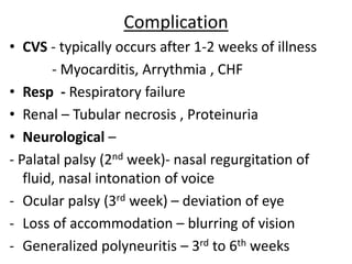
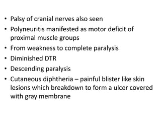
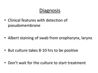
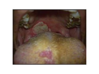
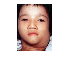
DIPHTHERIA
Introduction
• Diphtheria is a acute bacterial infection caused
by Corynebacterium diphtheriae
• A gram positive bacillus
• It secretes a potent exotoxin – major
determinant of the pathogenicity
• Diphtheria is endemic in India
• Common below 15 years
• Mostly in winter and autumn seasons
• Both sexes are equally affected
Pathogenesis
• Spreads through droplet infection during
coughing, sneezing , talking
• Once infected  remains infected till virulent
bacilli are present in lesions, usually 2-4 weeks
• Diphtheria is a rapidly developing acute febrile
illness with both local and systemic pathology
• Primary lesion in upper respiratory tract  necrosis
of epithelium  injury leads to plasma leak, fibrin
network formation with bacteria  pseudo-
membrane formation adherent to underlying
tissue (nose, pharynx, larynx, tonsils) scraping
leads to bleeding
• At this site they produce toxins that is absorbed and
disseminate to whole body
• Exotoxin affects the heart, kidney, liver, spleen,
muscle, peripheral nerves, adrenals
Clinical features
• Incubation period is 2-5 days
• Onset with fever, malaise, sore throat, headache,
weakness
• Nasal diphtheria – serosanguineous discharge
• Tonsillar diphththeria – dysphagia, sore throat,
cervical lymph node enlargement
• Laryngeal diphtheria – cough, hoarseness of
voice, inspiratory stridor, dyspnea
• Respiratory distress, retarction, cyanosis,
wheezing , nasal regurgitation of fluids
• Toxic looking patient with difficulty in breathing
• Hallmark is thick, gray, leathery membrane over
palate, pharynx, larynx, tonsils, uvula
• Regional lymph nodes are enlarged
• Extensive enlargement of anterior cervical and
submandibular lymph nodes  bull’s neck
appearance
Complication
• CVS - typically occurs after 1-2 weeks of illness
- Myocarditis, Arrythmia , CHF
• Resp - Respiratory failure
• Renal – Tubular necrosis , Proteinuria
• Neurological –
- Palatal palsy (2nd week)- nasal regurgitation of
fluid, nasal intonation of voice
- Ocular palsy (3rd week) – deviation of eye
- Loss of accommodation – blurring of vision
- Generalized polyneuritis – 3rd to 6th weeks
• Palsy of cranial nerves also seen
• Polyneuritis manifested as motor deficit of
proximal muscle groups
• From weakness to complete paralysis
• Diminished DTR
• Descending paralysis
• Cutaneous diphtheria – painful blister like skin
lesions which breakdown to form a ulcer covered
with gray membrane
Diagnosis
• Clinical features with detection of
pseudomembrane
• Albert staining of swab from oropharynx, larynx
• But culture takes 8-10 hrs to be positive
• Don’t wait for the culture to start treatment
Management
• In any suspected diphtheria immediately start
treatment with antitoxin and antibiotics
• Antitoxin is the mainstay of treatment to
neutralize the circulating toxin that is not bound
to tissue
• Antibiotics used to eradicate the organism and to
prevent spread
• Supportive treatment
• Mechanical ventilation if needed
• Airway obstruction and myocarditis are the main
cause of death
• Dose of antitoxin depends on the site and extent of
disease
• Antibiotics – Penicillin / Erythromycin
• Treatment of carriers
Prevention
• Isolation of patients – till two cultures from nose
and throat are negative
• Care of contacts – kept under observation for at
least 7 days. No role of prophylactic antitoxins
• Active immunization
• Repalce TT with Td
PERTUSIS
Introduction
• Pertusis or whooping cough is caused by a gram
negative coccobacillus Bordetella pertusis
• Highly communicable disease
• It is a disease of infants and young children
• Females are more predisposed than males
• More in malnourished patients, overcrowding,
low socioeconomic status groups
• Common in winter and spring
• Disease is characterized by paroxysmal bouts of
cough ending with a whoop lasting for months
• Intense spasmodic cough
• Spreads through droplet infection via respiratory
secretions
• Highly contagious disease with secondary attack
rate of 100%
Clinical features
• Incubation period 7-14 days
• Natural course of disease is in 3 phases
• Catarrhal phase  cough, cold, running nose,
mild fever lasting for 10 days
• Paroxysmal phase  bouts of cough ending
with a whoop, face is congested, vomiting
- Classical whoop is the rescue inspiration at the
end of coughing
- Lasts for 2 weeks
• Convalescent phase  if no complication then the
disease progresses to this phase
• Relapse of cough is common
• So called as cough of 100 days
• Cough is exhaustive and scaring
• Can lead to apnea, respiratory difficulty, cyanosis
Complications
• Subconjunctival hemorrhage
• Nose bleeds
• Hemoptysis
• Intracranial hemorrhage is rare
• Development of hernias
• Pneumothorax
• Secondary infections
• CNS complications due to hypoxia/toxins can
lead to seizures and encephalopathy
Management
• Diagnosis is through typical history and swab
culture from nasopharynx
• Treated with antibiotics like Erythromycin/
Azithromycin/ Clarithromycin/Trimethoprim-
sulfamethoxazole for 2 weeks
• Supportive treatment – maintain hydration,
nutrition, hygiene
- Avoid provoking paroxysms of cough
- Comfort during paroxysms of cough
• Clearing airway during cough to prevent
aspiration
• Early treatment of complications
• Treatment of contacts with antibiotic
Erythromycin for 2 weeks
• Active immunization
Thank u

Diptheria

  • 1.
  • 2.
    Introduction • Diphtheria isa acute bacterial infection caused by Corynebacterium diphtheriae • A gram positive bacillus • It secretes a potent exotoxin – major determinant of the pathogenicity • Diphtheria is endemic in India • Common below 15 years • Mostly in winter and autumn seasons • Both sexes are equally affected
  • 3.
    Pathogenesis • Spreads throughdroplet infection during coughing, sneezing , talking • Once infected  remains infected till virulent bacilli are present in lesions, usually 2-4 weeks • Diphtheria is a rapidly developing acute febrile illness with both local and systemic pathology
  • 4.
    • Primary lesionin upper respiratory tract  necrosis of epithelium  injury leads to plasma leak, fibrin network formation with bacteria  pseudo- membrane formation adherent to underlying tissue (nose, pharynx, larynx, tonsils) scraping leads to bleeding • At this site they produce toxins that is absorbed and disseminate to whole body • Exotoxin affects the heart, kidney, liver, spleen, muscle, peripheral nerves, adrenals
  • 5.
    Clinical features • Incubationperiod is 2-5 days • Onset with fever, malaise, sore throat, headache, weakness • Nasal diphtheria – serosanguineous discharge • Tonsillar diphththeria – dysphagia, sore throat, cervical lymph node enlargement • Laryngeal diphtheria – cough, hoarseness of voice, inspiratory stridor, dyspnea • Respiratory distress, retarction, cyanosis, wheezing , nasal regurgitation of fluids
  • 6.
    • Toxic lookingpatient with difficulty in breathing • Hallmark is thick, gray, leathery membrane over palate, pharynx, larynx, tonsils, uvula • Regional lymph nodes are enlarged • Extensive enlargement of anterior cervical and submandibular lymph nodes  bull’s neck appearance
  • 7.
    Complication • CVS -typically occurs after 1-2 weeks of illness - Myocarditis, Arrythmia , CHF • Resp - Respiratory failure • Renal – Tubular necrosis , Proteinuria • Neurological – - Palatal palsy (2nd week)- nasal regurgitation of fluid, nasal intonation of voice - Ocular palsy (3rd week) – deviation of eye - Loss of accommodation – blurring of vision - Generalized polyneuritis – 3rd to 6th weeks
  • 8.
    • Palsy ofcranial nerves also seen • Polyneuritis manifested as motor deficit of proximal muscle groups • From weakness to complete paralysis • Diminished DTR • Descending paralysis • Cutaneous diphtheria – painful blister like skin lesions which breakdown to form a ulcer covered with gray membrane
  • 9.
    Diagnosis • Clinical featureswith detection of pseudomembrane • Albert staining of swab from oropharynx, larynx • But culture takes 8-10 hrs to be positive • Don’t wait for the culture to start treatment
  • 12.
    Management • In anysuspected diphtheria immediately start treatment with antitoxin and antibiotics • Antitoxin is the mainstay of treatment to neutralize the circulating toxin that is not bound to tissue • Antibiotics used to eradicate the organism and to prevent spread • Supportive treatment
  • 13.
    • Mechanical ventilationif needed • Airway obstruction and myocarditis are the main cause of death • Dose of antitoxin depends on the site and extent of disease • Antibiotics – Penicillin / Erythromycin • Treatment of carriers
  • 14.
    Prevention • Isolation ofpatients – till two cultures from nose and throat are negative • Care of contacts – kept under observation for at least 7 days. No role of prophylactic antitoxins • Active immunization • Repalce TT with Td
  • 15.
  • 16.
    Introduction • Pertusis orwhooping cough is caused by a gram negative coccobacillus Bordetella pertusis • Highly communicable disease • It is a disease of infants and young children • Females are more predisposed than males • More in malnourished patients, overcrowding, low socioeconomic status groups • Common in winter and spring
  • 17.
    • Disease ischaracterized by paroxysmal bouts of cough ending with a whoop lasting for months • Intense spasmodic cough • Spreads through droplet infection via respiratory secretions • Highly contagious disease with secondary attack rate of 100%
  • 18.
    Clinical features • Incubationperiod 7-14 days • Natural course of disease is in 3 phases • Catarrhal phase  cough, cold, running nose, mild fever lasting for 10 days • Paroxysmal phase  bouts of cough ending with a whoop, face is congested, vomiting - Classical whoop is the rescue inspiration at the end of coughing - Lasts for 2 weeks
  • 19.
    • Convalescent phase if no complication then the disease progresses to this phase • Relapse of cough is common • So called as cough of 100 days • Cough is exhaustive and scaring • Can lead to apnea, respiratory difficulty, cyanosis
  • 20.
    Complications • Subconjunctival hemorrhage •Nose bleeds • Hemoptysis • Intracranial hemorrhage is rare • Development of hernias • Pneumothorax • Secondary infections • CNS complications due to hypoxia/toxins can lead to seizures and encephalopathy
  • 21.
    Management • Diagnosis isthrough typical history and swab culture from nasopharynx • Treated with antibiotics like Erythromycin/ Azithromycin/ Clarithromycin/Trimethoprim- sulfamethoxazole for 2 weeks • Supportive treatment – maintain hydration, nutrition, hygiene - Avoid provoking paroxysms of cough - Comfort during paroxysms of cough
  • 22.
    • Clearing airwayduring cough to prevent aspiration • Early treatment of complications • Treatment of contacts with antibiotic Erythromycin for 2 weeks • Active immunization
  • 23.