Diphtheria
Dr. Shubhangi S. Kshirsagar
Assistant professor
Department of Swasthavritta &Yoga
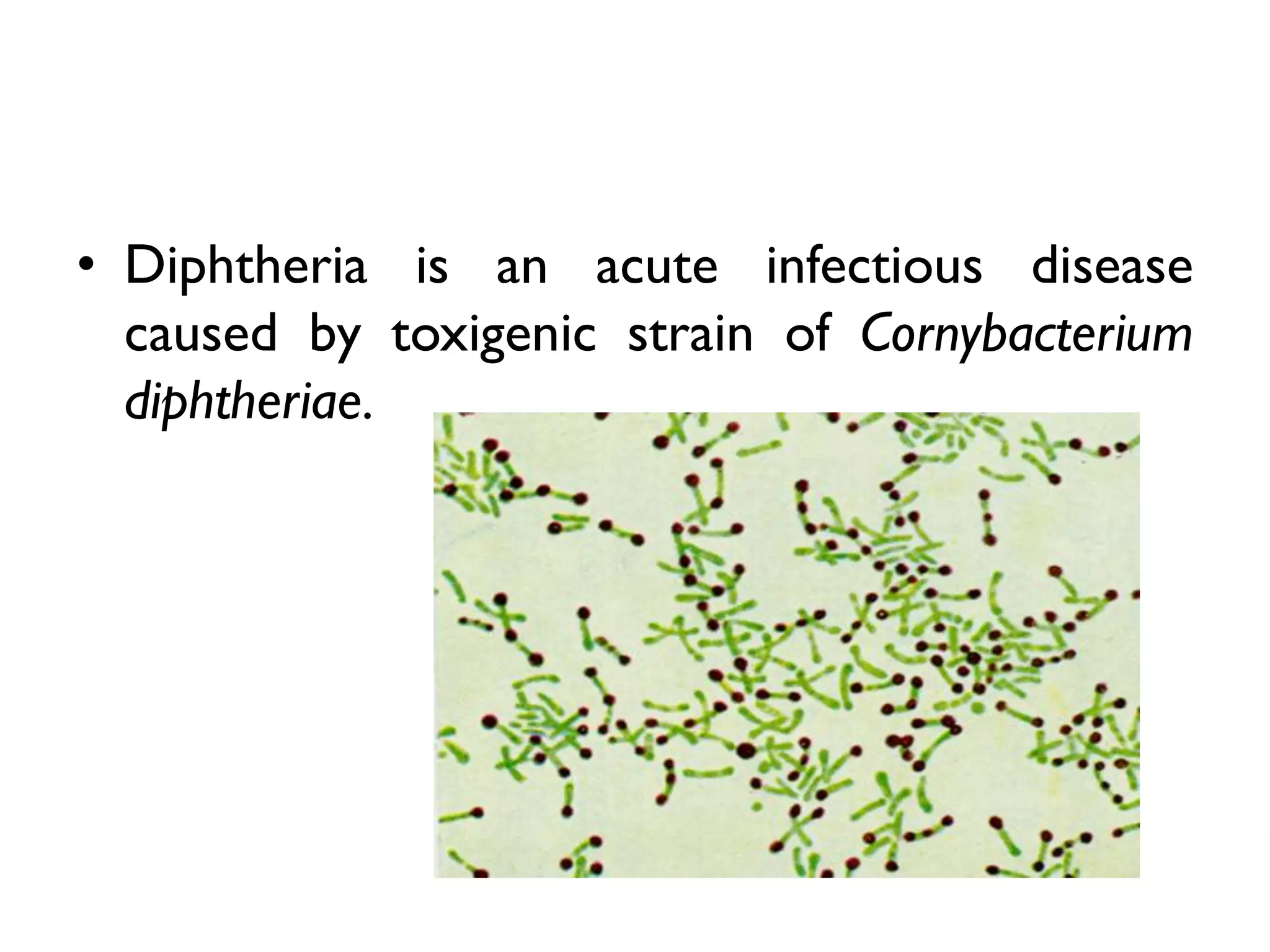
• Diphtheria is an acute infectious disease
caused by toxigenic strain of Cornybacterium
diphtheriae.
• The bacilli multiply locally, usually in the throat and
elaborate a powerful exotoxin which is responsible
for -
a. The formation of a grayish or yellowish
membrane ( false membrane) commonly cover
the tonsils, pharynx, larynx with well defined edges
and membrane can not be wiped away.
b. Marked congestion, oedema or local tissue
destruction
c. Enlargement of the regional lymph nodes
d. Signs and symptoms of toxaemia.
Epidemiological
determinant
Agent factors
1. Agent -
 Corynebacterium diphtheriae is a gram positive, non-
motile organism.
 It has no invasive power, but produces a powerful
exotoxin.
2. Source of infection
a. Case – subclinical to clinical
b. Carriers – more common source of infection.
95 carriers for 5 subclinical case
• Carriers may be temporary or chronic; Nasal or
throat carriers.
• Nasal carriers are particularly dangerous source of
infection because of frequent shedding of organism
into the environment than throat carriers.
• Temporary carriers state may last for about a
month.
• Chronic carriers may persist for a year or
so, unless the patient is treated.
3. Infective material
• Nasopharangeal secretion
• Discharges from skin lesion
• Contaminated fomites
• Infected dust
4. Period of infectivity
• Vary from 14-28 days from the onset of disease.
• But carriers may remain infective for much
longer periods.
Host factors
1. Age – particularly affects children aged 1-5.
2. Sex – both sexes affected
3. Immunity – Infant born of immune mothers
are relatively immune during first few weeks
or months of life.
Environmental factors
• It occurs in all seasons, although winter month favour
its spread.
• In Kolkata, the highest incidence was reported in
August;in Mumbai in winter months; and Delhi,
during August to October.
Mode of transmission
• Mainly by Droplet infection
• It can be also transmitted directly to
susceptible person from infected cutaneous
lesions.
Portal of entry
a. Respiratory tract – common
b. Non- respiratory route – skin where cuts,
wounds and ulcers, umbilicus in new born.
Incubation period
• 2-6days
• Occasionally longer
Clinical features
• Respiratory tract forms of diphtheria consist
of -
a. Pharyngotonsillar diphtheria
b. Laryngotracheal diphtheria
c. Nasal diphtheria
d. Combination of these
1. Pharyngotonsillar diphtheria
• Sore throat
• Difficulty in swallowing
• Low grade fever
• Examination of throat may shows only mild erythema,
localized exudate, or a pseudo-membrane.
• Membrane may localized or a patch of the posterior
pharynx or tonsil, may cover the entire tonsil, or less
frequently may spread to cover the soft and hard palate and
posterior portion of pharynx.
• In early stage, pseudomembrane may be
whitish and may be wipe off easily.
• The membrane may be extend to become
thick, blue white to gray black and adherent.
• Attempts to remove the membrane results in
bleeding.
• Patient with sever disease may have marked
oedema of the submandibular area and
anterior portion of the neck, along with
lymphadenopathy, giving a characteristic “bull-
necked” appearance
2. Laryngotracheal diphtheria
• Preceded by pharyngotonsillar disease
• Fever, hoarseness and croupy cough at presentation
• If the infection extend into bronchial tree, it is the
most severe form of disease.
• Diphtheria bacilli within the membrane continue to
produce toxin actively.
• Toxin is absorbed and result in distant toxic damage.
• in later stage, difficulty in vision, speech, swallowing
or movement of arms or legs.
• Toxin also produce nerve damage and resulting in
paralysis of the soft palate, eye, muscles or
extremities.
3. Nasal diphtheria
• It is the mildest form of respiratory diphtheria.
• Usually localized to the septum or turbinates
of one side of the nose.
• Occasionally a membrane may extend into the
pharynx.
Cutaneous diphtheria –
• Common in tropical areas.
• It often appears as a secondary infection of a
previous skin abrasion or lesion.
• The presenting lesion, often an ulcer, may be
surrounded by erythema and covered with
membrane.
• Non respiratory mucosal surface i.e.
conjunctivae and genitals may also be sites of
infection.
Control of diphtheria
1. Case and carriers
2. Contacts
3. Community
1. Case and carriers
1. Early detection of case and carriers
2. Isolation of all cases and carriers in hospital for 14 days
or until proved free of infection.
3.Treatment of cases and carriers
A. Cases -
a. Diptheria antitoxin - IM/IV in doses ranging from 20000
to 1,00,000 units or more depending upon the severity
of the case, after preliminary test dose of 0.2ml
Subcutaneously to detect sensitization to horse serum.
b. Penicillin or erythromycin for 5-6days
2. Carriers – treat with Erythromycin for 10 days.
Case Diptheria antitoxin dose
Mild early pharangeal or
laryngeal
20,000-40,000 units
Moderate nasopharangeal 40,000- 60,000 units
Severe, extensive or late
( 3days or more)
80,000-1,00,000 units
2. Contacts
• Where primary immunization or booster dose was
received within previous 2 years, no further action
needed.
• Where primary course or booster dose of diphtheria
toxoid was received more than 2 years before, only a
booster dose of diphtheria toxoid need be given.
• Non-immunized close contacts should receive
prophylactic penicillin or erythromycin and 1000-2000
units of diphtheria antitoxin
• Contacts should be placed under medical surveillance
and examined daily for at least a week.
3. Community
• Active immunization with diphteria
toxoid of all infants as early in life as
possible, as scheduled, with subsequent
booster doses every 10 years thereafter.
Diptheria

Diptheria

  • 1.
    Diphtheria Dr. Shubhangi S.Kshirsagar Assistant professor Department of Swasthavritta &Yoga
  • 2.
    • Diphtheria isan acute infectious disease caused by toxigenic strain of Cornybacterium diphtheriae.
  • 3.
    • The bacillimultiply locally, usually in the throat and elaborate a powerful exotoxin which is responsible for - a. The formation of a grayish or yellowish membrane ( false membrane) commonly cover the tonsils, pharynx, larynx with well defined edges and membrane can not be wiped away. b. Marked congestion, oedema or local tissue destruction c. Enlargement of the regional lymph nodes d. Signs and symptoms of toxaemia.
  • 4.
  • 5.
    Agent factors 1. Agent-  Corynebacterium diphtheriae is a gram positive, non- motile organism.  It has no invasive power, but produces a powerful exotoxin. 2. Source of infection a. Case – subclinical to clinical b. Carriers – more common source of infection. 95 carriers for 5 subclinical case
  • 6.
    • Carriers maybe temporary or chronic; Nasal or throat carriers. • Nasal carriers are particularly dangerous source of infection because of frequent shedding of organism into the environment than throat carriers. • Temporary carriers state may last for about a month. • Chronic carriers may persist for a year or so, unless the patient is treated.
  • 7.
    3. Infective material •Nasopharangeal secretion • Discharges from skin lesion • Contaminated fomites • Infected dust 4. Period of infectivity • Vary from 14-28 days from the onset of disease. • But carriers may remain infective for much longer periods.
  • 8.
    Host factors 1. Age– particularly affects children aged 1-5. 2. Sex – both sexes affected 3. Immunity – Infant born of immune mothers are relatively immune during first few weeks or months of life.
  • 9.
    Environmental factors • Itoccurs in all seasons, although winter month favour its spread. • In Kolkata, the highest incidence was reported in August;in Mumbai in winter months; and Delhi, during August to October.
  • 10.
    Mode of transmission •Mainly by Droplet infection • It can be also transmitted directly to susceptible person from infected cutaneous lesions.
  • 11.
    Portal of entry a.Respiratory tract – common b. Non- respiratory route – skin where cuts, wounds and ulcers, umbilicus in new born.
  • 12.
  • 13.
    Clinical features • Respiratorytract forms of diphtheria consist of - a. Pharyngotonsillar diphtheria b. Laryngotracheal diphtheria c. Nasal diphtheria d. Combination of these
  • 14.
    1. Pharyngotonsillar diphtheria •Sore throat • Difficulty in swallowing • Low grade fever • Examination of throat may shows only mild erythema, localized exudate, or a pseudo-membrane. • Membrane may localized or a patch of the posterior pharynx or tonsil, may cover the entire tonsil, or less frequently may spread to cover the soft and hard palate and posterior portion of pharynx.
  • 15.
    • In earlystage, pseudomembrane may be whitish and may be wipe off easily. • The membrane may be extend to become thick, blue white to gray black and adherent. • Attempts to remove the membrane results in bleeding. • Patient with sever disease may have marked oedema of the submandibular area and anterior portion of the neck, along with lymphadenopathy, giving a characteristic “bull- necked” appearance
  • 18.
    2. Laryngotracheal diphtheria •Preceded by pharyngotonsillar disease • Fever, hoarseness and croupy cough at presentation • If the infection extend into bronchial tree, it is the most severe form of disease. • Diphtheria bacilli within the membrane continue to produce toxin actively. • Toxin is absorbed and result in distant toxic damage. • in later stage, difficulty in vision, speech, swallowing or movement of arms or legs. • Toxin also produce nerve damage and resulting in paralysis of the soft palate, eye, muscles or extremities.
  • 19.
    3. Nasal diphtheria •It is the mildest form of respiratory diphtheria. • Usually localized to the septum or turbinates of one side of the nose. • Occasionally a membrane may extend into the pharynx.
  • 20.
    Cutaneous diphtheria – •Common in tropical areas. • It often appears as a secondary infection of a previous skin abrasion or lesion. • The presenting lesion, often an ulcer, may be surrounded by erythema and covered with membrane.
  • 21.
    • Non respiratorymucosal surface i.e. conjunctivae and genitals may also be sites of infection.
  • 22.
    Control of diphtheria 1.Case and carriers 2. Contacts 3. Community
  • 23.
    1. Case andcarriers 1. Early detection of case and carriers 2. Isolation of all cases and carriers in hospital for 14 days or until proved free of infection. 3.Treatment of cases and carriers A. Cases - a. Diptheria antitoxin - IM/IV in doses ranging from 20000 to 1,00,000 units or more depending upon the severity of the case, after preliminary test dose of 0.2ml Subcutaneously to detect sensitization to horse serum.
  • 24.
    b. Penicillin orerythromycin for 5-6days 2. Carriers – treat with Erythromycin for 10 days. Case Diptheria antitoxin dose Mild early pharangeal or laryngeal 20,000-40,000 units Moderate nasopharangeal 40,000- 60,000 units Severe, extensive or late ( 3days or more) 80,000-1,00,000 units
  • 25.
    2. Contacts • Whereprimary immunization or booster dose was received within previous 2 years, no further action needed. • Where primary course or booster dose of diphtheria toxoid was received more than 2 years before, only a booster dose of diphtheria toxoid need be given. • Non-immunized close contacts should receive prophylactic penicillin or erythromycin and 1000-2000 units of diphtheria antitoxin • Contacts should be placed under medical surveillance and examined daily for at least a week.
  • 26.
    3. Community • Activeimmunization with diphteria toxoid of all infants as early in life as possible, as scheduled, with subsequent booster doses every 10 years thereafter.