
More Related Content
Similar to Digestive-System.pptx
Similar to Digestive-System.pptx (20)
Recently uploaded
Recently uploaded (20)
Digestive-System.pptx
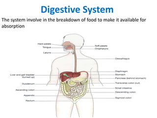
- 1. Digestive System The system involve in the breakdown of food to make it available for absorption
- 2. • The digestive system is the collective name • Describe the alimentary canal, accessory organs and variety of process • Prepare food for absorption • The alimentary canal begins at mouth • Passes through the thorax, abdomen and pelvis • End at anus • The complex of digestive processes gradually breaks down the foods • Chemical substances or enzymes effect these
- 3. Process of Digestion • The activities in the digestive system can be grouped as • Ingestion • This is the process of taking food into the alimentary tract • Propulsion • This moves the contents along the alimentary tract • Digestion consists of • Mechanical breakdown of food by, e.g. mastication (chewing) • Chemical digestion of food by enzymes produced by glands • Absorption • This is the process by which digested food enter into the blood and lymph capillaries • Elimination • Undigested substances are excreted by the bowel as faeces
- 4. ORGANS OF THE DIGESTIVE SYSTEM • Alimentary tract • Long tube through which food passes • Start at mouth and terminates at the anus • Various Part Include • Mouth Pharynx • Oesophagus Stomach • Small intestine Large intestine • Rectum Anal canal • The length of the GI tract is about 5–7 meters
- 5. Accessory organs • Various secretions poured by glands in the lining membrane • Gastric juice by stomach • Glands situated outside their secretions carry by ducts to • Include • 3 Pairs of salivary glands • Pancreas • Liver and the biliary tract • These organs and glands are linked physiologically as well as anatomically
- 6. Basic Structure Of The Alimentary Canal • Alimentary canal follow consistent pattern from the oesophagus onwards • Not apply so obviously to mouth and the pharynx • Modification in structure found for special functions • The walls of the alimentary tract are formed by four layers of tissue • Adventitia or Outer covering • Muscle layer • Submucosal layer • Mucosa(Inner lining)
- 7. • Adventitia (outer covering) • In thorax it consists of loose fibrous tissue • In abdomen it covered to peritoneum • Peritoneum is the largest serous membrane • A closed sac containing a small amount of serous fluid • Physical barrier to local spread of infection • Consist of two layers • Parietal layer: Lines the abdominal wall • Visceral layer: Covers the organs within the abdominal and pelvic cavities
- 8. Muscle layer • Consists of two layers of smooth muscle • The outer layer arranged longitudinally • Inner layer encircle the wall of the tube • Contain blood vessels lymph vessels • Plexus of sympathetic and parasympathetic nerves • Involve in contraction and relaxation • A waves like pattern of contraction Peristalsis • Contraction mixes food with the digestive juices • Onward movement of the content • Form valves preventing backflow
- 9. Muscularis mucosae Lamina propria Epithelium SUBMUCOSA Glands in Submucosa MUSCULARIS: Circular muscle Longitudinal muscle Myenteric plexus Areolar connective tissue
- 10. • Submucosa • Consists of loose connective tissue with some elastic fibers • It contain plexuses of • Blood vessels • Nerve • Arterioles • Venules • Capillaries • Nerve plexus • Sympathetic and parasympathetic
- 11. Mucosa Consists of three layers of tissue: • Mucous membrane The innermost Formed by columnar epithelium Three main functions: Protection, secretion and absorption • Lamina propria Consisting of loose connective tissue Provide supports contain blood vessel • Muscularis mucosa Thin outer layer of smooth muscle
- 12. Mouth • Referred to as oral or buccal cavity • It is a space extends from • Gums and teeth to the fauces (throat) • Formed by the • Cheeks • Hard and soft palates • Tongue • Covered externally by skin • Internally by a mucous membrane • Composed of nonkeratinized stratified squamous epithelium • Buccinator muscles & connective tissue
- 13. • Labia surrounding the opening of the mouth • Orbicularis oris • labial frenulum • The oral vestibule is a space b/w cheeks and lips • Internally by gums and teeth • Palate separates the oral cavity from the nasal cavity • Hard palate form the anterior portion • Soft palate forms the posterior portion
- 14. Salivary Glands • Secrete saliva into the oral cavity • Keep the mucous membranes pharynx moist • Cleanse the mouth and teeth • When food enters the mouth its secretion increases • Lubricates dissolves and begins the chemical breakdown of the food • The mucous membrane of the mouth and tongue contains • many small salivary glands that open directly, or indirectly via • There are three pairs of major salivary glands • Parotid • Submandibular • Sublingual glands
- 15. Composition of Saliva • Chemically, saliva is 99.5% water and 0.5% solutes • Immunoglobulin A, the lysozyme and salivary amylase • Chloride ions in the saliva activate salivary amylase • Bicarbonate and phosphate ions buffer acidic foods • Immunoglobulin A (IgA) prevents attachment of microbes • Enzyme lysozyme kills bacteria
- 16. • Salivation • The secretion of saliva • Controlled by the autonomic nervous system • Amounts of saliva secreted daily 1000–1500 ml • Sympathetic stimulation during stress resulting in dryness of the mouth • Impulses from the taste buds to salivary nuclei in the brain stem • Return to parasympathetic stimulation of facial glossopharyngeal Functions of saliva Chemical digestion of polysaccharides Lubrication of food Cleansing and lubricating Non-specific defence Taste
- 17. Esophagus • The esophagus is a collapsible muscular tube • About 25 cm long • Lies posterior to the trachea • Begins at laryngopharynx reach to stomach • Esophageal hiatus • Hiatus hernia • Having nonkeratinized stratified squamous epithelium
- 18. • Near the stomach the mucosa contains mucous glands • Muscularis of the superior one third skeletal muscle • Middle portion is mixed • Lower one third is smooth muscle Nasopharynx Hard palate Soft palate Uvula Oropharynx Epiglottis Laryngopharynx Larynx Esophagus
- 19. • Forms two sphincters • Upper esophageal sphincter consists of skeletal muscle • Lower esophageal sphincter of Smooth muscle • The esophagus secretes mucus and transports food into stomach • It does not produce digestive enzymes • It does not carry on absorption Lower esophageal sphincter
- 20. Stomach • The stomach is a J-shaped enlargement of the GI tract • Inferior to the diaphragm • The stomach connects the esophagus to the duodenum • Stomach is to serve as a mixing chamber and holding reservoir • Stomach forces a small quantity of material into the first portion of the small intestine • In the stomach, digestion of starch continues, digestion of proteins and triglycerides begins • Semisolid bolus is converted to a liquid • Certain substances are absorbed • The stomach has four main regions • Cardia • Fundus • Body • Pylorus CARDIA FUNDUS BODY PYLORUS
- 21. • The stomach composed of the same basic layers as the rest of the GI tract • Mucosa is a layer of simple columnar epithelial cells • Gastric glands contain three types of exocrine gland cells • Secrete their products into the stomach lumen: • Mucous neck cells • Chief cells • Parietal cell • Mucous neck cells secrete mucus • Parietal cells produce intrinsic factor and hydrochloric acid • Chief cells secrete pepsinogen and gastric lipase
- 22. • Gastric juice is secretions of mucous, parietal, and chief cells • 2000–3000 mL of gastric juice is secreted per day • Gastric glands include G cell, in the pyloric antrum secretes the hormone gastrin into the bloodstream • Muscularis has three layers of smooth muscle • Outer longitudinal layer • Middle circular layer • Inner oblique layer
- 23. Mechanical and Chemical Digestion in the Stomach • Food is converted into chyme • Foods remain in the fundus for a hour without mixed with gastric juice • Strongly acidic fluid kills many microbes in food • Denature protein and its enzymatic digestion of proteins also begins in the stomach • Gastric lipase digest lipid • Small amount of nutrients are absorbed here
- 24. Small Intestine • Continuous with the stomach at the pyloric sphincter • Leads into the large intestine at the ileocaecal valve • About 5 meters long and lies in the abdominal cavity • Comprises of three main sections • Duodenum • About 25 cm long and curves around the head of the pancreas Receive secretions from the gall bladder and pancreas • Jejunum • The middle section • About 2 meters long • Ileum • Terminal section about 3 meters long • Ends at the ileocaecal valve
- 25. • The walls of the small intestine are composed of the four layers • Peritoneum • Mesentery • Double layer of peritoneum • Attaches the jejunum and ileum to the posterior abdominal wall • Mucosa • The surface area of mucosa increased by villi and microvilli • They promote mixing of chyme as it passes along
- 26. Chemical digestion in the small intestine • When chyme passes to small intestine • Mixed with pancreatic juice, bile and intestinal juice, • In the small intestine the digestion of all the nutrients is completed • Carbohydrates are broken down to monosaccharides • Proteins are broken down to amino acids • Fats are broken down to fatty acids and glycerol • Pancreatic juice • Pancreatic juice enters the duodenum and consists of • Water • Mineral salts • Enzymes: • Amylase • Lipase • Inactive enzyme precursors: • Trypsinogen • Chymotrypsinogen • Procarboxypeptidase • Pancreatic juice is alkaline (pH 8)
- 27. LARGE INTESTINE (COLON) • Terminal portion of the GI tract • Overall functions of the large intestine • Completion of absorption • Production of certain vitamins • Formation of feces • Expulsion of feces from the body
- 28. • It is about 1.5 m (5 ft) long and 6.5 cm (2.5 in) in diameter • Extends from the ileum to the anus • Four major regions of • Cecum • Colon • Rectum • Anal canal • Opening from the ileum guarded by ileocaecal sphincter • Allows materials from the small intestine to large intestine
- 29. • Cecum • Inferior to the ileocaecal valve • Small pouch about 6 cm (2.4 in.) long • Appendix attach to it • Colon • Long tube in which the cecum merge • Divided into • Ascending • Transverse • Descendin
- 30. • Ascending colon • Ascends on the right side of the abdomen • Reaches the inferior surface of the liver • Transverse colon • Continuation of the colon across the abdomen to the left side as • Curves beneath the inferior end of the spleen • Descending colon • Inferiorly to the level of the iliac crest • The sigmoid colon • Near the left iliac crest, • Projects medially to the midline and terminates as the rectum Ileocaecal sphincter Cecum Appendix Ascending colon Descending colon Transerve colon Sigmoid colon
- 31. • Rectum • The last portion • 20 cm (8 in.) of the GI tract • Anterior to the sacrum and coccyx • Anal canal • The terminal 2–3 cm (1 in.) of the rectum • The mucous membrane arranged in longitudinal folds called anal columns • Anus • Opening of the anal canal to the exterior • Guarded by an internal anal sphincter of smooth muscle and an external anal sphincter of skeletal muscle
- 32. Functions of the large intestine, rectum and anal canal • Absorption • Absorption of water continues • Absorption of mineral salts, vitamins and some drugs • Microbial activity • Heavily colonized by certain types of bacteria • Synthesize vitamin K and folic acid • Mass movement • Does not exhibit peristaltic movement • A strong wave along the transerve colon forcing content to descending and sigmoid colon known as mass movement • Often precipitated by the entry of food into the stomach • This combination of stimulus and response is called the gastrocolic reflex
- 33. Defaecation • When contents of the sigmoid colon enter into the rectum • Stimulation of nerve endings in its walls by stretch • Defaecation occurs by reflex • Pudendal nerve to the external anal sphincter • is under conscious control • Defaecation involves • Involuntary contraction of the muscle of the rectum • Relaxation of the internal anal sphincter • Contraction of the abdominal muscles • Lowering of the diaphragm increase intra-abdominal pressure