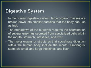
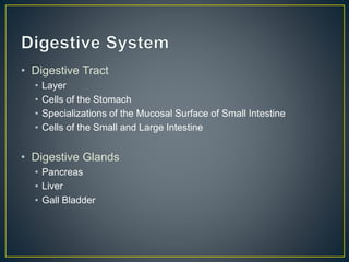
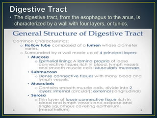
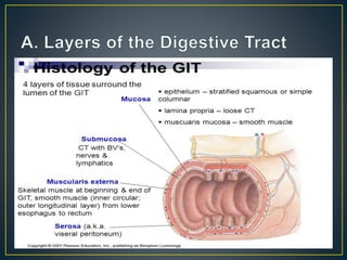
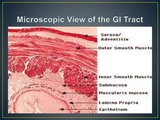
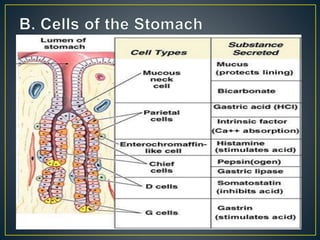
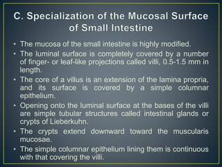
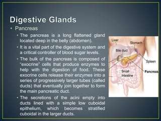
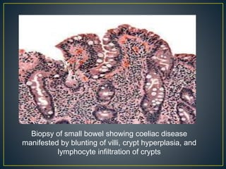
By: Yasmeen Cabrera
• In the human digestive system, large organic masses are
broken down into smaller particles that the body can use
as fuel.
• The breakdown of the nutrients requires the coordination
of several enzymes secreted from specialized cells within
the mouth, stomach, intestines, and liver.
• The major organs or structures that coordinate digestion
within the human body include the mouth, esophagus,
stomach, small and large intestines, and liver.
• Digestive Tract
• Layer
• Cells of the Stomach
• Specializations of the Mucosal Surface of Small Intestine
• Cells of the Small and Large Intestine
• Digestive Glands
• Pancreas
• Liver
• Gall Bladder
• The digestive tract, from the esophagus to the anus, is
characterized by a wall with four layers, or tunics.
• The mucosa of the small intestine is highly modified.
• The luminal surface is completely covered by a number
of finger- or leaf-like projections called villi, 0.5-1.5 mm in
length.
• The core of a villus is an extension of the lamina propria,
and its surface is covered by a simple columnar
epithelium.
• Opening onto the luminal surface at the bases of the villi
are simple tubular structures called intestinal glands or
crypts of Lieberkuhn.
• The crypts extend downward toward the muscularis
mucosae.
• The simple columnar epithelium lining them is continuous
with that covering the villi.
• Mucosal folds: The inner surface of the small intestine is
not flat, but thrown into circular folds. This not only
increases the surface area, but helps regulate the flow of
digested food through your intestine.
• Villi: The folds form numerous tiny projections which stick
out into the open space inside your small intestine (or
lumen), and are covered with cells that help absorb
nutrients from the food that passes through.
• Microvilli: The cells on the villi are packed full of tiny
hairlike structures called microvilli. This helps increase
the surface of each individual cell, meaning that 5.
Mucosal Folds Villi
Microvilli
• Small Intestine
• The epithelium of the villi is made up of tall
columnar absorptive cells called enterocytes,
and goblet cells, which secrete mucin, for
lubrication of the intestinal contents, and
protection of the epithelium.
• Enterocytes or absorptive cells - tall
columnar cells with microvilli and a basal
nucleus, specialized for the transport of
substances.
• Goblet cells- These mucus-secreting cells
• Paneth cells (at the base of the crypts) - they have a
defensive function, and stain intensely eosinophilic,
due to secretory granules of antimicrobial peptides
called defensins, as well as lysozyme and
phospholipase A. These cells last for several weeks.
• Endocrine cells, (also eosinophilic) which produce
secretin, somatostatin, enteroglucagon and
serotonin. One type of endocrine cell for each type
of hormone.
• Stem cells, found at the base of the crypts, which
divide continuously to replace enterocytes (every 2-3
days), goblet cells, paneth cells and neuroendocrine
cells.
• Intraepithelial lymphocytes (mostly T-cells).
• Large Intestine
• Goblet cells
• are scattered among the absorptive cells in the
epithelium of the small intestine and colon.
• These epithelial cells are specialized for
secretion of mucus, which facilitates passage of
material through the bowel.
• Enterocytes
• are the predominant epithelial cell type lining the
lumen of the small intestine and colon.
• These cells are specialized for absorption of
nutrients across the apical plasma membrane
and export of these same nutrients across the
basal plasma membrane.
• Chief Cells
• secret pepsinogen
• Stem Cell
• Stem cells of the intestinal mucosa line the walls
of the crypts and continually replenish the
intestinal epithelium
• completely replacing all the absorptive cells and
goblet cells approximate once every four days.
• These cells are inconspicuous when resting, but
mitotic figures (intensely basophilic condensed
chromatin) are common and easily noticed in the
crypts of the small intestine.
Microscopic View of Large Intestinal
Cells
• Pancreas
• The pancreas is a long flattened gland
located deep in the belly (abdomen).
• It is a vital part of the digestive system and
a critical controller of blood sugar levels.
• The bulk of the pancreas is composed of
“exocrine” cells that produce enzymes to
help with the digestion of food. These
exocrine cells release their enzymes into a
series of progressively larger tubes (called
ducts) that eventually join together to form
the main pancreatic duct.
• The secretions of the acini empty into
ducts lined with a simple low cuboidal
epithelium, which becomes stratified
cuboidal in the larger ducts.
• Liver
• The liver has a thin capsule of dense
connective tissue, and a visceral (inferior)
layer of peritoneal mesothelium, and is
divided into left and right lobes.
• It is a major metabolic organ, and is
important for degrading alcohol and
drugs.
• It stores glycogen, secretes glucose,
plasma proteins and lipoproteins into the
blood, and secretes bilirubin (by-produce
of haemoglobin), secretory IgA, and bile
salts (which emulsify fats) as components
of bile (endocrine secretion of the liver).
• Gall Bladder
• The gall bladder is a
simple muscular sac,
lined by a simple
columnar epithelium.
• It receives and stores
bile from the liver via the
hepatic and then cystic
duct, and can store
about 50 to 100ml in
humans.
• Celiac Disease
• is an autoimmune disorder of the small intestine that
occurs in genetically predisposed people of all ages
from middle infancy onward. Symptoms include pain and
discomfort in the digestive tract, chronic constipation
and diarrhoea, failure to thrive (in children), anaemia
and fatigue, but these may be absent, and symptoms in
other organ systems have been described. Vitamin
deficiencies are often noted in people with coeliac
disease owing to the reduced ability of the small
intestine to properly absorb nutrients from food.
Biopsy of small bowel showing coeliac disease
manifested by blunting of villi, crypt hyperplasia, and
lymphocyte infiltration of crypts
• Inflammatory Bowel Disease
• Inflammatory bowel disease (IBD) occurs when the small or large
intestine (bowel) becomes inflamed. When the intestine is inflamed, it
becomes swollen and painful. This causes symptoms such as
diarrhea, bloating, stomach cramps and, sometimes, weight loss.
• There are two main types of IBD: Crohn’s disease and ulcerative
colitis.
• Crohn’s​ disease​ can affect any part of the GI tract​. It causes the
walls of the affected part to thicken and develop a cobblestone-like
surface. Depending on where Crohn’s disease occurs, symptoms can
include stomach pain, vomiting, loss of appetite, bloody diarrhea, poor
growth and mouth ulcers.
• Ulcerative colitis is similar to Crohn’s disease, but it affects only the
colon (large intestine). When a person has ulcerative colitis, the lining
of their colon develops ulcers, or tears. This can lead to stomach
pain, bloody diarrhea, bleeding from tears around the anus, weight
loss and decreased appetite.
• Irritable Bowel Syndrome
• is a chronic (long-term) condition that affects the large and small
intestine. In IBS, food moves through the colon too slowly, too
quickly or unpredictably. As a result, the colon does not absorb
the right amount of water from food. This can lead to diarrhea or
constipation.

Digestive tract (Histology)

  • 1.
  • 2.
    • In thehuman digestive system, large organic masses are broken down into smaller particles that the body can use as fuel. • The breakdown of the nutrients requires the coordination of several enzymes secreted from specialized cells within the mouth, stomach, intestines, and liver. • The major organs or structures that coordinate digestion within the human body include the mouth, esophagus, stomach, small and large intestines, and liver.
  • 3.
    • Digestive Tract •Layer • Cells of the Stomach • Specializations of the Mucosal Surface of Small Intestine • Cells of the Small and Large Intestine • Digestive Glands • Pancreas • Liver • Gall Bladder
  • 4.
    • The digestivetract, from the esophagus to the anus, is characterized by a wall with four layers, or tunics.
  • 8.
    • The mucosaof the small intestine is highly modified. • The luminal surface is completely covered by a number of finger- or leaf-like projections called villi, 0.5-1.5 mm in length. • The core of a villus is an extension of the lamina propria, and its surface is covered by a simple columnar epithelium. • Opening onto the luminal surface at the bases of the villi are simple tubular structures called intestinal glands or crypts of Lieberkuhn. • The crypts extend downward toward the muscularis mucosae. • The simple columnar epithelium lining them is continuous with that covering the villi.
  • 9.
    • Mucosal folds:The inner surface of the small intestine is not flat, but thrown into circular folds. This not only increases the surface area, but helps regulate the flow of digested food through your intestine. • Villi: The folds form numerous tiny projections which stick out into the open space inside your small intestine (or lumen), and are covered with cells that help absorb nutrients from the food that passes through. • Microvilli: The cells on the villi are packed full of tiny hairlike structures called microvilli. This helps increase the surface of each individual cell, meaning that 5.
  • 10.
  • 11.
    • Small Intestine •The epithelium of the villi is made up of tall columnar absorptive cells called enterocytes, and goblet cells, which secrete mucin, for lubrication of the intestinal contents, and protection of the epithelium. • Enterocytes or absorptive cells - tall columnar cells with microvilli and a basal nucleus, specialized for the transport of substances. • Goblet cells- These mucus-secreting cells
  • 12.
    • Paneth cells(at the base of the crypts) - they have a defensive function, and stain intensely eosinophilic, due to secretory granules of antimicrobial peptides called defensins, as well as lysozyme and phospholipase A. These cells last for several weeks. • Endocrine cells, (also eosinophilic) which produce secretin, somatostatin, enteroglucagon and serotonin. One type of endocrine cell for each type of hormone. • Stem cells, found at the base of the crypts, which divide continuously to replace enterocytes (every 2-3 days), goblet cells, paneth cells and neuroendocrine cells. • Intraepithelial lymphocytes (mostly T-cells).
  • 14.
    • Large Intestine •Goblet cells • are scattered among the absorptive cells in the epithelium of the small intestine and colon. • These epithelial cells are specialized for secretion of mucus, which facilitates passage of material through the bowel. • Enterocytes • are the predominant epithelial cell type lining the lumen of the small intestine and colon. • These cells are specialized for absorption of nutrients across the apical plasma membrane and export of these same nutrients across the basal plasma membrane.
  • 15.
    • Chief Cells •secret pepsinogen • Stem Cell • Stem cells of the intestinal mucosa line the walls of the crypts and continually replenish the intestinal epithelium • completely replacing all the absorptive cells and goblet cells approximate once every four days. • These cells are inconspicuous when resting, but mitotic figures (intensely basophilic condensed chromatin) are common and easily noticed in the crypts of the small intestine.
  • 16.
    Microscopic View ofLarge Intestinal Cells
  • 17.
    • Pancreas • Thepancreas is a long flattened gland located deep in the belly (abdomen). • It is a vital part of the digestive system and a critical controller of blood sugar levels. • The bulk of the pancreas is composed of “exocrine” cells that produce enzymes to help with the digestion of food. These exocrine cells release their enzymes into a series of progressively larger tubes (called ducts) that eventually join together to form the main pancreatic duct. • The secretions of the acini empty into ducts lined with a simple low cuboidal epithelium, which becomes stratified cuboidal in the larger ducts.
  • 18.
    • Liver • Theliver has a thin capsule of dense connective tissue, and a visceral (inferior) layer of peritoneal mesothelium, and is divided into left and right lobes. • It is a major metabolic organ, and is important for degrading alcohol and drugs. • It stores glycogen, secretes glucose, plasma proteins and lipoproteins into the blood, and secretes bilirubin (by-produce of haemoglobin), secretory IgA, and bile salts (which emulsify fats) as components of bile (endocrine secretion of the liver).
  • 19.
    • Gall Bladder •The gall bladder is a simple muscular sac, lined by a simple columnar epithelium. • It receives and stores bile from the liver via the hepatic and then cystic duct, and can store about 50 to 100ml in humans.
  • 20.
    • Celiac Disease •is an autoimmune disorder of the small intestine that occurs in genetically predisposed people of all ages from middle infancy onward. Symptoms include pain and discomfort in the digestive tract, chronic constipation and diarrhoea, failure to thrive (in children), anaemia and fatigue, but these may be absent, and symptoms in other organ systems have been described. Vitamin deficiencies are often noted in people with coeliac disease owing to the reduced ability of the small intestine to properly absorb nutrients from food.
  • 21.
    Biopsy of smallbowel showing coeliac disease manifested by blunting of villi, crypt hyperplasia, and lymphocyte infiltration of crypts
  • 22.
    • Inflammatory BowelDisease • Inflammatory bowel disease (IBD) occurs when the small or large intestine (bowel) becomes inflamed. When the intestine is inflamed, it becomes swollen and painful. This causes symptoms such as diarrhea, bloating, stomach cramps and, sometimes, weight loss. • There are two main types of IBD: Crohn’s disease and ulcerative colitis. • Crohn’s​ disease​ can affect any part of the GI tract​. It causes the walls of the affected part to thicken and develop a cobblestone-like surface. Depending on where Crohn’s disease occurs, symptoms can include stomach pain, vomiting, loss of appetite, bloody diarrhea, poor growth and mouth ulcers. • Ulcerative colitis is similar to Crohn’s disease, but it affects only the colon (large intestine). When a person has ulcerative colitis, the lining of their colon develops ulcers, or tears. This can lead to stomach pain, bloody diarrhea, bleeding from tears around the anus, weight loss and decreased appetite.
  • 25.
    • Irritable BowelSyndrome • is a chronic (long-term) condition that affects the large and small intestine. In IBS, food moves through the colon too slowly, too quickly or unpredictably. As a result, the colon does not absorb the right amount of water from food. This can lead to diarrhea or constipation.