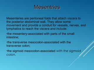
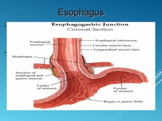
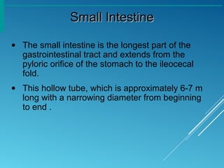
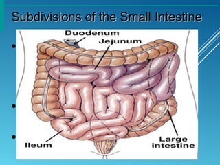
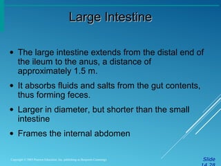
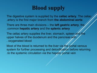
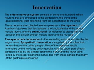
Anatomy Digestive systemAnatomy Digestive system
Organs of the Digestive SystemOrgans of the Digestive System
• Two main groups
• Alimentary canal or gastrointestinal tract –
continuous coiled hollow tube from mouth to
anus(5-7 meter)
• Accessory digestive organs: teeth ,tongue
,salivary gland ,liver ,gallbladder ,and
pancreas
Organs of the Digestive SystemOrgans of the Digestive System
Organs of the Alimentary CanalOrgans of the Alimentary Canal
• Mouth
• Pharynx
• Esophagus
• Stomach
• Small intestine
• Large intestine
• Anus
Layers of Alimentary Canal OrgansLayers of Alimentary Canal Organs
• Mucosa
• Innermost layer
• Moist membrane
1. Surface epithelium : secretion and
absorbtion,renew every 5-7 days also
contain enteroendocrine cells
2. Small amount of connective tissue
(lamina propria): contain blood and
lymphatic vessele also contain MALT
3. Small smooth muscle layer
Layers of Alimentary Canal OrgansLayers of Alimentary Canal Organs
• Submucosa
•Just beneath the mucosa
•Soft connective tissue with blood vessels,
nerve endings, and lymphatics also contain
submucosal plexus
Layers of Alimentary Canal OrgansLayers of Alimentary Canal Organs
• Muscularis externa – smooth muscle
1. Inner circular layer
2. Outer longitudinal layer
Between them is myenteric plexus
• Serosa
• Outermost layer – visceral peritoneum
• Layer of serous fluid-producing cells
(mesothelium)
Layers of Alimentary Canal OrgansLayers of Alimentary Canal Organs
• A thin membrane (the peritoneum) lines the walls of the
abdominal cavity and covers much of the viscera.
• The parietal peritoneum lines the walls of the cavity
and the visceral peritoneum covers the viscera.
• Between the parietal and visceral layers of peritoneum
is a potential space (the peritoneal cavity).
• The peritoneal cavity is subdivided further into
The greater sac
The omental bursa .
PeritoneumPeritoneum
• The omenta consist of two layers of
peritoneum, which pass from the
stomach and the first part of the
duodenum to other viscera. There are
two:
The greater omentum
The lesser omentum
OmentumOmentum
OmentumOmentum
Mesenteries are peritoneal folds that attach viscera to
the posterior abdominal wall. They allow some
movement and provide a conduit for vessels, nerves, and
lymphatics to reach the viscera and include:
•the mesentery-associated with parts of the small
intestine;
•the transverse mesocolon-associated with the
transverse colon;
•the sigmoid mesocolon-associated with the sigmoid
colon.
MesentriesMesentries
EsophagusEsophagus
• The esophagus is a muscular, collapsible tube
about 25 cm long that joins the pharynx to the
stomach.
• The esophagus enters the abdomen through an
opening in the right crus of the diaphragm
• Passageway for food only (respiratory system
branches off after the pharynx)
• Conducts food by peristalsis
• The upper third is skeletal muscle (voluntary),
middle third is mixed, and lower third is smooth
muscle (involuntary).
• esophagogastric junction is located
approximately at the level of the diaphragm.
Contractions of the diaphragm create sphincter-like
effects, preventing reflux of stomach acids and
content. The esophagogastric junction is a
functional, not anatomical, sphincter.
EsophagusEsophagus
Stomach AnatomyStomach Anatomy
• Located on the left side of the abdominal
cavity
• Food enters at the cardioesophageal
sphincter
• It is roughly J-shaped and has two openings,
the cardiac and pyloric
Stomach AnatomyStomach Anatomy
• Regions of the stomach
•Cardiac region – near the heart
•Fundus
•Body
•Phylorus – funnel shaped terminal end
Stomach AnatomyStomach Anatomy
• Other features of the stomach include:
• The greater curvature
• The lesser curvature
• The cardial notch
• The angular incisure
Small IntestineSmall Intestine
• The small intestine is the longest part of the
gastrointestinal tract and extends from the
pyloric orifice of the stomach to the ileocecal
fold.
• This hollow tube, which is approximately 6-7 m
long with a narrowing diameter from beginning
to end .
Subdivisions of the Small IntestineSubdivisions of the Small Intestine
• Duodenum(25cm)
•Attached to the stomach
•Curves around the head of the pancreas
•Fixed retroperitoneal structure
• Jejunum (2.5m)
•Attaches anteriorly to the duodenum
• Ileum (3.5m)
•Extends from jejunum to large intestine
Large IntestineLarge Intestine
SlideCopyright © 2003 Pearson Education, Inc. publishing as Benjamin Cummings
• The large intestine extends from the distal end of
the ileum to the anus, a distance of
approximately 1.5 m.
• It absorbs fluids and salts from the gut contents,
thus forming feces.
• Larger in diameter, but shorter than the small
intestine
• Frames the internal abdomen
Structures of the Large IntestineStructures of the Large Intestine
SlideCopyright © 2003 Pearson Education, Inc. publishing as Benjamin Cummings
• Cecum
• Colon
•Ascending
•Transverse
•Descending
•S-shaped sigmoidal
• Rectum
• Anus – external body opening
The digestive system is supplied by the celiac artery. The celiac
artery is the first major branch from the abdominal aorta.
There are three main divisions – the left gastric artery, the
common hepatic artery and the splenic artery.
The celiac artery supplies the liver, stomach, spleen and the
upper halves of the duodenum and the pancreas with
oxygenated blood.
Most of the blood is returned to the liver via the portal venous
system for further processing and detoxification before returning
to the systemic circulation via the hepatic portal vein.
Blood supply
Innervation
The enteric nervous system consists of some one hundred million
neurons that are embedded in the peritoneum, the lining of the
gastrointestinal tract extending from the oesophagus to the anus.
These neurons are collected into two plexuses - the myenteric (or
Auerbach's) plexus that lies between the longitudinal and the smooth
muscle layers, and the submucosal (or Meissner's) plexus that lies
between the circular smooth muscle layer and the mucosa.
Parasympathetic innervation to the ascending colon is supplied by the
vagus nerve. Sympathetic innervation is supplied by the splanchnic
nerves that join the celiac ganglia. Most of the digestive tract is
innervated by the two large celiac ganglia, with the upper part of each
ganglion joined by the greater splanchnic nerve and the lower parts
joined by the lesser splanchnic nerve. It is from these ganglia that many
of the gastric plexuses arise.

Digestion anatomy

  • 1.
  • 2.
    Organs of theDigestive SystemOrgans of the Digestive System • Two main groups • Alimentary canal or gastrointestinal tract – continuous coiled hollow tube from mouth to anus(5-7 meter) • Accessory digestive organs: teeth ,tongue ,salivary gland ,liver ,gallbladder ,and pancreas
  • 3.
    Organs of theDigestive SystemOrgans of the Digestive System
  • 4.
    Organs of theAlimentary CanalOrgans of the Alimentary Canal • Mouth • Pharynx • Esophagus • Stomach • Small intestine • Large intestine • Anus
  • 5.
    Layers of AlimentaryCanal OrgansLayers of Alimentary Canal Organs • Mucosa • Innermost layer • Moist membrane 1. Surface epithelium : secretion and absorbtion,renew every 5-7 days also contain enteroendocrine cells 2. Small amount of connective tissue (lamina propria): contain blood and lymphatic vessele also contain MALT 3. Small smooth muscle layer
  • 6.
    Layers of AlimentaryCanal OrgansLayers of Alimentary Canal Organs • Submucosa •Just beneath the mucosa •Soft connective tissue with blood vessels, nerve endings, and lymphatics also contain submucosal plexus
  • 7.
    Layers of AlimentaryCanal OrgansLayers of Alimentary Canal Organs • Muscularis externa – smooth muscle 1. Inner circular layer 2. Outer longitudinal layer Between them is myenteric plexus • Serosa • Outermost layer – visceral peritoneum • Layer of serous fluid-producing cells (mesothelium)
  • 8.
    Layers of AlimentaryCanal OrgansLayers of Alimentary Canal Organs
  • 9.
    • A thinmembrane (the peritoneum) lines the walls of the abdominal cavity and covers much of the viscera. • The parietal peritoneum lines the walls of the cavity and the visceral peritoneum covers the viscera. • Between the parietal and visceral layers of peritoneum is a potential space (the peritoneal cavity). • The peritoneal cavity is subdivided further into The greater sac The omental bursa . PeritoneumPeritoneum
  • 10.
  • 11.
    • The omentaconsist of two layers of peritoneum, which pass from the stomach and the first part of the duodenum to other viscera. There are two: The greater omentum The lesser omentum OmentumOmentum
  • 12.
  • 13.
    Mesenteries are peritonealfolds that attach viscera to the posterior abdominal wall. They allow some movement and provide a conduit for vessels, nerves, and lymphatics to reach the viscera and include: •the mesentery-associated with parts of the small intestine; •the transverse mesocolon-associated with the transverse colon; •the sigmoid mesocolon-associated with the sigmoid colon. MesentriesMesentries
  • 14.
  • 15.
    EsophagusEsophagus • The esophagusis a muscular, collapsible tube about 25 cm long that joins the pharynx to the stomach. • The esophagus enters the abdomen through an opening in the right crus of the diaphragm • Passageway for food only (respiratory system branches off after the pharynx) • Conducts food by peristalsis
  • 16.
    • The upperthird is skeletal muscle (voluntary), middle third is mixed, and lower third is smooth muscle (involuntary). • esophagogastric junction is located approximately at the level of the diaphragm. Contractions of the diaphragm create sphincter-like effects, preventing reflux of stomach acids and content. The esophagogastric junction is a functional, not anatomical, sphincter. EsophagusEsophagus
  • 17.
    Stomach AnatomyStomach Anatomy •Located on the left side of the abdominal cavity • Food enters at the cardioesophageal sphincter • It is roughly J-shaped and has two openings, the cardiac and pyloric
  • 18.
    Stomach AnatomyStomach Anatomy •Regions of the stomach •Cardiac region – near the heart •Fundus •Body •Phylorus – funnel shaped terminal end
  • 19.
    Stomach AnatomyStomach Anatomy •Other features of the stomach include: • The greater curvature • The lesser curvature • The cardial notch • The angular incisure
  • 20.
    Small IntestineSmall Intestine •The small intestine is the longest part of the gastrointestinal tract and extends from the pyloric orifice of the stomach to the ileocecal fold. • This hollow tube, which is approximately 6-7 m long with a narrowing diameter from beginning to end .
  • 21.
    Subdivisions of theSmall IntestineSubdivisions of the Small Intestine • Duodenum(25cm) •Attached to the stomach •Curves around the head of the pancreas •Fixed retroperitoneal structure • Jejunum (2.5m) •Attaches anteriorly to the duodenum • Ileum (3.5m) •Extends from jejunum to large intestine
  • 22.
    Large IntestineLarge Intestine SlideCopyright© 2003 Pearson Education, Inc. publishing as Benjamin Cummings • The large intestine extends from the distal end of the ileum to the anus, a distance of approximately 1.5 m. • It absorbs fluids and salts from the gut contents, thus forming feces. • Larger in diameter, but shorter than the small intestine • Frames the internal abdomen
  • 23.
    Structures of theLarge IntestineStructures of the Large Intestine SlideCopyright © 2003 Pearson Education, Inc. publishing as Benjamin Cummings • Cecum • Colon •Ascending •Transverse •Descending •S-shaped sigmoidal • Rectum • Anus – external body opening
  • 24.
    The digestive systemis supplied by the celiac artery. The celiac artery is the first major branch from the abdominal aorta. There are three main divisions – the left gastric artery, the common hepatic artery and the splenic artery. The celiac artery supplies the liver, stomach, spleen and the upper halves of the duodenum and the pancreas with oxygenated blood. Most of the blood is returned to the liver via the portal venous system for further processing and detoxification before returning to the systemic circulation via the hepatic portal vein. Blood supply
  • 25.
    Innervation The enteric nervoussystem consists of some one hundred million neurons that are embedded in the peritoneum, the lining of the gastrointestinal tract extending from the oesophagus to the anus. These neurons are collected into two plexuses - the myenteric (or Auerbach's) plexus that lies between the longitudinal and the smooth muscle layers, and the submucosal (or Meissner's) plexus that lies between the circular smooth muscle layer and the mucosa. Parasympathetic innervation to the ascending colon is supplied by the vagus nerve. Sympathetic innervation is supplied by the splanchnic nerves that join the celiac ganglia. Most of the digestive tract is innervated by the two large celiac ganglia, with the upper part of each ganglion joined by the greater splanchnic nerve and the lower parts joined by the lesser splanchnic nerve. It is from these ganglia that many of the gastric plexuses arise.